V
Rev Bras Cir Cardiovasc | Braz J Cardiovasc Surg
Editorial
Screening of fetal congenital heart disease: the
challenge continues
Luciane Alves da Rocha
1,
MD; Edward Araujo Júnior
1, MD, PhD; Luciano Marcondes Machado
Nardozza
1, MD, PhD; Antonio Fernandes Moron
1, MD, PhD
DOI: 10.5935/1678-9741.20130048
Congenital heart disease (CHD) is the most common congenital malformation [1] in fetuses. It affects eight per 1,000 live births and is more common antenatally [2-5]. In beginning, cardiac evaluation was conined to pregnancies at increased risk of CHD, such as those with a family history of CHD or where extracardiac malformations had been detected. However, up to 86% of CHD occurs in pregnancies where there are no known high risk features [6], emphasizing the need for an effective fetal cardiac screening program for all pregnancies [7,8]. For this reason, in the mid 80’s started the idea of teaching the obstetrician to assess the heart in a simpliied form during routine obstetric scanning [9,10].
Four chamber view scanning became an integral part of the fetal anatomical survey in many countries by the end of the 1980s [9,10]. However, prenatal screening based on visualization of the four-chamber view has much lower sensitivity [6,11]. This is partly because the four-chamber view may appear normal in cases of many anomalies, such as transposition of the great vessels, tetralogy of Fallot, double outlet right ventricle, truncus arteriosus, pulmonary or aortic stenosis/atresia and coarctation of the aorta. Anomalies of the great vessels are associated with an abnormal four-chamber view in 30% of cases [12].
When four-chamber and great vessels view are examined, the sensitivity of ultrasound screening for congenital heart defects increases from approximately 30% to 69–83% [6,11,13]. Therefore, we support the idea of evaluation both the four-chamber view and the outlow tracts (Figure 1). Then, we could improve the rate of prenatal detection of congenital heart disease.
In 2006, the International Society of Ultrasound in Obstetrics and Gynecology (ISUOG) published a guideline in which they described the “basic” and “extended basic” cardiac ultrasound examinations [14]. The intention was to standardize the assessment and to maximize the detection of heart anomalies during the second-trimester scan (Figure 1). However, we agree that a comprehensive fetal echocardiography should be performed when heart anomalies are suspected. One of the problems to follow this guideline is the dificulty of obtaining images of the outlow tracts.
This happens because unlike the four-chamber view, the aorta and pulmonary artery do not lie in a single axis. In
1 Federal University of São Paulo (UNIFESP), Department of Obstetrics, São Paulo, SP, Brazil. E-mail: [email protected]
Fig. 1 - Images for evaluating the fetal cardiac screening by two-dimensional ultrasound. A: four-chamber view of the fetal heart. B: left ventricular outlow tract (LVOT). C: right ventricular outlow tract (RVOT). D: three-vessel view of the fetal heart. LV: left ventricle. RV: right ventricle. LA: left atrium. RA: right atrium. P: pulmonary artery. Ao: aorta. SVC: superior vena cava
VI
Rev Bras Cir Cardiovasc | Braz J Cardiovasc Surg
compliance to DeVore study [15], we state that it is necessary experienced hands to view the outlow tract. Accordingly, it is important additional training.
The development of new technologies is gaining strength as they; theoretically, facilitate the evaluation of the fetal heart exam including the outlow tracts. Four-dimensional ultrasonography (4DUS) with spatio-temporal image correlation (STIC) technology allows the acquisition of a volume dataset from the fetal heart, and displays a cineloop of a complete single cardiac cycle in motion. Moreover, STIC offers the advantage of ofline assessment of cardiac structures, connections, and functions [16-18] (Figure 2).
REFERENCES
1. Lee K, Khoshnood B, Chen L, Wall SN, Cromie WJ, Mittendorf RL. Infant mortality from congenital malformations in the United States, 1970-1997. Obstet Gynecol. 2001;98(4):620-7.
2. Hoffman JI, Christianson R. Congenital heart disease in a cohort of 19,502 births with long-term follow-up. Am J Cardiol. 1978;42(4):641-7.
3. Mitchell SC, Korones SB, Berendes HW. Congenital heart disease in 56,109 births. Incidence and natural history. Circulation. 1971;43(3):323-32.
4. Hoffman JI. Incidence of congenital heart disease: II. Prenatal incidence. Pediatr Cardiol. 1995;16(4):155-65.
5. Gerlis LM. Cardiac malformations in spontaneous abortions. Int J Cardiol. 1985;7(1):29-46.
6. Achiron R, Glaser J, Gelernter I, Hegesh J, Yagel S. Extended fetal echocardiographic examination for detecting cardiac malformations in low risk pregnancies. BMJ. 1992;304(6828):671-4.
7. Robinson JN, Simpson LL, Abuhamad AZ. Screening for fetal heart disease with ultrasound. Clin Obstet Gynecol. 2003;46(4):890-6.
8. Smythe JF, Copel JA, Kleinman CS. Outcome of prenatally detected cardiac malformations. Am J Cardiol. 1992;69(17):1471-4.
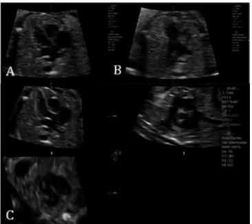
Fig. 2 - Images for evaluating the fetal cardiac screening by four-dimensional ultrasonography with spatiotemporal image correlation (STIC) by multiplanar mode. A: four-chamber view of the fetal heart. B: three-vessel view of the fetal heart. C: left ventricular outlow tract (LVOT) and right ventricular outlow tract (RVOT)
The STIC technology has several tools to analyze all cardiac structures: dynamic multiplanar mode, render mode, omni view, M mode, volume analysis, tomographic ultrasound imaging (TUI). These are new technologies that are still in development and in study.
Despite the great efforts made so far, the STIC is still considered an additional method in echocardiography and, rarely, they are available in daily practice. There are many inherent dificulties in obtaining adequate cardiac images in a STIC [16], for example: the fetal position may vary during the exam; fetal movement during the acquisition of the volume; higher maternal body mass indices or oligohydramnios may make images more dificult and acquiring the images of the heart can be time consuming.
However, we believe that the new methods will be very useful for tracking in the future. Technology tends to advance and sensitivity of this new method will continue improving. Thereby, the 3D/4D technology allows for a more comprehensive and practical assessment of CHD in the fetus.
VII
Rev Bras Cir Cardiovasc | Braz J Cardiovasc Surg 9. Allan L. Antenatal diagnosis of heart disease. Heart.
2000;83(3):367.
10. Allan L. Prenatal diagnosis of structural cardiac defects. Am J Med Genet Part C Semin Med Genet. 2007;145C(1):73-6.
11. Xu Y, Hu YL, Ru T, Gu Y, Yang Y, Dai CY. Importance of “Guidelines for performing fetal cardiac scan” in prenatal screening for fetal congenital heart disease. Zhonghua Fu Chan Ke Za Zhi. 2009;44(2):103-7.
12. Paladini D, Rustico M, Todros T, Palmieri S, Gaglioti P, Benettoni A, et al. Conotruncal anomalies in prenatal life. Ultrasound Obstet Gynecol. 1996;8(4):241-6.
13. Carvalho JS, Mavrides E, Shinebourne EA, Campbell S, Thilaganathan B. Improving the effectiveness of routine prenatal screening for major congenital heart defects. Heart. 2002;88(4):387-91.
14. International Society of Ultrasound in Obstetrics & Gynecology.
Cardiac screening examination of the fetus: guidelines for performing the ‘basic’ and ‘extended basic’ cardiac scan. Ultrasound Obstet Gynecol. 2006;27(1):107-13.
15. DeVore GR. The aortic and pulmonary outflow tract screening examination in the human fetus. J Ultrasound Med. 1992;11(7):345-8.
16. DeVore GR, Falkensammer P, Sklansky MS, Platt LD. Spatio-temporal image correlation (STIC): new technology for evaluation of the fetal heart. Ultrasound Obstet Gynecol. 2003;22(4):380-7.
17. Viñals F, Poblete P, Giuliano A. Spatio-temporal image correlation (STIC): a new tool for the prenatal screening of congenital heart defects. Ultrasound Obstet Gynecol. 2003;22(4):388-94.