Positive Selection Drives the Evolution
of
rhino,
a Member of the Heterochromatin
Protein 1 Family in
Drosophila
Danielle Vermaak1, Steven Henikoff1,2, Harmit S. Malik1*
1Fred Hutchinson Cancer Research Center, Seattle, Washington, United States of America,2Howard Hughes Medical Institute, Basic Sciences Division, Fred Hutchinson Cancer Research Center, Seattle, Washington, United States of America
Heterochromatin comprises a significant component of many eukaryotic genomes. In comparison to euchromatin, heterochromatin is gene poor, transposon rich, and late replicating. It serves many important biological roles, from gene silencing to accurate chromosome segregation, yet little is known about the evolutionary constraints that shape heterochromatin. A complementary approach to the traditional one of directly studying heterochromatic DNA sequence is to study the evolution of proteins that bind and define heterochromatin. One of the best markers for heterochromatin is the heterochromatin protein 1 (HP1), which is an essential, nonhistone chromosomal protein. Here we investigate the molecular evolution of five HP1 paralogs present in Drosophila melanogaster. Three of these paralogs have ubiquitous expression patterns in adultDrosophilatissues, whereasHP1D/rhinoandHP1Eare expressed predominantly in ovaries and testes respectively. The HP1 paralogs also have distinct localization preferences in Drosophilacells. Thus, Rhino localizes to the heterochromatic compartment inDrosophilatissue culture cells, but in a pattern distinct from HP1A and lysine-9 dimethylated H3. Using molecular evolution and population genetic analyses, we find thatrhinohas been subject to positive selection in all three domains of the protein: the N-terminal chromo domain, the C-terminal chromo-shadow domain, and the hinge region that connects these two modules. Maximum likelihood analysis ofrhinosequences from 20 species of Drosophilareveals that a small number of residues of the chromo and shadow domains have been subject to repeated positive selection. The rapid and positive selection of rhinois highly unusual for a gene encoding a chromosomal protein and suggests thatrhinois involved in a genetic conflict that affects the germline, belying the notion that heterochromatin is simply a passive recipient of‘‘junk DNA’’ in eukaryotic genomes.
Citation: Vermaak D, Henikoff S, Malik HS (2005) Positive selection drives the evolution ofrhino, a member of the heterochromatin protein 1 family inDrosophila. PLoS Genet 1(1): e9.
Introduction
Repetitive DNA sequences can constitute large parts of many genomes (approximately 30% in human and fly genomes) and are involved in fundamental cellular processes [1–3]. For example, centromeres in higher eukaryotes consist of large, repetitive regions required for accurate chromo-some segregation during each cell division [4]. Heterochro-matin flanks the centromere and is also essential for segregation [5–7]. It is composed largely of repetitive DNA and transposable elements and their relics, but can contain genes important for fertility and viability [8,9]. Transcrip-tionally silent heterochromatin can influence the expression of not only mobile elements embedded in heterochromatin, but also euchromatic genes [6,10–12]. Given the importance of heterochromatin, it is not surprising that perturbation of heterochromatic proteins is associated with cancer and other diseases [13,14].
The study of repetitive heterochromatic DNA lags far behind that of euchromatic regions because heterochromatin is hard to sequence and manipulate experimentally. Even when DNA sequence is available, the underlying evolutionary forces that shape patterns of rapidly changing repetitive sequences and chromosomal architecture are hard to discern. A complementary approach is to study the evolution of protein components that associate with repetitive DNA
instead of studying the DNA directly. These protein components have been well studied, especially inDrosophila genomes [15–18]. Using a similar strategy, the discovery of positive selection acting on the proteins that bind centro-meric DNA has led to the centromere-drive hypothesis that may account for the sequence complexity of centromeres [19–21].
Here, we examine the evolutionary pressures that shape proteins that bind heterochromatic DNA. Heterochromatin protein 1 (HP1) is a ubiquitous component of heterochro-matin that is the best available surrogate to study hetero-chromatin complexity. HP1 was first identified in flies [18,22] and is present in most eukaryotes where it is required for
Received February 7, 2005; Accepted May 13, 2005; Published July 25, 2005 DOI: 10.1371/journal.pgen.0010009
Copyright:Ó2005 Vermaak et al. This is an open-access article distributed under the terms of the Creative Commons Attribution License, which permits unrestricted use, distribution, and reproduction in any medium, provided the original work is properly cited.
Abbreviations: dN, number of replacement changes per site; dS, number of synonymous changes per site; f, fixed between species; GFP, green fluorescent protein; HP1, heterochromatin protein 1; H3K4me, histone H3 dimethylated at lysine 4; H3K9me, histone H3 dimethylated at lysine 9; p, polymorphic within species; R, replacement; S, synonymous
maintenance of most aspects of the heterochromatic state [6,10,11,23]. HP1 consists of a N-terminal chromo domain, a hinge region, and a C-terminal chromo shadow (or simply ‘‘shadow’’) domain that structurally resembles the chromo domain and mediates homodimerization [16,22,24–26]. The chromo domain binds to histone H3 tails methylated at lysine 9 (H3K9me), a covalent modification associated with hetero-chromatin maintenance and transcriptional silencing [10,11,27,28] and can directly influence the targeting of HP1 in vivo [29].
Multiple HP1-like genes, which may have different func-tions, can be found in the same genome. In vertebrates, for example, there are at least threeHP1-like genes (HP1a, HP1b, and HP1c) that each encode proteins with distinct local-ization patterns, despite being about 65% identical [22,30– 33].Drosophila melanogastercontains five genes with HP1-like domain organization. We undertook a molecular evolu-tionary study of theseHP1paralogs in Drosophila,aiming to use them as a surrogate for studying heterochromatic DNA evolution.HP1A (orSu[Var]205) was the first of these to be identified. This HP1A gene encodes the prototypic HP1 protein required for heterochromatin maintenance [18,34]. The functions of the other four HP1 proteins are unknown. However, HP1B and HP1C differ from HP1A in their chromatin localization [35], suggesting that their function is not redundant with HP1A. The fourth HP1-like protein, HP1D/Rhino (hereafter referred to as‘‘Rhino’’), was discov-ered in a screen for female sterile mutants [36] whereas we identified the fifth, HP1E, using bioinformatic criteria in this study.
rhinomutants display a variety of late-stage eggshell defects, among them the fused dorsal appendages for which the gene was named [36]. Careful characterization of mutant egg chambers revealed several defects [36]. First, nurse cells failed to undergo a higher-order chromatin structure
reorganiza-tion from a‘‘five-blob’’state to a dispersed state at stage 5. Second, although transcript levels of several patterning genes were unaffected, transcripts of key patterning genes such as gurken and oskar were mislocalized. Furthermore, Gurken protein synthesis was delayed in early egg chambers and germaria, and Gurken protein showed aberrant accumulation in later egg chambers [36]. Unlike other HP1 proteins, Rhino is expressed predominantly during oogenesis [36]. Its unusual expression pattern suggested that the evolutionary con-straints onrhinomight more accurately reflect pressures on heterochromatin in the female germline, relatively free from constraints imposed during somatic expression.
In this report, we show that tagged Rhino protein localizes to distinct foci within the heterochromatic domain of tissue culture cells. Remarkably, we find that all three domains of Rhino show strong evidence of recurrent positive selection. Such positive selection implies that rhino is involved in a heritable and recurrent genetic conflict, raising the intrigu-ing possibility that heterochromatin itself might represent a paleontological record of this genetic conflict.
Results
HP1 Paralogs inDrosophilaGenomes
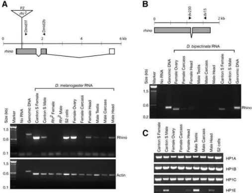
D. melanogastercontains fiveHP1-like genes, defined as such because they all encode an N-terminal chromo domain and a C-terminal shadow domain (Figure 1A). Four of these paralogs have been identified in previous analyses [36,37], whereas HP1E is newly identified in this report. These paralogs show differences in their conservation across
Drosophila species. HP1A, HP1B, and HP1C are highly
conserved, even between D. melanogaster and the more distantly related D. pseudoobscura (Figure 1A). In contrast, rhino differs significantly in size and amino acid sequence betweenD. melanogasterandD. simulans. In addition, theHP1E gene appears to have degenerated in the D. pseudoobscura genome, whereasD. pseudoobscurapossessesHP1F,a novel HP1 that theD. melanogastergenome lacks altogether.
Among the HP1 paralogs, theHP1D/rhinogene appears to be particularly rapidly evolving. In phylogenetic analyses, both therhinochromo and shadow domains appear to have evolved far more rapidly (Figure 1B–C) than their counter-parts in other HP1s in Drosophila (compare branch lengths between D. melanogaster, D. erecta, and D. pseudoobscura orthologs, which have bold branches).HP1E also appears to evolve rapidly in its chromo domain but is not preserved in D. pseudoobscura. Thus,rhinoappears unique among theHP1 -like genes in being well conserved yet evolving rapidly. Becauserhinois evolving so rapidly, orthologs are not likely to be unambiguously identified in other organisms.
rhinoIs Expressed Predominantly in Ovaries
Previous Northern blot analysis had detected a 1.6 kbrhino mRNA in female flies, early embryos, and ovary, but not in male flies andrhinomutants [36]. In situ analysis showed that therhinotranscript was present both within the germline and somatic cells of the ovary [36]. However, an abundant and much larger band on the Northern blot did not show the same restricted expression pattern. This band was also present in RNA made fromrhi2 mutant flies suggesting that it did not contain rhino transcript. In order to further delineate the expression pattern of this unusualHP1gene, we Positive Selection ofrhino
Synopsis
Eukaryotic genomes are organized into good and bad neighbor-hoods. In fruit fly genomes, most genes are found in euchromatin— good neighborhoods that tend to be amenable to gene expression and deficient in selfish mobile elements. Conversely, heterochro-matic regions are deficient in genes but chock full of mobile genetic elements, both dead and alive. Cells expend considerable effort to maintain this organization, to prevent bad neighborhoods from exerting their negative influence on the rest of the genome. At the forefront of this organization are the HP1 proteins, which are involved in the compaction and silencing of heterochromatic sequences. First discovered inDrosophila, HP1 proteins have been subsequently found in virtually all fungi, plants, and animals.
used RT-PCR to assess the presence ofrhinomRNA in male or female flies and in different tissues, because it provides a more sensitive assay that complements the previous Northern analysis (Figure 2).
We confirmed the predominant expression of rhino in D. melanogasterovaries, although low levels of transcript could also be detected in testis, head, and faintly in carcass, likely below detection limits for Northern analysis (Figure 2A). Endogenous rhino transcript was also present in S2 tissue culture cells that were used for our localization studies. Furthermore, the absence of any rhino transcript from rhi2 mutant flies by RT-PCR confirms that the large cross-reacting band seen on previous Northern analysis [36] does not contain rhino transcript. We have extended this finding to show that the predominant expression ofrhinois restricted to ovaries in another distantly related species, D. bipectinata (Figure 2B). In contrast, we found thatHP1A, -B,and-Cgenes were abundantly expressed in all gross adult tissues that we examined (Figure 2C). Interestingly, HP1E showed an expression pattern restricted predominantly to the male testis, suggesting that two of the five HP1 paralogs in D. melanogasterare each devoted predominantly to testes and ovaries respectively. This may highlight the fact that chromatin structure is likely to be inherently different in somatic versus germline cells, that may have spurred this specialization.
Rhino Localization inD. melanogasterCells
The localization of protein products of three HP1 genes
have been tested so far inDrosophilatissue culture cells. Only HP1A was found to localize predominantly to heterochro-matin, whereas HP1C localized to euchromatin and HP1B to both euchromatin and heterochromatin [35]. Therefore, we decided to first study the localization pattern of Rhino to determine whether it localized to heterochromatin.Drosophila S2 interphase cells have a DAPI-dense staining area that helps demarcate cytological boundaries of heterochromatin, although it is worth noting that DAPI does not stain all heterochromatic DNA, owing to sequence-dependent DNA-binding preference [38]. H3K4me is an excellent cytological marker for euchromatin, whereas H3K9me marks hetero-chromatin [10,11]. The localization patterns of green fluo-rescent protein (GFP) fused to HP1A, HP1B, or HP1C and expressed in tissue culture cells were previously shown to be faithful representations of the localization of the endogenous proteins by antibody staining [35]. We therefore expressed rhinoas a C-terminal GFP fusion protein inDrosophilaS2 cells, followed by immunostaining with antibodies to HP1A, HP1B, HP1C, or specific modifications of histone H3 (Figure 3) for comparison of localization patterns.
The localization pattern of Rhino-GFP differed from that of HP1A, -B, and -C in interphase tissue culture cells (Figure 3 and Figure S1). Rhino-GFP formed distinct foci that occupied a limited area in the nucleus. These Rhino-GFP foci were located in the heterochromatic compartment as defined by the absence of H3K4me staining. Strikingly, Rhino-GFP also did not directly overlap with common Figure 1.HP1 Paralogs inDrosophila
(A) Proteins encoded byD. melanogaster HP1s and selected orthologs (obtained by PCR from syntenic locations) are drawn to scale (indicated at bottom) with a dark rectangle resembling the N-terminal chromo domain and a lighter rectangle the C-terminal chromo shadow domain. TheHP1E open reading frame is no longer preserved inD. pseudoobscura,andD. melanogasterdoes not containHP1F. The hinge regions and N- and C-terminal extensions cannot be aligned between different HP1 types, for example HP1A versus HP1B. HP1D/Rhino contains a very long hinge region that is poorly conserved between species.
(B) A neighbor-joining phylogenetic tree based on an alignment of selected HP1 chromo and (C) shadow domains. The monophyletic vertebrate HP1 paralogs are shown for comparison.rhinoevolution is clearly distinct from vertebrate or otherDrosophila HP1s.HP1orthologs betweenD. melanogaster, D. erecta,andD pseudoobscura are shown connected by bold branches (HP1Eis not conserved inD. pseudoobscura). The divergence times for D. melanogaster–D. erectaandD. melanogaster–D. pseudoobscuraare approximately 9 and 25 million years respectively, whereas those for mouse– human are approximately 80 million years. Clearly, therhinochromo and shadow domains are far more divergent between theseDrosophilaspecies than the chromo domains ofHP1A, -B,and-C.
markers of heterochromatin, HP1A or H3K9me, rather appearing interspersed with, or surrounding these signals. Thus, unlike HP1A, we expect that the Rhino chromo domain does not bind H3K9me. The Rhino-GFP localization pattern was not an artifact of GFP-tagging because it was also observed with a Rhino protein that was N-terminally tagged with a biotinylated peptide (Figure S1). We conclude that among HP1 paralogs, Rhino-GFP has a unique localization pattern within the heterochromatic domain in tissue culture cells. Its localization pattern in oocytes is currently unknown.
Molecular Evolution ofrhino: Positive Selection of the Hinge and Chromo Shadow Domains
The indication that rhino may be a rapidly evolving HP1 (see Figure 1), its predominant expression in ovaries (see Figure 2), and its interesting cytological localization pattern (Figure 3), led us to investigate its evolutionary history in further detail. Uncovering evolutionary constraints under which differentHP1genes evolve can provide insight into the evolutionary forces that shape heterochromatin. To study the molecular evolution of HP1 proteins in Drosophila, we obtained DNA sequence for HP1 orthologs in the closely related D. simulans species (diverged from D. melanogaster about 2.5 million years ago) by PCR.
Rapid evolution of HP1s may be attributed to relaxed constraint, allowing sequence changes to accumulate, especially if different gene copies are functionally redundant. Alterna-tively, amino acid replacement changes may confer a selective
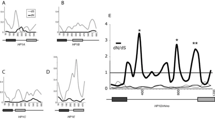
advantage, in which case they would be expected to accumulate at a rate higher than expected under neutral evolution (positive selection). To evaluate whether any of the HP1s are undergoing such positive selection between the closely relatedD. melanogaster andD. simulansspecies, we performed a 100-bp sliding window analysis of the number of replacement changes per site (dN) compared to the number of synonymous changes per site (dS) (Figure 4).HP1B, HP1CandHP1Ehad dN,dS in all windows, consistent with purifying selection, as expected for structural proteins evolving under strict constraints. To our surprise, we found dN . dS for several windows in the rhino gene corresponding to the hinge region of the encoded protein (Figure 4E). We used Monte Carlo simulations in the K-estimator program to show that three of these windows were statistically significant (dN . dS, p , 0.02, indicated by asterisks), consistent with positive selection ofrhinobetweenD. melanogasterandD. simulans. We also found two windows with dN .dS for theHP1A-encoded hinge with borderline significance (p-value approximately 0.05), but further detailed analysis including a population study ofD. melanogasterandD. simulans, and dN/dS comparisons among several other pairs of closely relatedDrosophilaspecies (D. Vermaak, H. S. Malik, unpublished data) led to the conclusion that there was no positive selection ofHP1A. Thus, no otherHP1homolog other thanrhinoshowed any evidence of positive selection, suggesting thatHP1D/rhinois again unique in this respect, not just among Drosophila HP1 paralogs, but also among allHP1genes identified so far. Figure 2.RT-PCR Analyses of the Various HP1 Paralogs
(A) Therhinogene fromD. melanogasteris drawn to scale. Exons are boxed (grey fill indicates coding sequence) and lines indicate introns. The position of aP[lacZ, ryþ
](PZ) element in therhi2mutant is shown (triangle; not to scale). Dmid1f and Dmid2b RT-PCR primers span the firstrhinointron. RT-PCR was carried out on roughly equivalent amounts of RNA using a primer set forrhinoor actin-42A (primer sequences in Table S2). Control reactions contained no RNA orD. melanogastergenomic DNA.
(B) Therhinogene fromD. bipectinatais schematized and primers used for RT-PCR indicated. RT-PCR analysis shows thatrhinois specifically expressed in ovaries.D. bipectinataseparated fromD. melanogasterapproximately 13 million years ago.
(C) RT-PCR reactions carried out for the other HP1 paralogs inD. melanogaster.HP1A, -B,and-Care ubiquitously expressed in adult tissues whereasHP1E expression appears to be predominantly restricted to the male testes.
DOI: 10.1371/journal.pgen.0010009.g002
Sliding window dN/dS analyses suggest thatrhinois subject to positive selection. To follow up on this initial observation, we undertook a more detailed study in D. melanogaster and D. simulans. We used PCR to obtain rhinosequence from 17 strains ofD. melanogasterand 11 strains of D. simulans. DNA sequence changes were categorized as replacement (R) or synonymous (S) (Table S1). Changes were further classified as either fixed between species (f) or polymorphic within species (p) (Table S1). Under a neutral evolutionary model, the ratio of replacement to synonymous changes that have been fixed between species (Rf:Sf) is expected to be roughly the same as the ratio for polymorphic changes (Rp:Sp) (McDonald-Kreit-man test) [39]. We did not find a significant deviation from neutrality when the entire rhino sequence was considered (Table 1, entire coding region,p¼0.13). However, a sliding
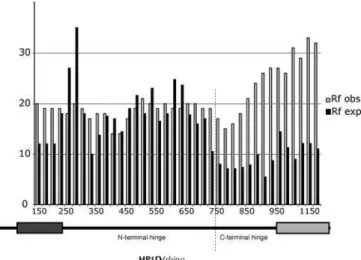
window analysis clearly showed that the observed fixed replacement changes far exceeded those expected under neutral evolution in the C terminal part of the protein (Figure 5). Indeed, the shadow domain had a highly significant deviation from neutrality (p , 0.01), suggesting that this domain has been subject to strong positive selection (Table 1, shadow). We used parsimony to assign each DNA sequence change within the shadow domain to either themelanogasteror simulanslineage by polarizing the changes to outgroup species D. teissieriandD. yakuba(Table S1, changes in themelanogaster lineage [m] or simulans lineage [s]). We concluded that the shadow domain has been subject to positive selection in the D. simulanslineage (Table 1, shadowD. simulansonly,p,0.05), but there were not enough polymorphisms to reach a similar conclusion for the D. melanogaster lineage despite a strong Rf:Sf ratio. Although the complete hinge alone does not reject neutrality, separating the long hinge domain into N-and C-terminal segments suggests that the C-terminal region of the hinge, abutting the shadow domain, has been subject to positive selection (p,0.01). We could not determine whether the positive selection in the hinge was lineage specific because of ambiguity in the alignment with outgroup species. Despite this strong signal for positive selection, we were unable to detect evidence of recent adaptive‘‘sweeps’’using Fu and Li [40] or Tajima [41] tests, suggesting that any such sweeps were not recent enough to result in standing single polymor-phisms. Thus, both the hinge and shadow domains of the protein encoded byrhinoshow strong evidence for relatively old episodes of positive selection between theD. melanogaster andD. simulanslineages.
rhinoEvolution in OtherDrosophilaSpecies
Is the positive selection ofrhinolimited to themelanogaster species group? To address this question, we identified D. pseudoobscura rhino by synteny withD. melanogaster; rhinois contained within an intron of another gene in both species. We used RT-PCR to confirm the predicted splice sites for rhinofrom theobscuraspecies group.D. pseudoobscura rhinois very different in length (317 vs. 418 encoded amino acids) and sequence fromD. melanogaster rhino(see Figure 1). In fact, the hinge region of the Rhino protein is changing so rapidly that it is unrecognizable in a BLAST comparison between D. melanogasterandD. pseudoobscura(e-value.1,000). To trace the evolution ofrhinobeyond themelanogasterspecies group, we obtainedrhinosequence from intervening species between D. melanogasterand D. pseudoobscura. Despite the evolutionary distance of D. melanogaster from D. pseudoobscura, we could identify noncoding conserved sequences both upstream and downstream of therhinogene, allowing us to design primers to amplifyrhinofrom 12 additionalDrosophilaspecies, shown schematically in Figure 6A.
We find thatrhinois evolving at an unprecedented rate for anHP1.The hinge regions cannot be unambiguously aligned between different species groups or in some cases not even within the same species group. We did not detect any significant similarity of the Rhino hinge regions to other proteins or motifs, yet all the hinge regions share certain sequence features, most noticeably long runs of serines as well as proline- and glutamine-rich sequences (Figure 6A). In some instances, we found clear evidence of positive selection (dN/dS . 1) for alignable segments of hinge regions of closely related pairs, D. yakuba versus D. teissieri, D. erecta
Figure 3.Rhino-GFP Localizes to Distinct Foci in the Heterochromatic Domain
A C-terminal GFP fusion protein ofrhinowas transiently expressed in Drosophilatissue culture cells (green in merge). Nuclei were stained with DAPI that stains DNA (blue in merge) and antibodies (red in merge) to HP1A, HP1B, HP1C, H3K9me, H3K4me, or fibrillarin (a nucleolar protein). H3K4me stains euchromatin whereas HP1A, H3K9me, Rhino-GFP, and bright DAPI staining all fall within heterochromatin. Rhino-GFP does not overlap with any of the antibody staining patterns, but appears to localize adjacent to HP1A and H3K9me within the heterochromatic domain.
versusD. orena,D. lutescensversus D prostipennis,D. bipectinata versusD. parabipectinata,andD. pseudoobscuraversusD. miranda (representative examples shown in Figure 6B). Our ability to detect instances of dN/dS . 1 within the hinge region for multiple species pairs within a small sampling ofDrosophila species suggests that positive selection of the hinge is a common feature inrhinoevolution.
Positive Selection ofrhinoChromo Domain
Phylogenetic analyses (see Figure 1B and 1C) suggested that not just the hinge region, but also the chromo and shadow domains of Rhino are diverging more rapidly than similar domains of other HP1s. For the hinge and shadow domains, we have already presented evidence that this rapid evolution is not due to lack of selective constraint, but rather due to positive selection. However, we were unable to detect positive selection within the chromo domain using dN/dS or McDonald-Kreitman tests, nor were we able to detect significant evidence of an adaptive sweep using standard tests (Fu and Li [40], Hudson-Kreitman-Aguade [42], Tajima’s D [41]). We reasoned that it may be hard to detect positive selection of the chromo domain because the majority of its codons are likely to be functionally constrained and therefore under purifying selection. However, such purifying selection may be masking positive selection of a small number of codons within the chromo domain. We therefore used a codon by codon maximum likelihood test, PAML [43], to ask
if we could detect any codons that have been under repeated, strong positive selection.
We used a DNA sequence alignment of the rhino gene corresponding to the encoded chromo domain from
differ-ent Drosophila species. The corresponding amino acid
sequence alignment is shown in Figure 7A. We note that a tree based upon this amino acid alignment is in agreement with the acceptedDrosophila phylogeny [44], suggesting that we are considering strict orthologs. Remarkably, models that Figure 4.Comparison ofD. melanogasterandD. simulans HP1s
(A–E) DifferentD. melanogasterandD. simulansHP1 coding DNA sequences were aligned (indels and unalignable sequences were removed) and dN (black line) and dS (grey line) values were calculated using K-estimator [75] with a sliding window of 100 bases and a 35-bp step size. The domain structure of each HP1 is shown schematically and to scale beneath each plot, with the dark rectangle representing the chromo domain and the grey rectangle the chromo shadow domain. ForHP1A,dN exceeds dS in the hinge region, but dS is very low in these windows. In contrast, forrhino,dN is higher throughout and exceeds dS in several windows corresponding to the hinge region (dN/dS values are also plotted forrhino). Windows in which statistically significant values for positive selection were obtained (dN/dS.1,p,0.02), are indicated by asterisks and map to the hinge region. DOI: 10.1371/journal.pgen.0010009.g004
Table 1.McDonald-Kreitman Test of rhinoinD. melanogaster and D. simulans
Domain ofrhino Rf Sf Rp Sp G-Valuea p-Value
Entire coding 88 35 49 31 2.301 0.13
Chromo 10 9 0 1 NA 1b
Hinge 56 17 46 22 1.427 0.23
N-terminal hingec 34 15 31 12 0.079 0.78
C-terminal hingec 22 2 15 10 6.827 0.009
Shadow 20 8 1 6 7.289 0.007
Shadow (D. melanogasteronly) 10 2 1 1 0.677 0.41
Shadow (D. simulansonly) 8 6 0 5 NA 0.044b
aG-value (with Williams correction) for test of 232 independence.
bp-Value calculated using Fisher’s exact test because the G-value calculation does not permit zeros. cWe arbitrarily define the N- and C-terminal hinge boundaries at position 750 in therhinoalignment based on Figure 5.
DOI: 10.1371/journal.pgen.0010009.t001
allow codons to evolve under positive selection (M8 and M2) fit the data significantly better than associated models that do not permit positive selection (M7 and M1) (p,0.001 in all cases, Table 2). Just a few codons account for this positive selection. In particular, three codons repeatedly show highly significant posterior probabilities (1E, 9L, and 25S in Table 2; arrows Figure 7A). The Rhino chromo domain structure is likely to be similar to that of known HP1 chromo domains, so we show the likely positions of the three adaptively evolving amino acids of the Rhino chromo domain on the known structure of Drosophila HP1A chromo domain bound to H3K9me peptide (Figure 7A; [45]). Position 1 is in close proximity to the groove that binds the methylated peptide, suggesting that this amino acid may be driven to adapt to a constantly changing substrate of Rhino. We cannot rule out that positions 9 and 25 may also be adapting to a substrate binding in the same position, because they could influence the overall conformation and thus binding specificity of the chromo domain. However, positions 9 and 25 are expected to be solvent accessible on the opposite side of the Rhino chromo domain and may represent an additional, potentially novel, interaction surface.
We have already shown that the shadow domain is under strong positive selection between D. melanogaster and D. simulans. To find out if some codons of the shadow domain have also been under continuous positive selection, we carried out a PAML analysis. A tree based on the shadow domain amino acid alignment also recapitulates Drosophila phylogeny (Figure 7B). We found significant evidence for positive selection, with most of the signal coming from just three codons (Table 2; Positions S31, I33, and I59 in Figure 7B). On the structure of a mouse HP1bshadow domain dimer [46], positions 31 and 33 are on the same side of the shadow
dimer and should be available for protein–protein inter-actions. The vertebrate HP1 chromo shadow domain dime-rization site is known to bind to many proteins through their PxVxL motif [47–49]. It is unclear if these interactions are conserved in Rhino, but rapid evolution in this region (including position 59 identified in our PAML analysis) certainly has the potential to easily influence protein–protein interactions.
PAML analyses like these are very useful to highlight codons that have been repeatedly subject to positive selection [43,50]; however, they do run a risk of false positives. This is somewhat ameliorated in our dataset because the tree lengths are of moderate value (Table 2). Similar tree lengths have been shown by simulations to have a significantly lower risk of false positives [51]. Nonetheless, the true test for the significance of these positively selected residues will come from functional assays on Rhino function and localization.
Discussion
In this paper, we have undertaken an evolutionary study of HP1-like proteins, with the ultimate aim of discerning the selective pressures that act on heterochromatin. We have found that Rhino, the only HP1 paralog that is expressed predominantly in ovaries, encodes a protein that has a unique localization pattern in S2 cells. Although it is excluded from the euchromatic compartment, the Rhino protein does not overlap with HP1A or H3K9me. This immediately suggests that H3K9me or HP1A does not mark all Drosophila heterochromatin, and that Rhino has a uniquely different specificity for a previously unappreciated compartment in heterochromatin.
It has not been easy to discern the molecular function of rhinofrom mutant phenotypes in eggshell defects. Possibilities range from a role for Rhino in gross chromatin structural changes, to transcriptional or translational regulation and even microtubule organization in the oocyte [36]. Despite our current lack of knowledge about the molecular function of rhino, the fact that mutations are female sterile, point to its importance to proper oogenesis [36]. HP1A, which is far better understood, is an essential gene. Such chromosomal proteins serving crucial functions are expected to be under strong evolutionary constraints and purifying selection. Although this is true for four of the five D. melanogaster HP1s including HP1A (see Figure 4), we find that all three domains ofrhinohave evolved under positive selection using multiple criteria, including dN/dS ratios, McDonald-Kreit-man, and PAML analyses [39,43]. What could be driving this positive selection of such an important HP1 protein?
Can co-evolutionary pressures explain the positive selec-tion acting onrhino? For instance,rhinomight be continually ‘‘catching up’’to mutations in interacting proteins required for its function. We believe this is unlikely, because mutations that compromise a required interaction are likely to be culled out of the population by purifying selection, long before a chance compensatory mutation inrhinocan occur. A second possibility is that the positive selection ofrhinomay be driven by changes in the regulation of key genes between two species. Although we cannot formally rule out such a scenario, it appears unlikely to explain the relatively constant positive selection that we have seen for approximately 25 million years ofDrosophilaevolution.
Figure 5.Population Genetics of HP1D/rhinobetweenD. melanogaster andD. simulans
Replacement changes that have been fixed betweenD. melanogaster (17 strains) andD. simulans(11 strains) (Rf obs [observed], open bars) were calculated with a 300-nucleotide sliding window, 25-nucleotide step size. The number of expected replacement changes for each window (Rf exp; solid bars) were calculated from the neutral expectation of the McDonald-Kreitman test (Rf:Sf’Rp:Sp). Rf obs exceeds Rf exp in the C-terminal part ofrhino(the C-terminal part of the hinge and the shadow domain as shown beneath), consistent with positive selection (also see Table 1). The chromo and chromo shadow domains are represented by dark and light rectangles, respectively.
Positive selection need not involve rhino’s ‘‘normal’’ function, whatever that may be, but rather underlie a second and unrelated‘‘defense’’function ofrhino. In such a scenario the positive selection onrhinowould be driven by a recurrent intracellular conflict that yields a selection advantage to the ‘‘winner.’’Genes encoding proteins involved in direct host– parasite interactions are often subject to positive selection. In this case, changes that are beneficial for the parasite (to evade
interactions for instance) will be followed by selection favoring changes in the host proteins (that restore inter-actions). Thus, two antagonistic entities locked in genetic conflict face repeated episodes of positive selection, only to arrive at the same quasi-steady state, a scenario formalized as the‘‘Red Queen’’hypothesis [52].rhinomay be subject to the same kind of genetic conflict that occurs intracellularly. It is especially intriguing that the only HP1 we have found to be Figure 6.Positive Selection ofrhinoin 25 Million Years ofDrosophilaEvolution
(A)rhinowas PCR amplified and sequenced from the indicatedDrosophilaspecies. Predicted protein sequences are drawn to scale with amino acid length shown on the right. The chromo and chromo shadow domains are relatively conserved and are indicated by the large dark and light rectangles, respectively. The hinge regions are rapidly evolving. They differ dramatically in size and sequence and cannot be aligned between different species groups (indicated on the left) and sometimes not even within the same species group, for example theD. bipectinataversusD. ananassaehinge. Within themelanogasterspecies group,D. melanogaster rhinoappears to have undergone large deletions up to 50 codons in the hinge region compared with its closest relativeD. simulans(indicated by slanted lines). These deletions are adjacent to the adaptively evolving hinge region identified between D. melanogasterandD. simulans. A 58 amino acid duplication present in theananassaespecies group is indicated by grey arrows. Thin black rectangles indicate runs of serine ranging between 70% and 100% serine.
(B) dN and dS calculations forrhinofrom alignments of two pairs of closely related species from themelanogasterandtakahashiispecies groups show multiple windows in which dN exceeds dS, indicative of positive selection.
DOI: 10.1371/journal.pgen.0010009.g006
subject to positive selection is expressed predominantly in ovaries ([36] and Figure 2), where such a competitive advantage has directly heritable consequences. We consider two models of genetic conflict to explain rhino’s positive selection.
Under the first model, rhino participates in suppressing ‘‘selfish’’ behavior of centromeres, which can compete to maximize their transmission advantage in female meiosis, where only one of four meiotic products is destined to
become the egg [21]. We have previously proposed that this kind of drive can have deleterious consequences for male meiosis and is likely to be suppressed either by centromeric proteins altering their DNA-binding specificity [19,20] or by heterochromatin proteins evolving to limit centromere boundaries, and thereby limiting‘‘strength’’[4,21,53]. Similar selective pressures have been previously proposed to result in deleterious mutations in the nod chromokinesin in D. melanogaster [54]. rhino may represent another repressor Figure 7.Positive Selection of the Chromo and Chromo Shadow Domains ofrhino
(A) An amino acid alignment of the chromo domain of differentDrosophilaspecies is shown with the distantly relatedHP1Aand humanHP1achromo domains for comparison. The neighbor-joining tree based on this alignment (shown on the left) recapitulates knownDrosophilaphylogeny. Amino acids of theHP1Achromo domain that are involved in binding to H3K9me are color coded: Blue amino acids form an aromatic cage that recognize K9me, and pink amino acids form a complementary surface for recognition of the H3 peptide [45]. The corresponding DNA sequence alignment was used in a PAML analysis. Three codons that have been under repeated and strong positive selection are indicated by arrows. The corresponding positions (red) are indicated on the known structure of theDrosophila HP1Achromo domain (light blue) bound to H3K9me (purple) [45].
(B) Amino acid alignment of representative chromo shadow domains ofrhinoorthologs fromDrosophila. The neighbor-joining tree based on this alignment also recapitulatesDrosophilaphylogeny. Amino acids of mouseHP1bknown to be involved in dimerization are shown in pink and those required for the shadow fold in blue [46]. We use arrows to indicate codons identified as being under positive selection by our PAML analysis. Corresponding positions of the mouseHP1bchromo shadow domain are indicated (red) on one of the shadow domains (light blue) of the dimer [46]. These positions are shown in yellow on the other shadow domain (green).
of the drive by directly or indirectly influencing centromere strength.
A second model is that positive selection onrhinois a direct result of genetic conflict betweenrhino and mobile genetic elements. Although we have no evidence to support this hypothesis, it is attractive for several reasons. Transposable elements can evolve rapidly and differ significantly between Drosophila species, including D. melanogaster and D. simulans [55,56]. Rhino-GFP localizes to the heterochromatic region of the nucleus (see Figure 3), which is highly enriched in transposable elements [57]. Finally, genome-bound trans-posable elements can only increase their genomic copy number by transposing in the germline, increasing selective pressures on host proteins that act as suppressors of germline transposition. Rhino may either interact with the integration machinery of transposons to direct their integration into transcriptionally silent heterochromatin, or it may directly bind and transcriptionally repress transposable elements that are newly introduced into heterochromatin. Some trans-posable elements are known to be major in vivo targets of HP1A, apparently involving the RNAi (RNA interference) pathway [1,11,58–62]. Similarly,rhinomay be under continual selection to directly bind transposable elements.
Whatever is driving the positive selection of rhino, mutations in any of Rhino’s three domains appear to be selected to giverhinothe upper hand in the current round of competition. The chromo and related shadow domains are very versatile interaction domains that can influence binding to DNA, RNA, and proteins [63]. The hinge domain can also strongly influence localization of HP1-like proteins [64,65]. Future experiments will address the functional role of the three amino acids under recurrent positive selection in the chromo and shadow domains (Figure 7) and help to
distinguish between our models of what drives the positive selection of rhino. These experiments promise to reveal insights into the organization of a substantial portion of Drosophilagenomes. It is probably not a coincidence that we have found positive selection only in an HP1-family member that is expressed predominantly in ovaries. Indeed, a restricted expression pattern may have allowed detection of a previously unremarked conflict that shapes at least a fraction of Drosophila heterochromatin, via the positive selection of rhino. Such a signal may have been masked for other HP1s due to their constrained roles in other tissues.
Our results complement previous findings that other proteins that bind heterochromatin appear to be among the most rapidly evolving proteins in an unbiased screen in Drosophila [67–68], although this does not appear to be the result of positive selection [69]. Polymorphisms in hetero-chromatin-binding proteins can have direct effects on non-disjunction frequencies [54,70,71]. Similarly, although HP1A, -B, and -C appear to be conserved and evolving under purifying selection, HP1 evolution (in both sequence and gene copy number; see Figure 1) in general appears quite rapid for a chromosomal protein with a highly conserved function in most eukaryotes. Thus, rapid changes in the genomic landscape may underlie rapid diversification of genes encoding HP1s and chromosomal proteins in general.
Materials and Methods
Sequences fromDrosophilaand databases and RT-PCR.Drosophila
species and strains (Table S1) were obtained from the Drosophila
stock center (currently in Tucson, Arizona) and genomic DNA was
prepared by standard methods [19]. Therhinolocus was amplified
using PCR Supermix High Fidelity (Invitrogen, Carlsbad, California, United States) with the primers indicated in Table S2. PCR products were either sequenced directly or following Topo-TA cloning
Table 2.PAML Analyses ofrhinoChromo and Shadow Domains inDrosophila
Domain Analysis 2(lnk) Degrees
of Freedom
p-Value dN/dS Proportion of Sites
Sites Identified (Posterior Probabilities)
Chromo
(branch length, S¼11–16)
M0 vs. M3 106.00 4 p,0.0001 3.25 5.4% 1 E (0.91)
9 L (1.00) 25 S (1.00)
M1 vs. M2 35.156 2 p,0.0001 8.25 5.6% 1 E (0.98)
9 L (1.00) 25 S (1.00)
M7 vs. M8 17.938 2 p,0.0002 2.49 7.3% 1 E (0.90)
9 L (0.99) 25 S (1.00) Shadow
(branch length, S¼10–11)
M0 vs. M3 45.816 4 p,0.0001 2.09 8% 18 N (0.76)
31 S (1.00) 33 I (0.99) 39 N (0.51) 59 I (0.99)
M1 vs. M2 17.918 2 p,0.0002 4.34 5.8% 31 S (1.00)
33 I (0.98) 59 I (0.98)
M7 vs. M8 9.014 2 p,0.012 2.38 6% 31 S (0.98)
33 I (0.92) 59 I (0.93)
Codon positions are as defined inD. melanogaster.krefers to log likelihood. Branch length, S, refers to the number of nucleotide substitutions per codon, and can vary from one model to another (range shown). Analyses shown were carried out using the F61 model of codon frequencies, but similar results were obtained with the F334 model.
DOI: 10.1371/journal.pgen.0010009.t002
(Invitrogen). RNA was prepared from whole male or female flies or different tissues (head, ovary, testis, or carcass) using a kit (Qiagen RNeasy; Qiagen, Valencia, California, United States) and cleared of genomic DNA by DNase I digestion (Ambion DNA-free; Ambion, Austin, Texas, United States). RNA concentrations were measured from various tissues, and the same amount of total RNA was used as template in the RT-PCR analysis. RT-PCR (Invitrogen) to evaluate
the presence of rhino mRNA was carried out using Dmid1f and
Dmid2b primers (Table S2) that span therhino intron, along with
actin-42Aprimers [72] as a control. For D. bipectinata, primers dv15
and dv230 that span the rhino intron were used. RT-PCR and
sequencing was carried out to confirm the predicted splice-site positions for rhino from D. simulans (strain 2), D. bipectinata, and D. miranda. Splice sites forrhinofrom other species were predicted using Berkeley Drosophila Genome Project Splice site predictor (http://www.fruitfly.org/seq_tools/splice.html). All sequences have been deposited in Genbank (accession numbers AY944308– AY944358, Table S2).
Sequence analysis.Sequences were assembled using DNA Strider [73]. Clustal_X [74] was used to obtain pairwise or multiple alignments and to generate formatted files for further analysis. Pairwise sequence alignments used for dN/dS analysis were hand edited, using the amino acid sequence as a guide to place indels. For instance, there is an 80 amino acid length difference between the D. melanogasterandD. simulanshinge regions. These regions cannot be compared in tests for positive selection. Pairwise dN and dS comparisons and confidence values were calculated using the K-estimator software [75,77]. Sliding window size was arbitrarily chosen as 100 bases with 35 base steps for all pairwise dN/dS comparisons. Confidence interval estimates were calculated using Monte Carlo simulations, taking into account (1) dN and dS values, (2) the number of codons, (3) transition: transversion ratio, and (4) GC content and amino acid composition. Thus, K-estimator [75] at least takes into account most of the confounding variables that are known to give false positives in terms of dN/dS. We also present a dN/dS analysis using the reconstructed hypothetical ancestors to all the D. melanogasterandD. simulans rhinosequences (Figure S2).
The DNASP software package [77] was used to perform several tests for positive selection using genomic sequence ofrhinofrom 17 strains ofD. melanogasterand 11 strains ofD. simulans. The Fu and Li [40], Tajima’s D [41], and Hudson-Kreitman-Aguade [42] tests were carried out on the complete sequence, including the intron, whereas the McDonald-Kreitman test [39] was carried out on coding regions only (1,209 total positions with indels removed). Fixed replacement changes in the chromo and chromo shadow domains were polarized usingD. yakubaandD. teissierisequences as outgroups, but we could not unambiguously polarize all changes in the hinge region. The
expected fixed replacement changes (Rfexpected) shown in Figure 4B
were calculated from the ratio Rfexpected ¼ Sfobserved(Rpobserved/
Spobserved) according to the neutral expectation in the
McDonald-Kreitman test, where R¼replacement, S¼synonymous, f¼fixed
between population, p¼polymorphic within the population (similar
to the previously proposed‘‘Neutrality Index’’[78]). A sliding window of 300 nucleotides with step size of 25 was used for presentation purposes.
Neighbor-joining phylogenetic trees were constructed using the PAUP software, version 4.0b10 [79] and appropriate Clustal_X multiple alignments of either the chromo or chromo shadow domains. A total of 1,000 replicates were carried out for boot-strapping. Maximum likelihood analysis was performed with the PAML software package [43] in separate analyses for multiple alignments of the chromo domain and the shadow domains (the rapid evolution of the hinge in both size and sequence precluded its comparison in such a multiple alignment). Codons that were repeatedly subject to positive selection were identified using N sites models (M1, M7) that do not permit positive selection compared to models (M2, M8) that permit sites to evolve under positive selection. The strength of positive selection was calculated by comparing twice the log likelihood difference (M2 vs. M1, M8 vs. M7) in a chi-square test with two degrees of freedom. Codons that were identified as having evolved under positive selection with high posterior proba-bilities (p.0.95) were highlighted on a three-dimensional structure of the respective domains and visualized using the Cn3D software (version 4.0) [80].
Plasmid constructs.A plasmid for expressingrhinoas a C-terminal GFP fusion protein under control of the hsp70 heat shock promoter
(HSRhiGFP) was constructed as follows: rhino coding sequence
flanked by XbaI and NotI restriction enzyme sites was amplified by
RT-PCR fromD. melanogaster(Canton S) using primers KcRhiF and
KcRhiB (Table S1). The PCR product was digested and cloned into a
modified heat shock expression plasmid [81] that had been digested
with XbaI and EagI and phosphatase treated to yield therhinoopen
reading frame followed by a six amino acid linker and GFP. Correct cloning was verified by sequencing. An N-terminal fusion protein of a biotin recognition peptide (MAGGLNDIFEAQKIEWHEDTGGS) to rhino(BLRPRhi) was constructed as follows: Primers dv99 and dv100
were used to amplifyrhinocoding sequence with flanking NotI and
BamHI restriction enzyme sites from the HSRhiGFP plasmid. The PCR fragment was TA cloned and the sequence verified before digestion of the TA clone and subcloning of the gel-isolated fragment into a BLRP expression vector with a metallotheionine promoter [82,83]. A plasmid (pBirA) expressing theEscherichia colibiotin ligase enzyme (BirA) from a metallotheionine promoter was a gift from Takehito Furuyama.
Cell culture, transfection, and immunostaining. S2 cells gen, D-mel2) were maintained in serum-free insect media (Invitro-gen) supplemented with 90 ml/l of 200 mM L-Glutamine (Sigma, St. Louis, Missouri, United States). Twenty micrograms of the HSRhiGFP plasmid was transfected as previously described [81]. Cells were heat shocked for 1 h on the next day and allowed to recover for 2 h before
immunostaining [84]. In the case of the BLRPrhino construct, 10lg
of plasmid DNA were co-transfected with 10lg of pBirA plasmid that contains the biotin ligase under control of a metallotheionine promoter. After overnight incubation, cells were induced for 3 h
with 500 lM CuSO4, added directly to the media, followed by
immunostaining. HP1A, HP1B, and HP1C antibodies have been previously described [35]. Antibodies to H3K9me or H3K4me were purchased from Upstate Biotech (Waltham, Massachusetts, United States). Monoclonal mouse anti-Fibrillarin antibody was purchased from Encor Biotechnology Inc (Alachua, Florida, United States). All antibodies, including the secondary Texas-red fluorescently labeled goat anti-rabbit or anti-mouse antibodies (Amersham, Piscataway, New Jersey, United States), were used at a dilution of 1/200, with the exception of the anti-fibrillarin antibody that was used at 1/500. Images of nuclei were obtained and de-convolved using the Deltavision software (Applied Precision, Issaquah, Washington, United States).
Supporting Information
Figure S1. Rhino-GFP Localization inDrosophilaS2 Cells
These additional images of Rhino-GFP show a localization pattern that is distinct from HP1A, H3K4me, and H3K9me. In addition, an N-terminal biotinylated-tagged Rhino protein shows the same local-ization pattern as that of the C-terminal GFP-tagged Rhino protein. Found at DOI: 10.1371/journal.pgen.0010009.sg001 (5.2 MB PDF).
Figure S2. A Sliding Window dN/ dS Analysis
Only those changes that were found to have been fixed differences betweenD. melanogasterandD. simulanswere used. All intraspecific polymorphisms were eliminated for this analysis. Compared to Fig-ure 4, the signal for positive selection now appears concentrated exclusively in the C-terminal region ofrhino.
Found at DOI: 10.1371/journal.pgen.0010009.sg002 (203 KB PDF).
Table S1. All Polymorphisms within the Coding Region of therhino
Gene inD. melanogasterandD. simulansAre Shown
Changes are highlighted as being either fixed (f) between species or polymorphic (p) within species, as replacement (R) or synonymous (s) changes. Fixed changes were polarized using an outgroup species to changes along either theD. melanogaster(m) orD. simulans(s) lineages. Many changes could not be unambiguously polarized.
Found at DOI: 10.1371/journal.pgen.0010009.st001 (34 KB DOC).
Table S2. List of Primers Used and Accession Numbers of Sequences Obtained in This Study
Found at DOI: 10.1371/journal.pgen.0010009.st002 (36 KB XLS).
Accession Numbers
The Flybase (http://flybase.bio.indiana.edu) accession numbers of the
genes discussed in this paper are rhino (CG10683) and HP1E
(CG8120). New sequences obtained during the course of this study have been deposited in Genbank under the accession numbers AY944308–AY944358. The Molecular Modeling Database (MMDB; http://www.ncbi.nlm.nih.gov/Structure/MMDB/mmdb.shtml) accession numbers of the proteins discussed in this paper are H3K9me (19011,
Acknowledgments
We thank the Drosophila stock center (Tucson, Arizona) for various Drosophila species stocks and Judy O’Brien for maintenance of Drosophilastocks. We are grateful to Kami Ahmad and Celeste Berg for useful discussions throughout this project, and Jiro Yasuhara, Barbara Wakimoto, Sara Sawyer, and Julie Kerns for comments on the manuscript. We also gratefully acknowledge Terri Bryson for help
with maintenance of Drosophila tissue culture cells, and Takehito
Furuyama for the BLRP and pBirA plasmids. This work was supported by a Damon Runyon Cancer Postdoctoral Fellowship (DV), by the
Howard Hughes Medical Institute (SH), startup funds from the Fred Hutchinson Cancer Research Center, and a Scholar Award from the Sidney Kimmel Foundation (HSM). HSM is an Alfred P. Sloan Fellow in Computational and Evolutionary Molecular Biology.
Competing interests.The authors have declared that no competing interests exist.
Author contributions. DV and HSM conceived and designed the experiments. DV performed the experiments. DV and HSM analyzed the data. DV and SH contributed reagents/materials/analysis tools.
DV, SH, and HSM wrote the paper. &
References
1. Hodgetts R (2004) Eukaryotic gene regulation by targeted chromatin re-modeling at dispersed, middle-repetitive sequence elements. Curr Opin Genet Dev 14: 680–685.
2. Kazazian HH Jr (2004) Mobile elements: Drivers of genome evolution. Science 303: 1626–1632.
3. Deininger PL, Moran JV, Batzer MA, Kazazian HH Jr (2003) Mobile elements and mammalian genome evolution. Curr Opin Genet Dev 13: 651–658.
4. Malik HS, Henikoff S (2002) Conflict begets complexity: The evolution of centromeres. Curr Opin Genet Dev 12: 711–718.
5. Choo KH (2001) Domain organization at the centromere and neocen-tromere. Dev Cell 1: 165–177.
6. Dillon N (2004) Heterochromatin structure and function. Biol Cell 96: 631– 637.
7. Bernard P, Maure JF, Partridge JF, Genier S, Javerzat JP, et al. (2001) Requirement of heterochromatin for cohesion at centromeres. Science 294: 2539–2542.
8. Wakimoto BT, Hearn MG (1990) The effects of chromosome rearrange-ments on the expression of heterochromatic genes in chromosome 2L of
Drosophila melanogaster. Genetics 125: 141–154.
9. Hoskins RA, Smith CD, Carlson JW, Carvalho AB, Halpern A, et al. (2002) Heterochromatic sequences in aDrosophilawhole-genome shotgun assem-bly. Genome Biol 3: RESEARCH0085.
10. Richards EJ, Elgin SC (2002) Epigenetic codes for heterochromatin formation and silencing: Rounding up the usual suspects. Cell 108: 489– 500.
11. Grewal SI, Moazed D (2003) Heterochromatin and epigenetic control of gene expression. Science 301: 798–802.
12. Weiler KS, Wakimoto BT (1995) Heterochromatin and gene expression in
Drosophila. Annu Rev Genet 29: 577–605.
13. Kirschmann DA, Lininger RA, Gardner LM, Seftor EA, Odero VA, et al. (2000) Down-regulation of HP1Hsalpha expression is associated with the metastatic phenotype in breast cancer. Cancer Res 60: 3359–3363. 14. Luciani JJ, Depetris D, Missirian C, Mignon-Ravix C, Metzler-Guillemain C,
et al. (2004) Subcellular distribution of HP1 proteins is altered in ICF syndrome. Eur J Hum Genet 13: 41–51.
15. Li Y, Danzer JR, Alvarez P, Belmont AS, Wallrath LL (2003) Effects of tethering HP1 to euchromatic regions of theDrosophilagenome. Develop-ment 130: 1817–1824.
16. Li Y, Kirschmann DA, Wallrath LL (2002) Does heterochromatin protein 1 always follow code? Proc Natl Acad Sci U S A 99 (Suppl 4): 16462–16469. 17. Pal-Bhadra M, Leibovitch BA, Gandhi SG, Rao M, Bhadra U, et al. (2004)
Heterochromatic silencing and HP1 localization inDrosophilaare depend-ent on the RNAi machinery. Science 303: 669–672.
18. Eissenberg JC, Morris GD, Reuter G, Hartnett T (1992) The heterochro-matin-associated protein HP-1 is an essential protein in Drosophilawith dosage-dependent effects on position-effect variegation. Genetics 131: 345–352.
19. Malik HS, Henikoff S (2001) Adaptive evolution of Cid, a centromere-specific histone inDrosophila. Genetics 157: 1293–1298.
20. Talbert PB, Bryson TD, Henikoff S (2004) Adaptive evolution of centromere proteins in plants and animals. J Biol 3: 18.
21. Henikoff S, Ahmad K, Malik HS (2001) The centromere paradox: Stable inheritance with rapidly evolving DNA. Science 293: 1098–1102. 22. Eissenberg JC, Elgin SC (2000) The HP1 protein family: getting a grip on
chromatin. Curr Opin Genet Dev 10: 204–210.
23. Cowell IG, Aucott R, Mahadevaiah SK, Burgoyne PS, Huskisson N, et al. (2002) Heterochromatin, HP1 and methylation at lysine 9 of histone H3 in animals. Chromosoma 111: 22–36.
24. Aasland R, Stewart AF (1995) The chromo shadow domain, a second chromo domain in heterochromatin-binding protein 1, HP1. Nucleic Acids Res 23: 3168–3173.
25. Singh PB, Miller JR, Pearce J, Kothary R, Burton RD, et al. (1991) A sequence motif found in aDrosophilaheterochromatin protein is conserved in animals and plants. Nucleic Acids Res 19: 789–794.
26. Paro R, Hogness DS (1991) The Polycomb protein shares a homologous domain with a heterochromatin-associated protein ofDrosophila. Proc Natl Acad Sci U S A 88: 263–267.
27. Lachner M, O’Carroll D, Rea S, Mechtler K, Jenuwein T (2001) Methylation
of histone H3 lysine 9 creates a binding site for HP1 proteins. Nature 410: 116–120.
28. Bannister AJ, Zegerman P, Partridge JF, Miska EA, Thomas JO, et al. (2001) Selective recognition of methylated lysine 9 on histone H3 by the HP1 chromo domain. Nature 410: 120–124.
29. Platero JS, Hartnett T, Eissenberg JC (1995) Functional analysis of the chromo domain of HP1. EMBO J 14: 3977–3986.
30. Ma J, Hwang KK, Worman HJ, Courvalin JC, Eissenberg JC (2001) Expression and functional analysis of three isoforms of human hetero-chromatin-associated protein HP1 inDrosophila. Chromosoma 109: 536– 544.
31. Hayakawa T, Haraguchi T, Masumoto H, Hiraoka Y (2003) Cell cycle behavior of human HP1 subtypes: Distinct molecular domains of HP1 are required for their centromeric localization during interphase and metaphase. J Cell Sci 116: 3327–3338.
32. Minc E, Allory Y, Courvalin JC, Buendia B (2001) Immunolocalization of HP1 proteins in metaphasic mammalian chromosomes. Methods Cell Sci 23: 171–174.
33. Minc E, Courvalin JC, Buendia B (2000) HP1gamma associates with euchromatin and heterochromatin in mammalian nuclei and chromo-somes. Cytogenet Cell Genet 90: 279–284.
34. James TC, Elgin SC (1986) Identification of a nonhistone chromosomal protein associated with heterochromatin inDrosophilamelanogaster and its gene. Mol Cell Biol 6: 3862–3872.
35. Smothers JF, Henikoff S (2001) The hinge and chromo shadow domain impart distinct targeting of HP1-like proteins. Mol Cell Biol 21: 2555–2569. 36. Volpe AM, Horowitz H, Grafer CM, Jackson SM, Berg CA (2001)Drosophila
rhino encodes a female-specific chromo-domain protein that affects chromosome structure and egg polarity. Genetics 159: 1117–1134. 37. Adams MD, Celniker SE, Holt RA, Evans CA, Gocayne JD, et al. (2000) The
genome sequence ofDrosophilamelanogaster. Science 287: 2185–2195. 38. Mohan S, Yathindra N (1994) A study of the interaction of DAPI with DNA
containing AT and non-AT sequences—molecular specificity of minor groove binding drugs. J Biomol Struct Dyn 11: 849–867.
39. McDonald JH, Kreitman M (1991) Adaptive protein evolution at the Adh locus inDrosophila. Nature 351: 652–654.
40. Fu YX, Li WH (1993) Statistical tests of neutrality of mutations. Genetics 133: 693–709.
41. Tajima F (1989) Statistical method for testing the neutral mutation hypothesis by DNA polymorphism. Genetics 123: 585–595.
42. Hudson RR, Kreitman M, Aguade M (1987) A test of neutral molecular evolution based on nucleotide data. Genetics 116: 153–159.
43. Yang Z (1997) PAML: A program package for phylogenetic analysis by maximum likelihood. Comput Appl Biosci 13: 555–556.
44. Malik HS, Vermaak D, Henikoff S (2002) Recurrent evolution of DNA-binding motifs in theDrosophilacentromeric histone. Proc Natl Acad Sci U S A 99: 1449–1454.
45. Jacobs SA, Khorasanizadeh S (2002) Structure of HP1 chromodomain bound to a lysine 9-methylated histone H3 tail. Science 295: 2080–2083. 46. Brasher SV, Smith BO, Fogh RH, Nietlispach D, Thiru A, et al. (2000) The
structure of mouse HP1 suggests a unique mode of single peptide recognition by the shadow chromo domain dimer. EMBO J 19: 1587–1597. 47. Cowieson NP, Partridge JF, Allshire RC, McLaughlin PJ (2000) Dimerisation of a chromo shadow domain and distinctions from the chromodomain as revealed by structural analysis. Curr Biol 10: 517–525.
48. Thiru A, Nietlispach D, Mott HR, Okuwaki M, Lyon D, et al. (2004) Structural basis of HP1/PXVXL motif peptide interactions and HP1 localisation to heterochromatin. EMBO J 23: 489–499.
49. Smothers JF, Henikoff S (2000) The HP1 chromo shadow domain binds a consensus peptide pentamer. Curr Biol 10: 27–30.
50. Wong WS, Yang Z, Goldman N, Nielsen R (2004) Accuracy and power of statistical methods for detecting adaptive evolution in protein coding sequences and for identifying positively selected sites. Genetics 168: 1041– 1051.
51. Anisimova M, Bielawski JP, Yang Z (2002) Accuracy and power of Bayes prediction of amino acid sites under positive selection. Mol Biol Evol 19: 950–958.
52. Van Valen L (1973) A new evolutionary law. Evolutionary Theory 1: 1–30. 53. Sullivan BA (2002) Centromere round-up at the heterochromatin corral.
Trends Biotechnol 20: 89–92.
54. Zwick ME, Salstrom JL, Langley CH (1999) Genetic variation in rates of
nondisjunction: Association of two naturally occurring polymorphisms in the chromokinesin nod with increased rates of nondisjunction inDrosophila melanogaster. Genetics 152: 1605–1614.
55. Daniels SB, Peterson KR, Strausbaugh LD, Kidwell MG, Chovnick A (1990) Evidence for horizontal transmission of the P transposable element betweenDrosophilaspecies. Genetics 124: 339–355.
56. Kaminker JS, Bergman CM, Kronmiller B, Carlson J, Svirskas R, et al. (2002) The transposable elements of theDrosophila melanogastereuchromatin: A genomics perspective. Genome Biol 3: RESEARCH0084.
57. Moshkin YM, Belyakin SN, Rubtsov NB, Kokoza EB, Alekseyenko AA, et al. (2002) Microdissection and sequence analysis of pericentric heterochro-matin from the Drosophila melanogaster mutant Suppressor of Under-replication. Chromosoma 111: 114–125.
58. Greil F, van der Kraan I, Delrow J, Smothers JF, de Wit E, et al. (2003) Distinct HP1 and Su(var)3–9 complexes bind to sets of developmentally coexpressed genes depending on chromosomal location. Genes Dev 17: 2825–2838.
59. Sun LV, Chen L, Greil F, Negre N, Li TR, et al. (2003) Protein-DNA interaction mapping using genomic tiling path microarrays inDrosophila. Proc Natl Acad Sci U S A 100: 9428–9433.
60. Sun FL, Haynes K, Simpson CL, Lee SD, Collins L, et al. (2004) cis-Acting determinants of heterochromatin formation on Drosophila melanogaster
chromosome four. Mol Cell Biol 24: 8210–8220.
61. van Steensel B, Henikoff S (2000) Identification of in vivo DNA targets of chromatin proteins using tethered dam methyltransferase. Nat Biotechnol 18: 424–428.
62. Matzke MA, Matzke AJ (2004) Planting the seeds of a new paradigm. PLoS Biol 2: E133.
63. Eissenberg JC (2001) Molecular biology of the chromo domain: an ancient chromatin module comes of age. Gene 275: 19–29.
64. Badugu R, Yoo Y, Singh PB, Kellum R (2005) Mutations in the heterochromatin protein 1 (HP1) hinge domain affect HP1 protein interactions and chromosomal distribution. Chromosoma 113: 370–384. 65. Meehan RR, Kao CF, Pennings S (2003) HP1 binding to native chromatin in
vitro is determined by the hinge region and not by the chromodomain. EMBO J 22: 3164–3174.
66. Schmid KJ, Tautz D (1997) A screen for fast evolving genes fromDrosophila. Proc Natl Acad Sci U S A 94: 9746–9750.
67. Shareef MM, Badugu R, Kellum R (2003) HP1/ORC complex and heterochromatin assembly. Genetica 117: 127–134.
68. Cenci G, Siriaco G, Raffa GD, Kellum R, Gatti M (2003) TheDrosophila
HOAP protein is required for telomere capping. Nat Cell Biol 5: 82–84. 69. Schmid KJ, Nigro L, Aquadro CF, Tautz D (1999) Large number of
replacement polymorphisms in rapidly evolving genes of Drosophila.
Implications for genome-wide surveys of DNA polymorphism. Genetics 153: 1717–1729.
70. Afshar K, Scholey J, Hawley RS (1995) Identification of the chromosome localization domain of theDrosophilanod kinesin-like protein. J Cell Biol 131: 833–843.
71. Zhang P, Knowles BA, Goldstein LS, Hawley RS (1990) A kinesin-like protein required for distributive chromosome segregation inDrosophila. Cell 62: 1053–1062.
72. Tobin SL, Cook PJ, Burn TC (1990) Transcripts of individualDrosophila
actin genes are differentially distributed during embryogenesis. Dev Genet 11: 15–26.
73. Douglas SE (1994) DNA Strider. A Macintosh program for handling protein and nucleic acid sequences. Methods Mol Biol 25: 181–194.
74. Thompson JD, Gibson TJ, Plewniak F, Jeanmougin F, Higgins DG (1997) The CLUSTAL_X windows interface: flexible strategies for multiple sequence alignment aided by quality analysis tools. Nucleic Acids Res 25: 4876–4882.
75. Comeron JM (1999) K-Estimator: Calculation of the number of nucleotide substitutions per site and the confidence intervals. Bioinformatics 15: 763– 764.
76. Sawyer SL, Emerman M, Malik HS (2004) Ancient adaptive evolution of the primate antiviral DNA-editing enzyme APOBEC3G. PLoS Biol 2: E275. 77. Rozas J, Sanchez-DelBarrio JC, Messeguer X, Rozas R (2003) DnaSP, DNA
polymorphism analyses by the coalescent and other methods. Bioinfor-matics 19: 2496–2497.
78. Rand DM, Kann LM (1996) Excess amino acid polymorphism in mitochondrial DNA: Contrasts among genes from Drosophila,mice, and humans. Mol Biol Evol 13: 735–748.
79. Swofford DL (2001) PAUP*: Phylogenetic analysis using parsimony (*and other methods). Version 4: Sunderland (Massachusetts): Sinauer Associates. 80. Wang Y, Geer LY, Chappey C, Kans JA, Bryant SH (2000) Cn3D: Sequence
and structure views for Entrez. Trends Biochem Sci 25: 300–302. 81. Vermaak D, Hayden HS, Henikoff S (2002) Centromere targeting element
within the histone fold domain of Cid. Mol Cell Biol 22: 7553–7561. 82. de Boer E, Rodriguez P, Bonte E, Krijgsveld J, Katsantoni E, et al. (2003)
Efficient biotinylation and single-step purification of tagged transcription factors in mammalian cells and transgenic mice. Proc Natl Acad Sci U S A 100: 7480–7485.
83. Beckett D, Kovaleva E, Schatz PJ (1999) A minimal peptide substrate in biotin holoenzyme synthetase-catalyzed biotinylation. Protein Sci 8: 921– 929.