Isoform-Specific Manner
Jee-Hyun Um1, Julie S. Pendergast2, Danielle A. Springer3, Marc Foretz4,5, Benoit Viollet4,5, Alexandra Brown1, Myung K. Kim1, Shin Yamazaki2, Jay H. Chung1*
1Laboratory of Obesity and Aging Research, Genetics and Developmental Biology Center, National Heart Lung and Blood Institute, National Institutes of Health, Bethesda, Maryland, United States of America,2Department of Biological Sciences, Vanderbilt University, Nashville, Tennessee, United States of America,3Mouse Phenotyping Core Facility, National Heart Lung and Blood Institute, National Institutes of Health, Bethesda, Maryland, United States of America,4Institut Cochin, Universite´ Paris Descartes, CNRS (UMR 8104), Paris, France,5INSERM, U567, Paris, France
Abstract
Background: AMP protein kinase (AMPK) plays an important role in food intake and energy metabolism, which are synchronized to the light-dark cycle. In vitro, AMPK affects the circadian rhythm by regulating at least two clock components, CKIaand CRY1, via direct phosphorylation. However, it is not known whether the catalytic activity of AMPK actually regulates circadian rhythmin vivo.
Methodology/Principal Finding:The catalytic subunit of AMPK has two isoforms:a1 anda2. We investigate the circadian rhythm of behavior, physiology and gene expression in AMPKa12/2and AMPKa22/2mice. We found that botha12/2 anda22/2mice are able to maintain a circadian rhythm of activity in dark-dark (DD) cycle, buta12/2mice have a shorter circadian period whereas a22/2 mice showed a tendency toward a slightly longer circadian period. Furthermore, the circadian rhythm of body temperature was dampened ina12/2mice, but not ina22/2mice. The circadian pattern of core clock gene expression was severely disrupted in fat ina12/2mice, but it was severely disrupted in the heart and skeletal muscle ofa22/2mice. Interestingly, other genes that showed circadian pattern of expression were dysreguated in both a12/2anda22/2mice. The circadian rhythm of nicotinamide phosphoryl-transferase (NAMPT) activity, which converts nicotinamide (NAM) to NAD+, is an important regulator of the circadian clock. We found that the NAMPT rhythm was absent in AMPK-deficient tissues and cells.
Conclusion/Significance: This study demonstrates that the catalytic activity of AMPK regulates circadian rhythm of behavior, energy metabolism and gene expression in isoform- and tissue-specific manners.
Citation:Um J-H, Pendergast JS, Springer DA, Foretz M, Viollet B, et al. (2011) AMPK Regulates Circadian Rhythms in a Tissue- and Isoform-Specific Manner. PLoS ONE 6(3): e18450. doi:10.1371/journal.pone.0018450
Editor:Michael Nitabach, Yale School of Medicine, United States of America
ReceivedDecember 1, 2010;AcceptedMarch 1, 2011;PublishedMarch 31, 2011
This is an open-access article, free of all copyright, and may be freely reproduced, distributed, transmitted, modified, built upon, or otherwise used by anyone for any lawful purpose. The work is made available under the Creative Commons CC0 public domain dedication.
Funding:This work was supported by the Intramural Research Program, National Heart Lung and Blood Institute, National Institutes of Health. The funders had no role in study design, data collection and analysis, decision to publish, or preparation of the manuscript.
Competing Interests:Author Shin Yamazaki is an Academic Editor of PLoS ONE. * E-mail: chungj@nhlbi.nih.gov
Introduction
Most organisms exhibit physiological and behavioral rhythms that are controlled by the circadian clock in coordination with the light-dark cycle of the environment [1]. The self-sustained circadian clock consists of autoregulated transcriptional/translational feedback loops of clock genes and their protein products [1]. In mammals, the master circadian clock is located in the hypothalamic suprachiasmatic nuclei (SCN). This master clock is set by light, drives the circadian rhythm of behavior and synchronizes the peripheral clocks [2,3,4,5]. The peripheral clocks in non-light-sensitive organs can also be entrained by other stimuli such as daily feeding [6,7].
Food intake and energy metabolism are closely linked to the circadian clock. In mice, high fat diet changes the period of the locomotor activity rhythm and disrupts the expression and cycling of circadian clock genes [8]. Also, a number of transcriptional regulators that are primarily involved in metabolic regulation have been shown to play a role in clock function. In peroxisome proliferator-activated receptor a (PPARa)-deficient mice [9,10],
temporally restricted feeding caused a prolonged phase shift of clock gene expression and PPARaresponsive genes [11]. Deletion of a related PPAR, PPARc[12,13,14], in endothelial cells blunted
the cardiovascular rhythm [15]. The expression of nicotinamide phosphoryl-transferase (NAMPT), which catalyzes NAD+
biosyn-thesis from nicotinamide (NAM), and the levels of NAD+
follow a circadian rhythm [16,17]. The NAD+-dependent deacetylase
SIRT1, which is activated by energy-deprivation and mediates a diverse array of stress responses [18,19], regulates the circadian clock by deacetylating PER2 and modulating the activity of CLOCK:BMAL1 complex [20,21]. One of the targets of SIRT1 is PPARc coactivator-1a (PGC-1a), the master regulator of mitochondrial biogenesis [22,23]. Recently, PGC-1a has also been shown to regulate the circadian rhythm by stimulating the expression of the clock geneBmal1[24]. Conversely, the disruption of the circadian clock can lead to metabolic dysregulation.Clock
AMP protein kinase (AMPK) functions as a fuel gauge by sensing increased AMP/ATP ratio [27]. AMPK, when activated by conditions that deplete energy such as hypoxia, ischemia, glucose deprivation and exercise switches on catabolic pathways to generate ATP and to suppress ATP consuming processes. In the hypothalamus, AMPK activity stimulates food intake [28,29,30] and in the periphery, AMPK activity stimulates fatty acid uptake and oxidation in addition to glucose uptake [31]. The catalytic subunit of AMPK has two isoforms, a1 and a2, which have
different tissue expression patterns. Muscle expresses predomi-nantly a2 isoform [32], whereas fat and brain express predom-inantly a1 isoform [33–34] and liver expresses both a1 and a2
isoforms [35]. Mice deficient in either AMPKa1 [36] or AMPKa2
[37] are viable, but mice deficient in both a1 and a2 are not viable, indicating that the two isoforms have partially redundant functions.
Recently, we and others have shown that AMPK regulates circadian rhythms in vitro. AMPK directly phosphorylates and activates the clock component CKIe, which leads to PER2
degradation and a phase advance in the circadian expression of clock genes [38]. AMPK also regulates circadian rhythm by phosphorylating the clock component CRY1 and decreasing its stability [39]. In addition, diurnal shift in energy utilization is blunted in mice deficient in one of the isoforms of the AMP binding subunits (c3) of AMPK [40].
Although the twoaisoforms of AMPK have partially redundant
functions, their expression patterns are different and as a result, the two a isoforms may have different functions at the
whole-organism level. In this study, we investigate how the two a
isoforms of AMPK regulate circadian rhythms by investigating the circadian rhythms of behavior, physiology and gene expression in AMPKa12/2and AMPKa22/2mice.
Results
AMPK activity has a diurnal rhythm in the hypothalamus
The master pacemaker for rhythmic behavior is located in the suprachiasmatic nucleus (SCN) in the hypothalamus [41,42, 43,44]. Although AMPK activity in the hypothalamus is known to stimulate food intake [28,29,30], it is not known whether it has a diurnal rhythm in the hypothalamus. We examined AMPK activity by visualizing phosphorylation of T172 in the catalytic subunit of AMPK, in the hypothalamus of C57BL/6J mice during the 24 hr light-dark cycle (12 h light:12 h dark). As shown in Fig. 1, T172 phosphorylation, which reflects the activity of AMPK, displayed a rhythmic pattern, with the peak occurring 4 hr into the dark (active) phase and the nadir occurring 4 hr into the light
(inactive) phase. Thus, hypothalamic AMPK activity is higher during the active phase when food intake is highest. We note that the hypothalamus is a heterogeneous brain region, and different nuclei in the hypothalamus (e.g. SCN, Arcuate Nucleus, PVN, etc.) may have different phases of peak AMPK activity than the whole hypothalamus.
Cell autonomous role of AMPK in circadian rhythm generation
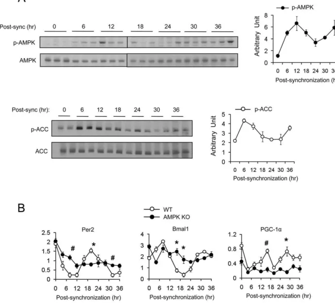
In live animals, the AMPK activity in the hypothalamus may be influenced by behavioral, physiological or metabolic fluctuations during the 24 hr cycle. Therefore, whether the AMPK cycle is cell-autonomous or not cannot be addressed in live animals. To test whether the cyclic pattern of AMPK activity is cell autono-mous, we examined T172 phosphorylation over a period 36 hr in wild type murine embryo fibroblasts (mefs) after synchronizing with forskolin [45]. AMPK phosphorylation in mefs exhibited an oscillatory pattern with a 24 hr period, with the maximal and minimal phosphorylation occurring approximately 12 hr and 24 hr after foskolin treatment, respectively (Fig 2A). AMPK-mediated phophorylation of ACC1 (S79) [46,47] also exhibited an oscillatory pattern that closely resembled the AMPK phosphory-lation pattern. These findings indicate that AMPK activity has cell-autonomous circadian rhythm.
AMPK is essential for circadian rhythm generation in mefs
In order to determine whether AMPK is required for circadian rhythm, we measured the mRNA levels of circadian genesPer2,
Bmal1 and PGC-1a in WT and AMPKa1/a22/2 mefs after forskolin synchronization. As shown in Fig. 2B, the oscillatory pattern ofPer2,Bmal1andPGC-1aexpression was not present in
AMPKa1/a22/2 mefs. These results indicate that not only is AMPK activity regulated by the circadian rhythm, but that it is also essential for circadian rhythm generation.
AMPKa12/2 and AMPKa22/2mice have an altered free-running period and feeding rhythm
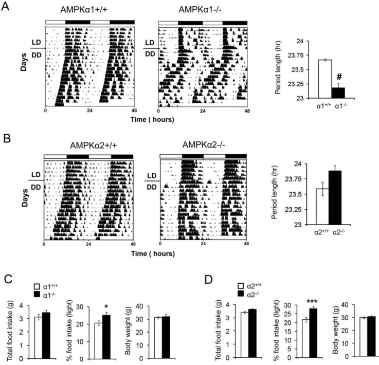
The SCN clock drives the circadian rhythm of locomotor activity. Since AMPK is essential for circadian rhythm generation in mefs, we investigated whether it is important for circadian rhythm generation in the SCN. We monitored the free-running locomotor activity in AMPKa12/2 and AMPKa22/2 mice
and their littermate controls. For this purpose, we used two different environmental conditions: A light-dark (LD) cycle, in which mice are exposed to 12 hr of light (6 am–6 pm) and 12 hr of
Figure 1. Circadian oscillation of AMPK activity in hypothalamus across the 24 hr light-dark cycle.Left, A representative Western blot showing the phosphorylation level of AMPK (T172) in extracts from mouse hypothalamus which were harvested at 4 hr intervals in a 12h light:12h dark (LD) cycle. The entire hypothalamus from 3 months old male C57BL/6J mice was used for this experiment. Right, Quantification of phosphorylated AMPKa(T172) is shown in arbitrary units (n = 3–4 per time point). Average AMPK activity (arbitrary unit) was calculated by densitometric quantification of phosphorylated proteins normalized to total proteins. Lights-on (6am; light) is indicated by a white bar and lights-off (6pm; dark) is indicated by a black bar. Results are means6S.E. * P,0.05 between 10 am and 10 pm.
doi:10.1371/journal.pone.0018450.g001
darkness (6 pm–6 am) and a dark-dark (DD) cycle, in which mice are exposed to constant darkness. The presence of circadian rhythmicity in DD is indicative of a functioning internal clock. As shown in Fig. 3A and B, both AMPKa12/2and AMPKa22/2
mice exhibited persistent circadian rhythmicity in DD, and the amplitudes of locomotor activity were similar to that of the WT littermates. The presence of circadian rhythmicity in the absence of daily light entrainment (i.e. DD) indicates that the SCN clock in AMPKa12/2 and AMPKa22/2 mice is largely intact.
However in the absence of light entrainment, the free running period of AMPKa12/2 mice was shorter than that of
AMPKa1+/+ littermates (23.2 hr vs. 23.7 hr, P = 0.0003). In contrast, the free running period of AMPKa2-/2was longer than
that of AMPKa2+/+littermates (23.9 hr vs. 23.6 hr), but this did not reach statistical significance (P = 0.07). The period lengths of
C57BL/6J, AMPKa1+/+ and AMPKa2+/+ mice were nearly identical (Figure S1).
Since AMPK activity in the hypothalamus stimulates food intake [28,29,30], it may play a role in the diurnal rhythm of food intake. To test this, we measured food intake of AMPKa12/2, AMPKa22/2and their littermate controls during the light and dark phases. The total food intake and body weight for AMPKa12/2, AMPKa22/2 and their littermate controls
was the same (Fig. 3 C, D). As nocturnal animals, mice consume most of their food during the dark phase. Both AMPKa12/2
and AMPKa22/2 mice ate more food during the light phase
than their wild-type littermates (20.6% vs. 25.1% for the AMPKa1 pair and 21.8% vs. 28% for the AMPKa2 pair)
(Fig. 3C), indicating that AMPK-deficient mice have blunted feeding rhythm.
Figure 2. Rhythmic expression of AMPK activity is cell autonomous.(A) WT mefs were synchronized by forskolin and harvested at the indicated time point. Phosphorylated AMPKa(T172) and phosphorylated-ACC (S79) were assessed by Western blot. Average AMPK and ACC activity (arbitrary unit) were calculated by densitometric quantification of phosphorylated proteins normalized to total proteins. Experiments were repeated at least three times. (B) Expression level (arbitrary units) of mPer2, Bmal1 and PGC-1ain WT and AMPKa1/a2 double knockout (AMPK KO) mefs after synchronization with forskolin. Results are means6S.E. * P,0.05,#P,0.001between WT and AMPK KO mefs.
AMPK is important for body temperature rhythm
We investigated whether AMPK is important for the circadian rhythmicity of metabolic parameters such as body temperature in light-dark (LD) as well as in constant darkness (DD) and oxygen consumption (VO2) (Fig 4). Compared to WT mice, AMPKa12/2
mice clearly had dampened circadian rhythm of core temperature (Fig. 4A). However, circadian rhythm of core temperature of AMPKa22/2appeared to be very similar to that of WT mice
(Fig. 4B). To better quantify the amplitude of the core temperature
rhythm, we performed cosinor analysis. A representative cosinor plot for each genotype in light-dark (LD) is shown in Fig. 4D. The calculations of the amplitude of the cosine curves indicate that AMPKa12/2 had lower amplitude in LD than either WT or
AMPKa22/2mice (Fig. 4C). The amplitude of the cosine curves of AMPKa12/2mice was also lower in DD, but the difference did not reach statistical significance.
We then compared VO2of WT and AMPKa12/2 mice in
LD. Although AMPKa12/2 mice tended to have higher VO2 Figure 3. Altered free-running period in AMPKa-deficient mice.(A–B) The activity rhythm was monitored by wheel running under light:dark
cycles 12 hr:12 hr (LD) or under constant darkness (DD). Activity records of representative AMPKa12/2and AMPKa22/2mice and their wild-type littermates are shown in double plotted actograms. Each horizontal line represents a 48 hr period and the vertical bars represent wheel running in 10-minute bins (n = 5–6).#P,0.0001, between AMPKa1+/+and AMPKa12/2mice. The free-running period was determined by using thex2 periodogram for days 1–14 in DD. The periods of AMPKa12/2, AMPKa22/2and their wild type littermate mice are shown in the right panel. (C,D) Total 24 hr food intake, % of food intake during the light period and body weight of AMPKa12/2and AMPKa22/2and their wild-type littermates (n = 10). Results are means 6 S.E. * P,0.05 between AMPKa1+/+and AMPKa12/2mice. *** P,0.001 between AMPKa2+/+and AMPKa22/2mice.
doi:10.1371/journal.pone.0018450.g003
than WT mice, they had similar amplitude of the circadian rhythm of VO2 (Fig. 4E). To assess energy utilization, we measured respiratory exchange ratio (RER) in AMPKa12/2
mice and WT mice. There was clear diurnal shift for energy utilization in both AMPKa12/2 mice. AMPKa12/2 mice showed a tendency toward the blunting of the shift, but it was not statistically significant. Taken together, these results indicate that AMPKa1 is required for generating normal amplitudes of core temperature rhythm, but not VO2or RER rhythms.
Expression patterns of Clock genes in peripheral tissues in AMPKadeficient mice
The circadian clock in peripheral tissues is self-sustained and can be entrained by food [6,7,31,48]. To further explore the role of AMPK in peripheral clock function, we examined the daily expression profile of circadian genes in the heart, gastrocnemius muscle and epididymal white fat in AMPKa12/2 and
AMPKa22/2mice. As shown in Fig. 5, the expression pattern
of the core clock genes (Per2, Bmal1, and Clock) exhibited a 24 hr rhythmicity in the heart, skeletal muscle and fat of WT mice, consistent with previous observations [49,50]. The heart and skeletal muscle express predominantly AMPKa2 and very little
AMPKa1. On the other hand, fat expresses predominantly
AMPKa1 and very little AMPKa2. Consistent with this expression
pattern of the two isoforms, the cyclic expression pattern of the core clock genes was preserved in the heart and skeletal muscle of AMPKa12/2mice (Fig. 5A), but they were significantly blunted
in AMPKa22/2 mice (Fig. 5B). Also as expected, the cyclic
expression pattern of clock genes was blunted in AMPKa12/2
fat (Fig. 5A) but not in AMPKa22/2fat (Fig. 5B). Surprisingly, the expression patterns ofPGC-1aand leptindid not fit this pattern.
The cyclic expression pattern ofPGC-1awas disrupted in the heart
and skeletal muscle of both AMPKa12/2 and AMPKa22/2
mice. Similarly, the cyclic expression pattern ofleptinwas disrupted
in the fat of both AMPKa12/2 and AMPKa22/2 mice.
Therefore, although the role of each isoform of AMPK in generating the cyclic expression pattern of core clock genes correlated with their relative abundance in peripheral tissues, the cyclic expression pattern ofPGC-1aand leptindid not.
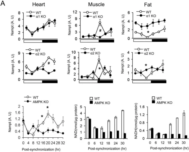
Circadian oscillation ofNamptmRNA and NAD+ production requires AMPK
AMPK has been shown to increase the expression of Nampt
and the product of its enzymatic reaction, NAD+ [51,52]. Since the circadian oscillation of NAD+ levels promotes circadian
Figure 5. Clock and clock related gene expression in heart, skeletal muscle and fat in (A) AMPKa12/2, (B) AMPKa22/2and WT mice.mRNA levels of mPer2, Bmal1, Clock, PGC-1aand leptin were measured by real-time PCR. The relative levels of mRNA are presented in arbitrary units. Light on is indicated by a white bar and light off is indicated by a black bar. The same WT data is plotted in A and B. Results are mean6S.E. * P,0.05,#P,0.001 between WT versus AMPKadeficient mice (n = 4–5 for each time point).
doi:10.1371/journal.pone.0018450.g005
rhythm generation via SIRT1 [17], we investigated whether AMPK is required for circadian oscillation ofNamptmRNA. As expected, Nampt mRNA levels displayed robust circadian oscillation in heart, skeletal muscle and fat in WT mice. However, circadian oscillation ofNamptmRNA was intact in the heart but not in skeletal muscle or fat of AMPKa12/2 mice (Fig. 6A). On the other hand, circadian oscillation of Nampt
mRNA was significantly blunted in all three tissues of AMPKa22/2 mice. Therefore, as was the case with PGC-1a
mRNA and leptin mRNA (Fig. 5), the circadian oscillation of
NamptmRNA expression in skeletal muscle and fat required both isoforms. To investigate whether circadian oscillation of Nampt
mRNA is cell-autonomous, we measuredNamptmRNA levels in WT and AMPKa1/a22/2 mefs after forskolin synchroniza-tion.NamptmRNA levels displayed circadian oscillation with the peak occurring 20 hrs after synchronization in WT mefs but not in AMPKa1/a22/2 mefs (Fig. 6B). Consistent with this,
intracellular NAD+ and NADH levels displayed circadian oscillation in WT mefs but not in AMPKa1/a22/2 mefs. Taken together, these results indicate that AMPK promotes circadian rhythm in part by generating the circadian oscillation of NAD+ production.
Discussion
AMPK regulates energy intake and expenditure to maintain cellular and whole body energy metabolism, which is coupled with daily light-dark cycles. To understand the intrinsic role of AMPK on circadian rhythmsin vivo, we studied the circadian behavior and physiology of mice deficient in AMPKa1 or AMPKa2. We found
that the circadian behavior of feeding and free-running period of AMPKa12/2mice and AMPKa22/2mice were dysregulated.
Circadian rhythms of core temperature were dysregulated in AMPKa12/2mice but not in AMPKa22/2mice. There was
no difference in the circadian rhythm of VO2between WT mice and AMPKa12/2mice.
In the hypothalamus, AMPK is a master regulator of food intake. Fasting increases AMPK activity and stimulates food intake, while refeeding suppress it [28,29]. It is intriguing that hypothalamic AMPK activity has a diurnal oscillation that peaks during the dark period when mice are active and eating. Thus, the timing of hypothalamic AMPK activity correlates with the timing of appetite.
We and others have previously demonstrated the effects of the molecular mechanisms of AMPK on circadian clockwork circuitry.
Figure 6. Circadian oscillation of NAD+and Nampt gene expression requires AMPK.(A) Nampt gene expression patterns of heart, skeletal muscle and fat tissue during 24 hr for AMPKa12/2, AMPKa22/2and WT mice. The relative levels of mRNA are presented in arbitrary units. Results are expressed as mean6S.E., * P,0.05,#P,0.001 between WT and AMPKadeficient mice. (n = 4–5 for each time point). The same WT data is plotted fora1 KO anda2 KO. (B) Nampt gene expression (arbitrary units) and cellular NAD+and NADH level in WT and AMPKa1/a2 KO mefs synchronized with forskolin. Results are expressed as mean6S.E., * P,0.05, between WT mefs and AMPKa1/a2 KO mefs (n = 3).
For example AMPK induces a phase advance of circadian expression of clock genes by degrading PER2 through phosphor-ylating Casein kinase IeSer389 [38] and AMPK contributes to metabolic entrainment of peripheral clocks by phosphorylating and destabilizing CRY1 [39]. In addition, the circadian rhythm of clock genes is absent in AMPKa1/a22/2mefs.
Thus it is possible that AMPK is a critical component or output of the central clock in the SCN hypothalamus. It is interesting to note that AMPKa12/2 mice exhibit a shortened period while AMPKa22/2mice tended to exhibit a longer period suggesting that thea1 anda2 isoform of AMPK may have distinct roles in
regulation of circadian period. Consistent with this, it has been observed that AMPKa2 activity is decreased in response to leptin injection in the hypothalamus but AMPKa1 activity is unchanged [28]. Further study is needed to evaluate the regulation of AMPK specifically on SCN and its neuronal network in hypothalamus.
In the peripheral tissue, AMPK controls energy metabolism by regulating the activity or expression of metabolic genes [31]. We found that AMPK regulates expression of peripheral clock genes in an isoform- and tissue-specific manner. One surprising discovery in this study was that the cyclic expression pattern of
PGC-1a, leptin and Nampt required the presence of both AMPK
isoforms even though only one isoform was predominantly expressed in the tissues we studied:a1 in fat anda2 in the heart
and skeletal muscle (Fig. 5). There may be several explanations for this that are not mutually exclusive. One explanation is that the two isoforms have non-overlapping functions even though the expression level of one isoform is significantly lower than the other. For example, in skeletal muscle, both AMPKa1 anda2 activities increased during treadmill running [53] but only AMPKa2 is required for glucose uptake after AICAR stimulation whereas AMPKa1 activation is required for glucose uptake after twitch
contraction [36,54]. Another explanation is that the cyclic expression pattern of PGC-1a, leptin and Nampt is more sensitive
to AMPK dosage than that of the core clock genes. Finally, the cyclic expression pattern of PGC-1a, leptin and Nampt may also
depend on extracellular signals. Circadian gene expression in peripheral tissues is intimately connected to feeding and the nutrient state [6,7,8,48]. Since the expression of leptin [55], PGC-1a [56] and Nampt [57] is regulated by food intake, and
the feeding rhythm is blunted in both AMPKa12/2 and
AMPKa22/2 mice (Fig. 3C), it is possible that the disruption of the expression pattern ofleptin, PGC-1aand NamptmRNA may,
at least in part, have resulted from feeding rhythm disruption. In addition, there may be cross-talks between fat and the heart or skeletal muscle that may be important for the cyclic expression pattern ofleptin and PGC-1ain which case the AMPK deficiency in
one tissue may affect the expression pattern ofleptin or PGC-1ain
another tissue.
The activity of SIRT1 and the NAD+salvage pathway regulate the circadian rhythm [20,21]. NAD+ level and Nampt, a rate limiting enzyme mediating NAD+biosynthesis, cycles with a 24-hour rhythm [17]. Recent studies showed that activation of AMPK enhances SIRT1 activity by increasing Nampt expression and intracellular NAD+levels, which induces deacetylation of SIRT1 targets such as PGC-1a[52,58]. Consistent with this we found that
rhythmic expression ofNampt and PGC-1awas abolished in both
AMPKa12/2and AMPKa22/2mice. It has been shown that PGC-1a is important for circadian rhythm generation in skeletal
muscle and liver [24] and is also an AMPK substrate. Our results indicate that the circadian regulation of the Nampt-SIRT1-PGC-1a
pathway is at least partially dependent on AMPKin vivo.
In summary, the role of AMPK in generating free-running period, metabolic rhythms and clock gene expression in peripheral
tissues is tissue- and isoform-specific. Furthermore AMPK is required for the cycling of NAD+level and circadian expression of
Nampt and PGC-1a. Thus, we demonstrated the importance of
AMPK in the circadian rhythms of behavior, energy metabolism and gene expression at the whole-organism level. Except for the gene expression changes (Fig. 5 and Fig. 6), the degree to which the circadian rhythm is disrupted in these AMPK-deficient mice is rather modest. However, this is not surprising since only one of the two isoforms is missing in each AMPK-deficient mouse, and it is well known that there is some functional redundancy between the two isoforms. Moreover, as it has been shown in skeletal muscle [36], deletion of one isoform can lead to a compensatory increase in the expression of the other isoform. Thus, conditional-knockout of both isoforms will be needed to fully demonstrate the role of AMPK in circadian rhythms.
Materials and Methods
Animals
Generation of AMPKa12/2 [36] and AMPKa22/2 [37] mice was previously described. AMPKa12/2and AMPKa22/2
mice and their littermates were generated by backcrossing AMPKa12/2 and AMPKa22/2 mice to C57BL/6J mice
(Jackson Laboratory, Bar Harbor, ME) for 6 generations followed by brother-sister mating. We used age-matched male mice for all our studies. For period length measurements and circadian rhythm of food intake studies, we used wile-type littermates as controls. For real-time PCR, indirect calorimetry and body temperature studies, we used C57BL/6J mice as wild-type controls. The mice were bred and group-housed in the animal facility in a 12 h-light/12 h-dark cycle (12L:12D) and provided food and water ad libitum. All experiments were approved by NHLBI ACUC (Animal Care and Use Committee).
Body weight and food intake measurements
Body weight and food intake were monitored during 5 days in four months old male AMPKa12/2, AMPKa22/2and their wild-type littermates exposed to a 12 hr:12 hr LD cycle.
Mouse wheel running activity
Voluntary mouse activity was measured by activity wheel running. Two months old male AMPKa12/2and AMPKa22/2mice and
their wild-type littermates were individually placed into cages with a running wheel and allowed to acclimate for one week prior to the experiment. Activity was measured with activity wheel counters (model 86061), an Animal Wheel Monitor Starter Interface (model 86056) and AWM software provided by Layfayette Instrument Company, Inc (Layfaytte, IN). Data were downloaded at 10 minute intervals during a 12 hr light/12 hr dark (LD) cycle for 10 days followed by a 24 hr constant darkness (DD) cycle for 30 days. Clock Lab (Actimetrics) Data Analysis software was used to produce the double-plotted actogram and period.
Indirect calorimetry
Metabolic rate (VO2) was determined by Oxymax chambers (Columbus Instruments). Energy expenditure was recorded by calculating the average energy expenditure for each 10 min time point during one week of LD 12 hr:12 hr cycle followed by three weeks of DD 12 hr:12 hr cycle. A few time points (,14 out of 1152) were found to have negative values. These outlier values were removed before calculation. Respiratory exchange ratio (RER) was calculated as the ratio between the CO2 production and the O2consumption.
Body temperature
For internal body temperature monitoring, mice were implanted with Data Science International (DSI) ETA-F20 transmitters. The transmitter was placed in the abdominal cavity and the ECG leads were sutured to the chest wall in a Lead II position. Animals were allowed to recover for two weeks prior to the initiation of monitoring using telemetric receivers. Body temperature was recorded under LD 12 hr:12 hr for one week followed by DD 12 hr:12 hr for three more weeks. Data were analyzed using DSI Ponemah software.
Cosinor Analysis
To analyze the amplitude of the core temperature rhythms, cosinor analysis was performed using Circadian Physiology software [59]. Cosine curves were fit to the data using a fixed 24-hr period. Prior to data analysis, data points showing a temperature below 35uC (,13 out of 7000 data points) were removed from each dataset as outlier data points.
Cell culture
AMPKa1/a2 double knockout and WT mouse embryo fibroblasts (mefs) [60] were maintained in high glucose Dulbecco’s Modified Eagle Medium (DMEM) with 10% fetal bovine serum. Confluent cells were synchronized with 10mM of forskolin (Calbiochem) treatment for 30 min.
Immunoblotting
Cells were lysed in RIPA buffer and subjected to Western blot. Dissected hypothalami from three months old male C57LB/6J mice were immediately frozen in liquid nitrogen and homogenized in lysis buffer [61]. Phosphorylation of AMPKaand ACC were determined by electrophoresis on 4–12% gradient SDS-polyacryl-amide gel followed by anti-phospho AMPKa (T172) and
anti-phospho ACC (S79) antibodies from Cell Signaling.
Real-time PCR
Heart, gastrocnemius muscle and epididymal white fat were isolated every 4 hr for a total 24 hr (mice maintained in 12L:12D) and pulverized in liquid nitrogen. Total RNA was isolated using the TRIzol reagent extraction kit (Invitrogen), according to
manufac-turer’s instructions. RNA was subsequently reverse-transcribed to cDNA by using the high capacity cDNA archive kit (ABI). The mRNA levels were measured by real time PCR using the TaqMan Gene Expression system and the ABI PRISMTM 7900HT Sequence Detection System (Applied Biosystem).
NAD+and NADH level measurement
The NAD+and NADH levels were measured from whole cell extracts of WT mefs and AMPKa1/a2 double knockout mefs
synchronized by with forskolin, by using the Biovision NAD/ NADH Quantification kit according to the manufacturer’s instruction.
Statistical analysis
Independent t tests (two-tailed) were used to compare two groups. For comparison between three groups, one-way ANOVA followed by Bonferroni post-test was used. Significance was accepted at P,0.05 unless indicated otherwise. Results are expressed as the mean6SEM
Supporting Information
Figure S1 Comparison of the free-running period of
wild-type controls. The free-running period of C57BL/6J (n = 5), AMPKa1+/+(n = 5) and AMPKa2+/+ (n = 7) mice are shown. The free-running period was determined by using thex2 periodogram for days 1–14 in DD. Result is expressed as means6
SEM. (TIF)
Acknowledgments
We thank Dalton Saunders for his assistance with animal management.
Author Contributions
Conceived and designed the experiments: JHU JHC SY. Performed the experiments: JHU JSP DAS SY. Analyzed the data: JHU JSP SY MKK DAS JHC. Contributed reagents/materials/analysis tools: MF BV. Wrote the paper: JHU JHC JSP SY AB.
References
1. Reppert SM, Weaver DR (2002) Coordination of circadian timing in mammals. Nature 418: 935–941.
2. Ibuka N, Inouye SI, Kawamura H (1977) Analysis of sleep-wakefulness rhythms in male rats after suprachiasmatic nucleus lesions and ocular enucleation. Brain Res 122: 33–47.
3. Ibuka N, Nihonmatsu I, Sekiguchi S (1980) Sleep-wakefulness rhythms in mice after suprachiasmatic nucleus lesions. Waking Sleeping 4: 167–173. 4. Welsh D, Richardson GS, Dement WC (1988) Effect of running wheel
availability on circadian patterns of sleep and wakefulness in mice. Physiol Behav 43: 771–777.
5. Lowrey PL, Takahashi JS (2004) Mammalian circadian biology: elucidating genome-wide levels of temporal organization. Annu Rev Genomics Hum Genet 5: 407–441.
6. Stokkan KA, Yamazaki S, Tei H, Sakaki Y, Menaker M (2001) Entrainment of the circadian clock in the liver by feeding. Science 291: 490–493.
7. Damiola F, Le Minh N, Preitner N, Kornmann B, Fleury-Olela F, et al. (2000) Restricted feeding uncouples circadian oscillators in peripheral tissues from the central pacemaker in the suprachiasmatic nucleus. Genes Dev 14: 2950–2961. 8. Kohsaka A, Laposky AD, Ramsey KM, Estrada C, Joshu C, et al. (2007) High-fat diet disrupts behavioral and molecular circadian rhythms in mice. Cell Metab 6: 414–421.
9. Leone TC, Weinheimer CJ, Kelly DP (1999) A critical role for the peroxisome proliferator-activated receptor alpha (PPARalpha) in the cellular fasting response: the PPARalpha-null mouse as a model of fatty acid oxidation disorders. Proc Natl Acad Sci U S A 96: 7473–7478.
10. Kersten S, Seydoux J, Peters JM, Gonzalez FJ, Desvergne B, et al. (1999) Peroxisome proliferator-activated receptor alpha mediates the adaptive response to fasting. J Clin Invest 103: 1489–1498.
11. Goh BC, Wu X, Evans AE, Johnson ML, Hill MR, et al. (2007) Food entrainment of circadian gene expression altered in PPARalpha-/- brown fat and heart. Biochem Biophys Res Commun 360: 828–833.
12. Barak Y, Nelson MC, Ong ES, Jones YZ, Ruiz-Lozano P, et al. (1999) PPAR gamma is required for placental, cardiac, and adipose tissue development. Mol Cell 4: 585–595.
13. Rosen ED, Sarraf P, Troy AE, Bradwin G, Moore K, et al. (1999) PPAR gamma is required for the differentiation of adipose tissue in vivo and in vitro. Mol Cell 4: 611–617.
14. Kubota N, Terauchi Y, Miki H, Tamemoto H, Yamauchi T, et al. (1999) PPAR gamma mediates high-fat diet-induced adipocyte hypertrophy and insulin resistance. Mol Cell 4: 597–609.
15. Wang N, Yang G, Jia Z, Zhang H, Aoyagi T, et al. (2008) Vascular PPARgamma controls circadian variation in blood pressure and heart rate through Bmal1. Cell Metab 8: 482–491.
16. Nakahata Y, Sahar S, Astarita G, Kaluzova M, Sassone-Corsi P (2009) Circadian control of the NAD+salvage pathway by CLOCK-SIRT1. Science 324: 654–657. 17. Ramsey KM, Yoshino J, Brace CS, Abrassart D, Kobayashi Y, et al. (2009) Circadian clock feedback cycle through NAMPT-mediated NAD+biosynthesis. Science 324: 651–654.
18. Bordone L, Guarente L (2005) Calorie restriction, SIRT1 and metabolism: understanding longevity. Nat Rev Mol Cell Biol 6: 298–305.
19. Dali-Youcef N, Lagouge M, Froelich S, Koehl C, Schoonjans K, et al. (2007) Sirtuins: the ‘magnificent seven’, function, metabolism and longevity. Ann Med 39: 335–345.
21. Nakahata Y, Kaluzova M, Grimaldi B, Sahar S, Hirayama J, et al. (2008) The NAD+-dependent deacetylase SIRT1 modulates CLOCK-mediated chromatin remodeling and circadian control. Cell 134: 329–340.
22. Puigserver P, Wu Z, Park CW, Graves R, Wright M, et al. (1998) A cold-inducible coactivator of nuclear receptors linked to adaptive thermogenesis. Cell 92: 829–839.
23. Wu Z, Puigserver P, Andersson U, Zhang C, Adelmant G, et al. (1999) Mechanisms controlling mitochondrial biogenesis and respiration through the thermogenic coactivator PGC-1. Cell 98: 115–124.
24. Liu C, Li S, Liu T, Borjigin J, Lin JD (2007) Transcriptional coactivator PGC-1alpha integrates the mammalian clock and energy metabolism. Nature 447: 477–481.
25. Turek FW, Joshu C, Kohsaka A, Lin E, Ivanova G, et al. (2005) Obesity and metabolic syndrome in circadian Clock mutant mice. Science 308: 1043–1045. 26. Yang S, Liu A, Weidenhammer A, Cooksey RC, McClain D, et al. (2009) The role of mPer2 clock gene in glucocorticoid and feeding rhythms. Endocrinology 150: 2153–2160.
27. Hardie DG (2007) AMP-activated/SNF1 protein kinases: conserved guardians of cellular energy. Nat Rev Mol Cell Biol 8: 774–785.
28. Minokoshi Y, Alquier T, Furukawa N, Kim YB, Lee A, et al. (2004) AMP-kinase regulates food intake by responding to hormonal and nutrient signals in the hypothalamus. Nature 428: 569–574.
29. Andersson U, Filipsson K, Abbott CR, Woods A, Smith K, et al. (2004) AMP-activated protein kinase plays a role in the control of food intake. J Biol Chem 279: 12005–12008.
30. Lopez M, Lage R, Saha AK, Perez-Tilve D, Vazquez MJ, et al. (2008) Hypothalamic fatty acid metabolism mediates the orexigenic action of ghrelin. Cell Metab 7: 389–399.
31. Kahn BB, Alquier T, Carling D, Hardie DG (2005) AMP-activated protein kinase: ancient energy gauge provides clues to modern understanding of metabolism. Cell Metab 1: 15–25.
32. Stapleton D, Mitchelhill KI, Gao G, Widmer J, Michell BJ, et al. (1996) Mammalian AMP-activated protein kinase subfamily. J Biol Chem 271: 611–614.
33. Lihn AS, Jessen N, Pedersen SB, Lund S, Richelsen B (2004) AICAR stimulates adiponectin and inhibits cytokines in adipose tissue. Biochem Biophys Res Commun 316: 853–858.
34. Cheung PC, Salt IP, Davies SP, Hardie DG, Carling D (2000) Characterization of AMP-activated protein kinase gamma-subunit isoforms and their role in AMP binding. Biochem J 346 Pt 3: 659–669.
35. Woods A, Salt I, Scott J, Hardie DG, Carling D (1996) The alpha1 and alpha2 isoforms of the AMP-activated protein kinase have similar activities in rat liver but exhibit differences in substrate specificity in vitro. FEBS Lett 397: 347–351. 36. Jorgensen SB, Viollet B, Andreelli F, Frosig C, Birk JB, et al. (2004) Knockout of the alpha2 but not alpha1 59-AMP-activated protein kinase isoform abolishes 5-aminoimidazole-4-carboxamide-1-beta-4-ribofuranosidebut not contraction-in-duced glucose uptake in skeletal muscle. J Biol Chem 279: 1070–1079. 37. Viollet B, Andreelli F, Jorgensen SB, Perrin C, Geloen A, et al. (2003) The
AMP-activated protein kinase alpha2 catalytic subunit controls whole-body insulin sensitivity. J Clin Invest 111: 91–98.
38. Um JH, Yang S, Yamazaki S, Kang H, Viollet B, et al. (2007) Activation of 59-AMP-activated kinase with diabetes drug metformin induces casein kinase Iepsilon (CKIepsilon)-dependent degradation of clock protein mPer2. J Biol Chem 282: 20794–20798.
39. Lamia KA, Sachdeva UM, DiTacchio L, Williams EC, Alvarez JG, et al. (2009) AMPK regulates the circadian clock by cryptochrome phosphorylation and degradation. Science 326: 437–440.
40. Vieira E, Nilsson EC, Nerstedt A, Ormestad M, Long YC, et al. (2008) Relationship between AMPK and the transcriptional balance of clock-related genes in skeletal muscle. Am J Physiol Endocrinol Metab 295: E1032–1037.
41. Saper CB, Scammell TE, Lu J (2005) Hypothalamic regulation of sleep and circadian rhythms. Nature 437: 1257–1263.
42. Weaver DR (1998) The suprachiasmatic nucleus: a 25-year retrospective. J Biol Rhythms 13: 100–112.
43. Ralph MR, Foster RG, Davis FC, Menaker M (1990) Transplanted suprachiasmatic nucleus determines circadian period. Science 247: 975–978. 44. Okamura H (2007) Suprachiasmatic nucleus clock time in the mammalian
circadian system. Cold Spring Harb Symp Quant Biol 72: 551–556. 45. Balsalobre A, Marcacci L, Schibler U (2000) Multiple signaling pathways elicit
circadian gene expression in cultured Rat-1 fibroblasts. Curr Biol 10: 1291–1294.
46. Ha J, Daniel S, Broyles SS, Kim KH (1994) Critical phosphorylation sites for acetyl-CoA carboxylase activity. J Biol Chem 269: 22162–22168.
47. Ruderman NB, Saha AK, Vavvas D, Witters LA (1999) Malonyl-CoA, fuel sensing, and insulin resistance. Am J Physiol 276: E1–E18.
48. Yoo SH, Yamazaki S, Lowrey PL, Shimomura K, Ko CH, et al. (2004) PERIOD2::LUCIFERASE real-time reporting of circadian dynamics reveals persistent circadian oscillations in mouse peripheral tissues. Proc Natl Acad Sci U S A 101: 5339–5346.
49. Yamamoto T, Nakahata Y, Soma H, Akashi M, Mamine T, et al. (2004) Transcriptional oscillation of canonical clock genes in mouse peripheral tissues. BMC Mol Biol 5: 18.
50. Ando H, Yanagihara H, Hayashi Y, Obi Y, Tsuruoka S, et al. (2005) Rhythmic messenger ribonucleic acid expression of clock genes and adipocytokines in mouse visceral adipose tissue. Endocrinology 146: 5631–5636.
51. Fulco M, Cen Y, Zhao P, Hoffman EP, McBurney MW, et al. (2008) Glucose restriction inhibits skeletal myoblast differentiation by activating SIRT1 through AMPK-mediated regulation of Nampt. Dev Cell 14: 661–673.
52. Canto C, Gerhart-Hines Z, Feige JN, Lagouge M, Noriega L, et al. (2009) AMPK regulates energy expenditure by modulating NAD+metabolism and SIRT1 activity. Nature 458: 1056–1060.
53. Jorgensen SB, Wojtaszewski JF, Viollet B, Andreelli F, Birk JB, et al. (2005) Effects of alpha-AMPK knockout on exercise-induced gene activation in mouse skeletal muscle. FASEB J 19: 1146–1148.
54. Jensen TE, Schjerling P, Viollet B, Wojtaszewski JF, Richter EA (2008) AMPK alpha1 activation is required for stimulation of glucose uptake by twitch contraction, but not by H2O2, in mouse skeletal muscle. PLoS One 3: e2102. 55. Saladin R, De Vos P, Guerre-Millo M, Leturque A, Girard J, et al. (1995)
Transient increase in obese gene expression after food intake or insulin administration. Nature 377: 527–529.
56. Nisoli E, Tonello C, Cardile A, Cozzi V, Bracale R, et al. (2005) Calorie restriction promotes mitochondrial biogenesis by inducing the expression of eNOS. Science 310: 314–317.
57. Arany Z, Lebrasseur N, Morris C, Smith E, Yang W, et al. (2007) The transcriptional coactivator PGC-1beta drives the formation of oxidative type IIX fibers in skeletal muscle. Cell Metab 5: 35–46.
58. Canto C, Jiang LQ, Deshmukh AS, Mataki C, Coste A, et al. (2010) Interdependence of AMPK and SIRT1 for metabolic adaptation to fasting and exercise in skeletal muscle. Cell Metab 11: 213–219.
59. Refinetti R (2006) Circadian physiology. Boca Raton: CRC Press/Taylor & Francis Group.
60. Laderoute KR, Amin K, Calaoagan JM, Knapp M, Le T, et al. (2006) 59 -AMP-activated protein kinase (AMPK) is induced by low-oxygen and glucose deprivation conditions found in solid-tumor microenvironments. Mol Cell Biol 26: 5336–5347.
61. Martin TL, Alquier T, Asakura K, Furukawa N, Preitner F, et al. (2006) Diet-induced obesity alters AMP kinase activity in hypothalamus and skeletal muscle. J Biol Chem 281: 18933–18941.