einstein. 2012;10(1):116-7
LEARNING BY IMAGES
Coronary heart disease patient with implantable cardioverter
desfibrilator and electrical storm submitted to
ventricular tachycardia ablation
Paciente coronariopata com cardiodesfibrilador e tempestade elétrica
submetido à ablação de taquicardia ventricular
Nilton José Carneiro da Silva1, Bruno Pereira Valdigem1, Christian Luize2, Fernando Lopes Nogueira2, Claudio Cirenza3,
Guilherme Fenelon3, Marcia Regina Pinho Makdisse1, Fátima Dumas Cintra3, Angelo Amato Vincenzo De Paola3
1 Hospital Israelita Albert Einstein – HIAE, Sao Paulo (SP), Brazil.
2 Universidade Federal de São Paulo – UNIFESP, São Paulo (SP), Brazil.
3 Universidade Federal de São Paulo – UNIFESP, São Paulo (SP), Brazil; Hospital Israelita Albert Einstein – HIAE, São Paulo (SP), Brazil.
Corresponding author: Nilton José Carneiro da Silva – Avenida Albert Einstein, 627 4ª andar, consultório 413 Bloco A – Morumbi – Zip code: 05651-901 – São Paulo (SP), Brazil – Phone.: (11) 2151-5143 – E-mail: niltonjose@einstein.br
Received on: Feb 28, 2011 – Accepted on: Feb 12, 2012
Figure 1. Electrocardiogram presenting clinical tachycardia at an Emergency Department Unit
Figure 2. Implantable cardioverter defibrillator (ICD) record of ventricular tachycardia and cardioversion
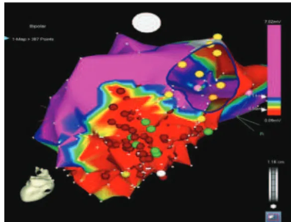
Figure 3. Electroanatomical map in posterior projection, mitral ring identified at 1 o’clock, red area corresponding to low voltage – “scar” and red circles are the sites where radiofrequency was applied
117
Coronary heart disease patient with ICD and electrical storm
einstein. 2012;10(1):116-7
A 69-year old diabetic patient with O2-dependent chronic obstructive pulmonary disease (COPD), history of myocardial revascularization in 1998 and one episode of ventricular tachycardia and placement of cardiac defibrillator in 2003. He was seen at the Emergency Department in February 2010 with palpitations and four shocks in the last two hours. The electrocardiogram showed wide QRS tachycardia with right bundle branch block (RBB) and superior axis (Figure 1). Arrhythmia reverted after sedation and eletrical cardioversion (ECV). The implantable cardioverter defibrillator (ICD) evaluation demonstrated appropriate therapy (shock for ventricular tachycardia), confirming the main hypothesis (Figure 2). He presented recurrence of tachycardia despite the optimized drug treatment, including intravenous amiodarone. The use of the antitachycardia pacing (ATP) (fast stimuli) through the ICD did not stop tachycardia; on the contrary, it would even increase it. Hence the patient was submitted to catheter ablation of ventricular tachycardia. Since there was more than one inducible tachycardia episode, and the patient did not tolerate arrhythmia for a prolonged period, it was decided to perform combined ablation of the arrhythmic substrate, guided by the electroanatomical map (CARTO) (Figure 3) to
assist with electrophysiological data (Figure 4). The procedure was successful and there have been no new ventricular tachycardia episodes in clinical follow-up.
The use of the electroanatomical map in electrophysiological procedures enabled managing complex arrhythmias in patients at high pre-procedure risk (1). Atrial fibrillation and ventricular tachycardia
related to structural cardiopathy have been addressed with this technology in most specialized centers, showing superior results than the conventional technique(2-3).
REFERENcES
1. Aliot EM, Stevenson WG, Almendral-Garrote JM. EHRA/HRS Expert Consensus on Catheter Ablation of Ventricular Arrhythmias: developed in a partnership with the European Heart Rhythm Association (EHRA), a Registered Branch of the European Society of Cardiology (ESC), and the Heart Rhythm Society (HRS); in collaboration with the American College of Cardiology (ACC) and the American Heart Association (AHA). Heart Rhythm. 2009;6(6):886-933.
2. Hsia HH, Lin D, Sauer WH, Callans DJ, Marchlinski FE. Anatomic characterization of endocardial substrate for hemodynamically stable reentrant ventricular tachycardia: identification of endocardial conducting channels. Heart Rhythm. 2006;3(5):503-12.