4 4 6 4 4 6 4 4 6 4 4 6 4 4 6
Universidade Federal de São Paulo – Escola Paulista de Medicina
Mailing address: Luiz Roberto Leite – Setor de Eletrofisiologia Clínica – Rua Napoleão de Barros, 593 – 04024-002 – São Paulo, SP, Brazil – E-mail: [email protected]
English version by Stela Maris C. e Gandour
Objective - To assess the impact of syncope during sustained ventricular tachycardia on total and cardiac mortality in patients with chronic chagasic heart disease.
Methods - We assessed 78 patients with sustained ventricular tachycardia and chronic Chagas' heart disease. The mean age was 53±10 years, 45 were males, and the mean ejection fraction was 49.6±13%. The patients were divided into 2 groups according to the pre-sence (GI=45) or abpre-sence (GII=33) of syncope during sus-tained ventricular tachycardia.
Results - After a mean follow-up of 49 months, total mortality was 35% (28 deaths), 22 deaths having a cardiac cause (78.6%). No difference was observed in total (33.3% x 39.4%) and cardiac (26.7% x 30.3%) mortality, or in nonfatal sustained ventricular tachycardia between GI and GII patients (57.6% x 54.4%, respectively). However, the presence of syncope during recurrences was significantly greater in those patients who had had the symptom from the beginning (65.4% x 18.1%, p<0.01).
Conclusion - Syncope during the presentation of sustained ventricular tachycardia is not associated with an increase in total or cardiac mortality in patients with chronic Chagas' heart disease. However, syncope during the recurrence ventricular tachycardia is greater in pati-ents experiencing syncope in the first episode, of sustained ventricular tachycardia.
Key words: syncope, sustained ventricular tachycardia, chronic Chagas' heart disease
Arq Bras Cardiol, volume 77 (nº 5), 446-52, 2001
Luiz Roberto Leite, Guilherme Fenelon, Ângela Tavares Paes, Angelo Amato Vincenzo de Paola
São Paulo, SP - Brazil
The Impact of Syncope During Clinical Presentation of
Sustained Ventricular Tachycardia on Total and Cardiac
Mortality in Patients with Chronic Chagasic Heart Disease
Identification of patients at high risk of cardiac death is fundamental in cardiological investigation. The importance of syncope during the clinical presentation of sustained ven-tricular tachycardia, as a prognostic factor, was extensively studied in patients with coronary artery disease, and it was considered a worse prognosis when compared with hemo-dynamically well-tolerated sustained tachyarrhythmias 1-5. In
chronic chagasic heart disease, however, the relevance of syncope during clinical presentation of sustained ventricu-lar tachycardia is not totally understood, and its incidence is variable according to reports published in the literature 6-10.
The objective of this study was to assess the impact of syncope during the clinical presentation of sustained ven-tricular tachycardia on total, cardiac, and sudden death in patients with chronic chagasic heart disease.
Methods
We consecutively assessed 78 patients with chronic Chagas' heart disease after an episode of sustained ventri-cular tachycardia in the clinical electrophysiology section of the Hospital São Paulo of the Universidade Federal de São Paulo. Forty-five patients were males and 33 were females, their mean age being 53±10 years (29 to 74). The mean left ventricular ejection fraction was 49.6% ± 13% (24 to 76), and 24 (30.8%) patients had ejection fraction < 40%. In this po-pulation, 11.5% of the patients had advanced heart failure (NYHA functional class III or IV).
4 4 7 4 4 7 4 4 7 4 4 7 4 4 7
The electrophysiological study with programmed ven-tricular stimulation was performed in all patients, according to stimulation techniques previously reported 10-13, using up
to 3 extrastimuli with a minimum coupling of 200ms, under 2 cycles of basal stimulation (600 and 450ms) 10-13.
The patients were classified according to the presence (group I) or absence (group II) of syncope during clinical presentation of sustained ventricular tachycardia.
To comply with the objectives of the study, the follo-wing definitions were used: 1) syncope, the sudden loss of consciousness followed by spontaneous recovery, conside-red present when the episode occurconside-red during electrocar-diographic monitoring or when, after regaining conscious-ness, the patient was in sustained ventricular tachycardia; 2) sustained ventricular tachycardia, which is the presence of consecutive ventricular beats with a heart rate >100bpm and duration >30s, or the need for immediate reversion, when he-modynamic impairment occurs; 3) sudden cardiac death, in the case of a witnessed death, occurring within 1 hour at most after symptom onset in a clinically stable patient, or in the case of an unwitnessed death, the patient should have been asymptomatic within the 24 hours preceding death; 4) car-diac death, the sum of sudden death and that resulting progression of heart failure or thromboembolic events; 5) recurrence of nonfatal sustained ventricular tachycardia, re-corded on an electrocardiogram or Holter, with no evolution to ventricular fibrillation or cardiopulmonary arrest.
After hospital discharge, the patients were assessed monthly during the first 6 months, and then bimonthly du-ring the first year. Dudu-ring follow-up, the following events were defined: death due to cardiac causes (sudden and nonsudden), to noncardiac causes, deaths after a maximum follow-up of 10 years, and recurrence of nonfatal sustained ventricular tachycardia. The initial treatment was conside-red the one used since hospital discharge, and maintenance treatment comprised the drugs used since the last contact. Continuous variables were presented as mean ± standard deviation. For comparison between the groups, the chi-square test or Fisher exact test, when appropriate, was used for qualitative variables, and Student t test was used for quan-titative variables. Survival curves were depicted according to the Kaplan-Meier method and compared by the log-rank test.
Results
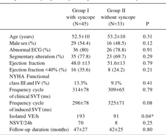
Of the 78 patients, 45 (57.7%) experienced syncope during clinical presentation of sustained ventricular tachy-cardia and comprised group I; the 33 (42.3%) patients who did not experience syncope comprised group II. Other symptoms present during sustained ventricular tachycar-dia were palpitation in 67 patients (GI=39 and GII=28, p=0.50) and dyspnea in 38 patients (GI=21 and GII=17, p=0.40). The clinical characteristics of both groups are sho-wn in table I. No difference between group I and II patients was observed in regard to age (52.5±10 and 53.2±10, p=0.31), sex, presence of advanced heart failure, left ventri-cular ejection fraction (48.1±13 and 51.6±13, p=0.8), number
of patients with ejection fraction <40% [GI=16 (35.6%) and GII=8 (24.2%), (p=0.21)], and segmentary alteration (77.8% and 69.7%, p=0.29). Density of isolated ventricular extrasys-toles per hour (193 vs 91, p=0.04) and nonsustained ventri-cular tachycardia in 24 hours (70 vs 8, p=0.25) recorded on the initial Holter was greater in the patients with syncope.
Most patients had electrocardiographic alterations, which were present in 80% (n=36) of the patients with syncope and 78.8% (n=26) of those without. The most com-mon electrocardiographic findings were as follows: right bundle-branch block associated with left anterosuperior di-visional block (29.5%), isolated right bundle-branch block (10.3%), isolated left bundle-branch block (10.3%), and left divisional anterosuperior block (11.5%). None of these alte-rations predominated in the patients experiencing or not ex-periencing syncope.
The frequency cycle of clinical sustained ventricular tachycardia was analyzed in 49 patients (GI=25, GII=24). The mean frequency cycle in GI was 314 ms [95% CI 282-347], and, in GII, it was 309 ms [95% CI 282-337], with no statisti-cally significant difference between the groups (p=0.79).
During electrophysiological study with programmed ventricular stimulation, sustained ventricular tachycardia was induced in 75 patients. The frequency cycle of induced sustained ventricular tachycardia was 296 ms [95% CI 273-319] in the patients experiencing syncope and 325 ms [95% CI 299-351] in those not experiencing syncope (tab. I). A ten-dency toward a shorter frequency cycle of induced sustai-ned ventricular tachycardia was observed in the patients experiencing syncope (p=0.08).
All chagasic patients with sustained ventricular ta-chycardia were successfully treated and were available for ambulatory follow-up. The cardiac drugs used during the last contact are listed in table II. At the end of follow-up, 71 patients were using amiodarone, 41 (91.1%) in the group of
Table I – Baseline characteristics of patients with sustained ventricular tachycardia and chronic chagasic heart disease according to the presence of syncope during clinical presentation of the arrhythmia
Group I Group II with syncope without syncope
(N=45) (N=33) P
Age (years) 52.5±10 53.2±10 0.31 Male sex (%) 29 (54.4) 16 (48.5) 0.12 Abnormal ECG (%) 36 (80) 26 (78.8) 0.91 Segmentary alteration (%) 35 (77.8) 23 (69.7) 0.29 Ejection fraction 48.0 ±13 51.6±13 0.79 Ejection fraction <40% (%) 16 (35.6) 8 (24.2) 0.21 NYHA Functional
class III and IV (%) 13.3% 9.1% 0.41 Frequency cycle 314±78 309±65 0.79 of clinical SVT (ms)
Frequency cycle 296±78 325±71 0.08 of induced SVT (ms)
Isolated VE/h 193 91 0.04*
NSVT/24h 70 8 0.25
Follow-up duration (months) 47±27 42±25 0.80
4 4 8 4 4 8 4 4 8 4 4 8 4 4 8
in group II, the mean follow-up was 46±32 months, and 5 (15.1%) patients were followed up for less than 24 months. Total mortality was 35% (28 deaths). No difference between the groups was observed in regard to total morta-lity; 15 (33.3%) patients died in group I, and 13 (39.4%) pati-ents died in group II (tab. III). The survival curve showed that, during the entire clinical follow-up, total mortality was similar in both groups (fig. 2).
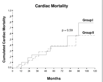
Of the deaths in the 78 patients, 28.2% had cardiac causes, accounting for 78.6% of deaths in this population (22 deaths). Figure 3 shows the curve of accumulated car-diac deaths in the 78 patients, which did not statistically dif-fer between groups I and II (p=0.64), 12 (26.7%) and 10 (30.3%) being the deaths from cardiac causes, respectively. Most cardiac deaths occurred suddenly. Of all cardiac dea-ths, 63.6% were considered sudden (14 deaths). Sudden death accounted for 50% (6/12) of the cardiac deaths in group I patients and for 80% (8/10) of the deaths in group II patients (p=0.24). Mean survival in group I was 83 months [95% CI 70-97], and in group II it was 75 months [95% CI 59-92] (tab. III).
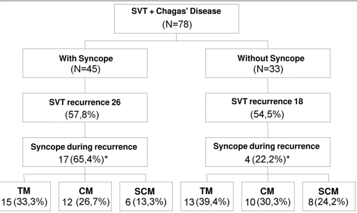
Recurrence of nonfatal sustained ventricular tachycar-dia was observed in 44 (56.4%) patients, 26 being in group I (57.8%) and 18 in group II (54.5%). No difference between the 2 groups was observed in the recurrence of nonfatal sustained ventricular tachycardia (fig. 4). Recurrences of sustained ventricular tachycardia were accompanied by syncope in 26.9% of the patients, being more frequent in those who already had the symptom prior to antiarrhythmic patients experiencing syncope and 30 (90.9%) in those not
experiencing syncope (p=0.21). Six patients were using sotalol, and 2 patients were not using antiarrhythmic drugs. Catheter ablation was performed in 34 patients, 18 (40%) pa-tients in group I and 16 (45%) papa-tients in group II (p=0.50). The use of a definitive pacemaker for bradyarrhythmias did not differ in the 2 groups. Six (13.3%) patients experiencing syncope and 7 (21.2%) not experiencing syncope used pa-cemakers implanted for bradyarrhythmias.
The mean follow-up time (fig. 1) was 49±33 months, and all patients were followed up for at least 1 year, except 1 patient in GI who was followed up for 10 months. In group I, the mean follow-up was 51±32 months, and 7 (15.5%) pati-ents in this group were followed up for less than 24 months;
Table II – Pharmacological and antiarrhythmic treatment
Group I Group II P with syncope without syncope
N=(45) N=(33)
Digitalis 10 (22.2%) 7 (21.2%) 1.00 ACE I 28 (62.2%) 24 (72.7%) 0.46 Diuretics 15 (33.3%) 17 (51.5%) 0.16 Amiodarone 41 (91.1%) 390.2±73.5 0.21 Dose média (mg) 30 (90.9%) 386.6±68.1 0.83
Sotalol 5 (15.1%) 1 (3%) 0.31
mean dose (mg) 224±35 160
Catheter ablation 18 (40%) 16 (45%) 0.50 Definitive pacemaker 6 (13.3%) 7 (21.2%) 0.75
ACE I - angiotensin-converting enzyme inhibitors.
Fig. 1 – Events during clinical follow-up of patients with chronic chagasic heart disease according to the presence of syncope during clinical presentation of sustained ventricular tachycardia. TM- total mortality; CM- cardiac mortality; SCM- sudden cardiac mortality.
TM
CM
SCM
TM
CM
SCM
With Syncope Without Syncope
SVT recurrence 26 SVT recurrence 18
Syncope during recurrence Syncope during recurrence
4 4 9 4 4 9 4 4 9 4 4 9 4 4 9
therapy. In the 26 group I patients with recurrence of nonfatal sustained ventricular tachycardia, 17 (65.4%) expe-rienced syncope; in the 18 group II patients with recurrence
of nonfatal sustained ventricular tachycardia, 4 (22.2%) experienced syncope (p<0.01).
To assess the importance of syncope associated with sustained ventricular tachycardia, the patients experien-cing syncope at any time during the study were compared with those not experiencing it. Therefore, syncope associa-ted with sustained ventricular tachycardia occurred in 4 of the 33 patients whose arrhythmia had initially occurred wi-thout syncope, adding to the 49 patients who experienced syncope associated with sustained ventricular tachycardia at any time during the study. When these 49 patients with syncope associated with sustained ventricular tachycardia were compared with the 29 without syncope, total (36.7% vs. 34.5%, p=0.91) and cardiac (24.5% and 34.5%) mortality did not differ at any time during the study.
Total and cardiac mortality was greater in the patients with left ventricular ejection fraction lower than 40%. Of the 28 deaths occurring during total clinical follow-up, 50% of the patients had an ejection fraction lower than 40% as compared with 30.8% of the total population and 29% of the patients without this event; this difference, however, was not statistically significant.
Discussion
Our study assessed the impact of syncope during the
Table III – Total, cardiac, and sudden mortality, and recurrence of nonfatal sustained ventricular tachycardia in patients with chronic chagasic heart disease
Total Cardiac Sudden SVT Survival Recurrence -free
Mortality mortality mortality recurrence (m) time (m)
(N=28) (N=22) (N=14) [95% CI] [95% CI]
Group I – with 15 (33.3%) 12 (26.7%) 6 (50%) 26 (57.8%) 83 [70; 97] 71 [59; 83] syncope (N=45)
Group II – without 13 (39.4%) 10 (30.3%) 8 (80%) 18 (54.5%) 75 [59; 92] 64 [50; 78] syncope (N=33)
P 0.43 0.59 0.24 0.82 0.50 0.59
Survival
Global Mortality
Group I Group II
Fig. 2 – Total survival curve of patients with chronic chagasic heart disease and sus-tained ventricular tachycardia according to the presence of syncope during clinical presentation of the arrhythmia. Group I- with syncope; group II- without syncope.
Months
Cumulated Cardiac Mortality
Group I
Group II
p = 0,59
Fig. 3 – Curve of accumulated cardiac mortality in patients with chronic chagasic heart disease and sustained ventricular tachycardia according to the presence of syncope during clinical presentation of the arrhythmia. Group I- with syncope; group II- without syncope.
Months
Cardiac Mortality
Fig. 4 – Recurrence-free survival of nonfatal sustained ventricular tachycardia in pa-tients with chronic chagasic heart disease and sustained ventricular tachycardia ac-cording to the presence of syncope during clinical presentation of the arrhythmia.
Group I Group II
p = 0,82
Non-Fatal SVT
Recurrence - free survival
4 5 0 4 5 0 4 5 0 4 5 0 4 5 0
clinical presentation of sustained ventricular tachycardia on total and cardiac mortality in patients with chronic chagasic heart disease. In our case series, the presence of syncope during sustained ventricular tachycardia, even though frequent (57.7%), did not influence total and cardiac mortality in the long run.
Sustained ventricular tachycardia in patients with structural heart disease is a potentially lethal arrhythmia. Previous studies reported that, in the presence of structural cardiac impairment, mainly in ischemic heart disease, when sustained ventricular tachycardia is accompanied by syn-cope or cardiopulmonary arrest, the risk of a fatal recurrence is greater than when this arrhythmia is hemodynamically well tolerated 1-5. In chronic chagasic heart disease,
howe-ver, the impact of the presence of syncope during clinical presentation of sustained ventricular tachycardia has been studied less 6-14.
Scanavacca et al 9, studied 35 patients with sustained
ventricular tachycardia and chronic chagasic heart disease treated with amiodarone. They showed that, despite recur-rence of arrhythmia in 30% of the patients, no death was observed in patients with an ejection fraction >30% and who were in functional class I or II during 27 months of cli-nical follow-up. In their study, cardiac mortality was 11.4%, occurring in patients with severe ventricular dysfunction. However, the authors did not evaluate mortality according to the clinical presentation of the arrhythmia. Mendoza et al 15
reported a mortality rate of 20% in patients with sustained ventricular tachycardia and chronic chagasic heart disease; however the importance of clinical presentation during the spontaneous arrhythmia in the evolution of these patients was not mentioned. In our study, total (33.3% vs. 39.4%, p=0.43) and cardiac (26.7% vs. 30.3%, p=0.59) mortality was similar in patients with and without syncope. These results may not be attributed to differences in clinical, electrophy-siological, or therapeutical characteristics, because they were similar for both groups, and most patients were recei-ving amiodarone. The higher mortality rate in our study as compared with that in other studies may have been due to the larger size of our sample and our longer follow-up.
It is important to emphasize that when we analyzed only the patients with ejection fraction below 40%, total, cardiac, and sudden deaths were similar in the patients with and without syncope. However, contrary to that which happens in coronary artery disease, but similar to that reported in other studies involving only patients with Chagas’ disease 6,15-18,
sustained ventricular tachycardia occurred in patients with mild ventricular dysfunction, as shown by the mean ejection fraction of 49.6%, and in only 30.8% of the patients with ejection fraction below 40%. Bestetti et al 6 reported
ventricu-lar dysfunction in 50% of the chagasic patients with sus-tained ventricular tachycardia studied with echocardio-graphy; only 10% had severe dysfunction. In patients un-dergoing catheter ablation, Sosa et al 16 reported a mean
ejec-tion fracejec-tion of 62%, measured on echocardiography; these results were similar to those reported by Mendoza et al 15,
studying 15 chagasic patients with sustained ventricular
tachycardia (mean ejection fraction of 56%). The small number of patients with severe ventricular dysfunction in our population was certainly a limitation in evaluating the importance of syncope associated with sustained ventricular tachycardia when the ejection fraction was reduced.
In our study, we could also observe a similar recur-rence rate of sustained ventricular tachycardia in patients with and without syncope (57.8% vs. 54.5%, p=0.82); syncopal sustained ventricular tachycardia, however, was more frequent in patients who had the symptom from the be-ginning [17/26 (65.4%) vs. 4/18 (22%), p<0.01]. Our data suggest that the presence of syncope during sustained ventricular tachycardia does not increase the probability of recurrence. Patients with previous episodes of syncope, however, more commonly experience this symptom during recurrences of sustained ventricular tachycardia. Likewise, Scanavacca et al 9 reported a 56% probability of recurrence
of sustained ventricular tachycardia in 36 months, which was similar for patients with and without syncope or resus-citated sudden death at the beginning of the study. No refe-rence to the presence or absence of syncope associated with recurrences was made in their study results.
Syncope in chronic chagasic heart disease may result from 3 factors: tachyarrhythmias, bradyarrhythmias, and autonomic dysfunction. Syncope associated with sustai-ned ventricular tachycardia has a variable incidence repor-ted in the literature 6-10. In the study by Bestetti et al 6, 3 out
of 15 chagasic patients with sustained ventricular tachycar-dia had syncope, but only 1 during arrhythmia. Scanavacca et al 9, in a similar population, reported syncope in 37% of the
35 patients studied. In patients with sustained ventricular tachycardia and chronic chagasic heart disease, who had undergone epicardial ablation, Sosa et al 16 reported a 60%
incidence of syncope and presyncope. This result was simi-lar to the 62.5% incidence reported by de Paola et al 10 in
chagasic patients with sustained ventricular tachycardia undergoing angiographic and electrophysiological studies, and to the 58% found in our study.
Even though syncope has been studied in the context of chronic chagasic heart disease, specifically during the cli-nical presentation of sustained ventricular tachycardia, its importance as a prognostic determinant is not totally kno-wn. Martinelli et al 13 carried out an electrophysiological
study in 53 chagasic patients with recurring syncope and reported that mortality was significantly greater in the pati-ents who had induced sustained ventricular tachycardia du-ring programmed ventricular stimulation. Mendonça et al 14
showed that inducibility of sustained ventricular tachycar-dia was greater in patients with nonsustained ventricular ta-chycardia and syncope, who evolved with cardiac death (45.4% x 14.2%, p<0.05). In these studies, however, no refe-rence was made to the presence of syncope associated with spontaneous sustained ventricular tachycardia. Bestetti et al 6,studying 74 patients with chagasic heart disease,
there-4 5 1 4 5 1 4 5 1 4 5 1 4 5 1 1. Fogoros RN, Fiedler SB, Elson JJ. The automatic implantable
cardioverter-defi-brillator in drug-refractory ventricular tachyarrhythmias. Ann Intern Med 1987; 107: 635-41.
2. Kadish AH, Buxton AE, Waxman HL, Flores B, Josephson ME, Marchlinski FE. Usefulness of electrophysiologic study to determine the clinical tolerance of arrhythmia recurrences during amiodarone therapy. J Am Coll Cardiol 1987; 10: 90-6.
3. Herre JM, Sauve MJ, Malone P, et al. Long-term results of amiodarone therapy in patients with recurrent sustained ventricular tachycardia or ventricular fibrilla-tion. J Am Coll Cardiol 1989; 13: 442-9.
4. Brugada P, Talajic M, Smeets J, Mulleneers R, Wellens HJ. The value of the clinical history to assess prognosis of patients with ventricular tachycardia or ventricu-lar fibrillation after myocardial infarction. Eur Heart J 1989; 10: 747-52. 5. Saxon LA, Uretz EF, Denes P. Significance of the clinical presentation in
ventri-cular tachycardia/fibrillation. Am Heart J 1989; 118: 695-701.
6. Bestetti RB, Santos CRF, Machado Jr OB, et al. Clinical profile of patients with Chagas’ disease before and during sustained ventricular tachycardia. Intern J Cardiol 1990; 29: 39-46.
7. Bestetti RB, Dalbo CMR, Arruda CA, Correia Fº, Freitas OC. Predictors of sudden cardiac death for patients with Chags’ disease: A hospital-derived co-hort study. Cardiology 1996; 87: 481-7.
8. Prata AR, Lopes ER, Chapadeiro E. Características da morte súbita tida como não esperada na doença de Chagas. Rev Soc Bras Med Trop 1986; 19: 9-12. 9. Scanavacca MI, Sosa EA, Lee JH, Bellotti G, Pileggi F. Empiric therapy with
amiodarone in patients with chronic Chagas cardiomyopathy and sustained ven-tricular tachycardia. Arq Bras Cardiol 1990; 54: 367-71.
10. de Paola AA, Melo WD, Tavora MZ, Martinez EE. Angiographic and
electrophy-References
siological substrates for ventricular tachycardia mapping through the coronary veins. Heart 1998; 79: 59-63.
11. Távora MZ, Mehta N, Silva RM, Gondim FA, Hara VM, de Paola AA. Characte-ristics and identification of sites of chagasic ventricular tachycardia by endocar-dial mapping. Arq Bras Cardiol 1999; 72: 451-74.
12. Silva RMFL, Távora MZP, Gondin FAA, Metha N, Hara VM, de Paola AAV. Valor preditivo das variáveis clínicas e eletrofisiológicas em pacientes com cardiopa-tia chagásica crônica e taquicardia ventricular não sustentada. Arq Bras Cardiol 2000; 75: 41-7.
13. Martinelli Filho M, Sosa E, Nishioka S, Scanavacca M, Bellotti G, Pileggi F. Clinical and electrophysiologic features of syncope in chronic chagasic heart disease. J Cardiovasc Electrophysiol 1994l; 5: 563-70.
14. Mendonça A, de Paola AAV, Hara MV, Metha N, Gondin FAA, Portugal OP. Va-riáveis clínicas e de função ventricular relacionada à mortalidade em pacientes com cardiopatia chagásica crônica e taquicardia ventricular não sustentada. Arq Bras Cardiol 1994; 63: 124.
15. Mendoza I, Camardo J, Molerio F, et al. Sustained ventricular tachycardia in chronic chagasic myocarditis: electrophysiologic and pharmacologic characte-ristics. Am J Cardiol 1986; 57: 423-7.
16. Sosa E, Scanavacca M, D´Avila A, et al. Endocardial and epicardial ablation guided by nonsurgical transthoracic epicardial mapping to treat recurrent ven-tricular tachycardia. J Cardiovasc Electrophysiol 1998; 9: 229-39. 17. Leite LR, Ponzi K, Fenelon G, Cintra F, Simões A, de Paola AAV. Preditores
clí-nicos e eletrofisiológicos de sobrevida em pacientes com taquicardia ventricular e cardiopatia chagásica crônica. Análise multivariada. Reblampa 2000; 13: 175. 18. Leite LR, de Paola AAV, Pereira KP, Luna Filho B. Clinical usefulness of electro-physiologic testing in patients with sustained ventricular tachycardia and
fore, the importance of the symptom when associated with the arrhythmia could not be determined.
The hemodynamic response to sustained ventricular tachycardia depends on several factors, which include ven-tricular dysfunction, atriovenven-tricular synchronism, the fre-quency cycle of ventricular tachycardia, and the response of the autonomous nervous system 19-20. However, the
response of blood pressure and severity of the symptoms may vary even in patients with similar ventricular function and ventricular tachycardia frequency cycles 19-23.
The frequency cycle of sustained ventricular tachy-cardia has been valued as 1 of the major markers of the hemo-dynamic response to ventricular tachycardia. Adhar et al 21
reported that the frequency cycle of ventricular tachycardia induced by electrophysiological study was shorter when clinical ventricular tachycardia manifested with syncope. However, this difference did not persist after multivariate analysis. On the other hand, Landolina et al 22 showed that
hemodynamic deterioration resulted from impaired barore-flex sensitivity, and not from the ejection fraction and the frequency cycle of tachycardia. In our study, the frequency cycle of clinical sustained ventricular tachycardia of the pa-tients with syncope (314 ms, 95% CI 282-347) did not signi-ficantly differ from that observed in patients without synco-pe (309 ms, 95% CI 282-337). However, a tendency (0.08) to-wards a shorter frequency cycle of induced sustained ventri-cular tachycardia was observed in patients in group I [296 ms (95% CI 273-319)] as compared with those in group II [325 ms (95% CI 299-351)]. Therefore, the presence of syncope in group I patients cannot be explained by the difference in the frequency cycle of sustained ventricular tachycardia.
Recently, Landolina et al 22 and Hamdan et al 23
repor-ted that impaired baroreflex sensitivity correlarepor-ted with he-modynamic deterioration during sustained ventricular ta-chycardia, and that the gain in arterial baroreflex significan-tly contributed to hemodynamic stability after initial dete-rioration. These studies were carried out in patients with coronary artery disease, in which severe impairment of the autonomous nervous system is an important prognostic factor, because it denotes an advanced stage of heart fai-lure, the major predictor of mortality. Chagas’ disease may affect the autonomous nervous system, independently from advanced cardiac impairment, or without advanced heart failure, and this may partially explain the mechanism of syncope in these patients, without resulting in an increase in mortality. However, because in our study no assessment of the autonomous nervous system was performed, no con-clusion can be drawn in regard to its role in the occurrence of syncope in this population.
4 5 2 4 5 2 4 5 2 4 5 2 4 5 2
chronic chagasic cardiomyopathy treated with amiodarone and sotalol. PACE 2000; 23: 714.
19. Hamer AW, Rubin SA, Peter T, Mandel WJ. Factors that predict syncope during ventricular tachycardia in patients. Am Heart J 1984; 107: 997-1005. 20. Steinbach KK, Merl O, Frohner K, et al. Hemodynamics during ventricular
ta-chyarrhythmias. Am Heart J 1994; 127: 1102-6.
21. Adhar GC, Larson LW, Bardy GH, Greene HL. Sustained ventricular
arrhyth-mias: differences between survivors of cardiac arrest and patients with recurrent sustained ventricular tachycardia. J Am Coll Cardiol 1988; 12: 159-65. 22. Landolina M, Mantica M, Pessano P, et al. Impaired baroreflex sensitivity is
cor-related with hemodynamic deterioration of sustained ventricular tachycardia. J Am Coll Cardiol 1997; 29: 568-75.