Case Report
Key Words
Cardiomyopathy, hypertrophic; tachycardia; pacemaker, artificial.
A 56-year-old man came to the Pacemaker Clinic for his regular pacemaker control. He had experienced a syncopal episode in the previous week. He had a previous diagnosis of non-obstructive hypertrophic cardiomyopathy. Due to a previous syncope and documented 2:1 infrahisian block, a dual-chamber permanent pacemaker had been implanted a few years before. The device was interrogated, showing several ventricular high rate episodes. A careful analysis of the stored intracardiac electrograms showed ventricular tachycardia (VT) with 2:1 ventriculoatrial conduction. The following presentation discusses the alternative diagnosis and clinical management in an unusual diagnosis of VT in the presence of non-obstructive hypertrophic cardiomyopathy.
Hypertrophic Cardiomyopathy and Tachyarrhythmias Detected by a
Pacemaker
Adrian Baranchuk
1, Syamkumar Divakaramenon
2, Sebastian Ribas
2, Carlos A. Morillo
2Arrhythmia Service, Kingston General Hospital, Queen’s University1; Arrhythmia Service, Hamilton General Hospital, McMaster University2
- Canada
Mailing address: Adrian Baranchuk•
76 Stuart St, K7L 2V7, Kingston - Canada E-mail: [email protected]
Manuscript received August 16, 2007; revised manuscript received Octubre 25, 2007; accepted January 24, 2008.
Case Report
A 56-year-old man came to the pacemaker clinic for his regular pacemaker control. The patient had presented a syncopal episode in the previous week, with no prodromes, while he was carrying heavy weight. He felt nauseated after recovering.
He had had a previous diagnosis of non-obstructive hypertrophic cardiomyopathy and the last echocardiogram performed in 2003 showed: IVS: 1.9; LV post wall: 1.0; LVDD: 4.8; LVSD: 2.6; LA: 5.0; Ejection Fraction: 46%. Due to a previous syncopal episode, an electrophysiology study was carried out in 2003, which showed: AH (nodal conduction): 155 ms; HV (distal conduction): 60 ms in conducted beats; 2:1 infrahisian block; non-inducible VT (with a protocol of 3 extra-stimuli). A cardiac angiogram showed normal coronary arteries. The patient was treated with 50 mg of Metoprolol, twice a day and subsequently, a dual-chamber permanent pacemaker was implanted (Kappa 931, Medtronic).
At the time of the consultation, the physical examination was unremarkable. His 12-lead ECG showed atrial sensing with ventricular pacing. The device was interrogated, showing
a pacemaker programmed in the DDD mode, with a lower rate of 60 bpm, pacing thresholds (atrial and ventricular) less
than 1 V, atrial impedance 599 Ω, ventricular impedance 550 Ω. Atrial and ventricular sensing were programmed in
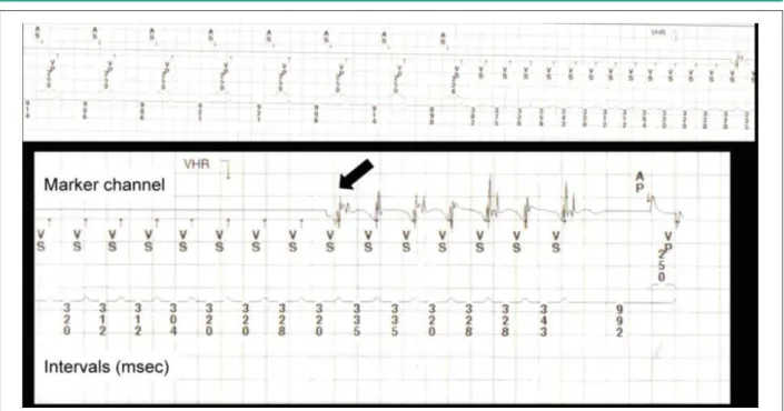
unipolar configuration. Several ventricular high rate episodes were detected with more than 300.000 premature ventricular beats (PVCs). It is remarkable that the ventricular rate during the episodes was almost double than the atrial rate. The stored intracardiac electrograms obtained during the episode are shown in Figure 1.
The diagnosis of VT with 2:1 ventriculo-atrial conduction was attained (Fig. 2) and the patient was referred for an upgrade to an implantable cardioverter-defibrillator (ICD) insertion. The ICD upgrade was performed with no complications. The old ventricular pacing lead was abandoned. The new ventricular lead and the old atrial lead were connected to the ICD.
Discussion
This is an interesting case that assesses the diagnostic possibilities of the new pacemakers. The diagnostic ability of the current pacemakers allows intracardiac electrograms to be stored for further analysis1. The perfect symptom-rhythm correlation (syncope during the ventricular high-rate episode) was paramount in the clinical decision-making.
The analysis of the intracardiac electrograms revealed initiation of the tachycardia with a PVC (Fig.1, black arrow), followed by a rapid ventricular rhythm at 320 ms. The ventriculo-atrial conduction (VA) is 2:1, thus supraventricular arrhythmias were unlikely (Fig. 2). The ventricular channel (Fig. 2; arrows) showed atrial activation following ventricular activation every other beat.
Atrial flutter with 2:1 conduction and conducted atrial fibrillation were ruled out because the tachycardia started with a PVC and there were more ventricular beats than atrial beats. In addition, conducted atrial fibrillation is usually irregular and this tachycardia was regular. The fact that the patient was, most of the time, in atrial sensing-ventricular pacing (due to high-degree AV block) did not completely rule out conducted supraventricular rhythms; however, it made this possibility less likely to occur.
Pacemaker tachycardia could not be suspected because there was not a paced rhythm during tachycardia (a necessary requisite to suspect it).
The decision to upgrade the pacemaker to an ICD was based on a series of facts: 1) The presence of structural heart disease (non-obstructive hypertrophic cardiomyopathy); 2)
Case Report
Baranchuk et al Tachyarrhythmias detected by a pacemaker
Arq Bras Cardiol 2009;92(3): e13-e15
Figure 1 -Intracardiac electrograms; AS - atrial sensing; AP - atrial pacing; VP - ventricular pacing; VS - ventricular sensing.
Figure 2 -Ventricular tachycardia with 2:1 VA conduction; VS - ventricular sensing; V - ventricular electrogram; A - atrial electrogram.
The clinical presentation (syncope) and 3) The documented sustained VT (through the analysis of the electrograms stored in the pacemaker).
In this clinical scenario, the presence of syncope alone increases the risk of sudden death by 5-fold2. In addition, VT was detected through the analysis of the intracardiac electrograms (increasing the risk of sudden death by almost two-fold). An electrophysiology study may not add up more relevant information. The ICD implantation is preferred over antiarrhythmic drugs for high-risk patients (HCM, syncope, VT)2.
Conclusion
The current generation of pacemakers facilitates the diagnosis of complicated clinical situations. Proper interpretation of the stored intracardiac electrograms may help in the diagnosis of life-threatening ventricular arrhythmias. The identification
of ventricular tachycardia in a patient with non-obstructive hypertrophic cardiomyopathy and syncope has helped in the decision-making of upgrading a pacemaker to an implantable cardioverter-defibrillator.
Potential Conflict of Interest
No potential conflict of interest relevant to this article was reported.
Sources of Funding
There were no external funding sources for this study.
Study Association
This study is not associated with any post-graduation program.
Case Report
Baranchuk et al
Tachyarrhythmias detected by a pacemaker
Arq Bras Cardiol 2009;92(3): e13-e15
References
1. Willems R, Morck ML, Exner DV, Rose SM, Gillis AM. Ventricular high-rate episodes in pacemaker diagnostics identify a high-risk subgroup of patients with tachy-brady syndrome. Heart Rhythm. 2004; 1: 414-21.
2. Elliott PM, Poloniecki J, Dickie S, Sharma S, Monserrat L, Varnava A, et al. Sudden death in hypertrophic cardiomyopathy: identification of high risk patients. J Am Coll Cardiol. 2000; 36: 2212-8.