61
Alternative method of smearing
samples on slides facilitate visualization
of hemoglobin H inclusion bodies in
alpha thalassemia
Método alternativo de dispersão das amostras no
esfregaço facilita a visualização dos corpos de
inclusão de hemoglobina H na talassemia alfa
Carta ao Editor / Letter to EditorCarlos F. Mendiburu1
Claudia R. Bonini-Domingos2
1Unesp – Ibilce, Depto. de Biologia, Laboratório de Hemoglobinas
e Genética das Doenças Hematológicas (LHGDH).
Trabalho realizado na Unesp, Campus de São José do Rio Preto -SP, Laboratório de Hemoglobinas e Genética das Doenças Hematológicas (LHGDH), Departamento de Biologia, Instituto de Biociências, Letras e Ciências Exatas.
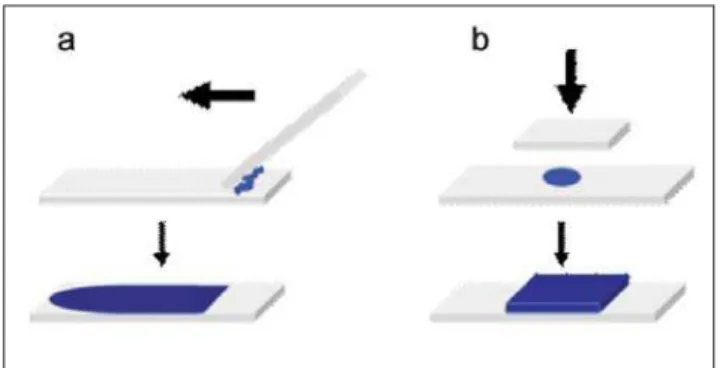
Figure 1. Routine (a) and Alternative (b) methods used in the obtaining of the smears
Figure 3. Different regions from the same sample film obtained with routine and alternative methods after 60 minutes of incubation Figure 2. Different regions from the same sample film obtained with routine and alternative methods after 30 minutes of incubation Sr. Editor
Alpha thalassemia syndromes are associated with reduced synthesis of α-globin chains and high β-globin chain concentrations; this imbalance induces the formation of beta chain tetramers denominated hemoglobin H (HbH).1
HbH can be detected in peripheral blood erythrocytes as inclusion bodies in cells stained with supravital oxidizing dyes, most commonly brilliant cresyl blue (BCB).2 The
number of cells that contain inclusions detected by BCB is related to the alpha thalassemia genotype, i.e. inclusion bodies are more numerous in syndromes with greater excesses of β-globin.3 This simple test is useful for the
diagnosis of alpha thalassemia, however, the identification of inclusion bodies is laborious and the results are highly observer dependent.4,5
In an attempt to improve the identification of inclusion bodies, we tested an alternative method of smearing samples on slides.
In this study, twenty blood samples of adult individuals with electrophoretic profiles suggestive of alpha thalassemia were analyzed. Each sample was incubated for 30 and 60 minutes with BCB as described previously6 and
smeared on slides using two methodologies: the classical method, in which the samples are smeared on the slides using a spreader slide7 (Figure 1a) and the "alternative"
method which consists in covering one drop of a sample on the slide using a cover-slip and spreading the sample by finger pressure, between two sheets of absorbent paper (Figure 1b). The blood smears were examined by light
62
Avaliação: Editor e dois revisores externos Conflito de interesse: não declarado
Recebido: 30/05/2007
Aceito após modificações: 14/09/2007
Correspondência: Carlos Fabián Mendiburu
LHGDH, Departamento de Biologia, Unesp-Ibilce Rua Cristóvão Colombo, 2265, Jardim Nazareth 15054-000 – São José do Rio Preto-SP
Fone: +55-17-32212392. Fax: +55-17-32212390 E-mail: fmendiburu@yahoo.com.ar
Rev. bras. hematol. hemoter. 2008;30(1):61-69 Carta ao Editor
alternative (left) methods, paired by incubation times of 30 and 60 minutes, respectability. With the slides obtained by the classical method, several larger granules were observed in the cytoplasm of some cells, but inclusion bodies could not be clearly identified. On the other hand, the alternative method allowed a clearer identification of the HbH inclusion bodies. With the smears obtained by the alternative method, the cells had a better definition and the HbH inclusion bodies were more evident and easily distinguishable both after 30 and 60 minutes of incubation, making a more accurate differentiation possible.
Identification of HbH inclusion bodies assists in the analysis of alpha thalassemia, making diagnosis conclusive in many cases. However, smears obtained by the classical method do not always clearly identify the corpuscles, hindering diagnosis. Our results showed that this alternative method is a useful option for screening HbH inclusion bodies in samples where the classical method does not always identify them.
Resumo
Nas talassemias alfa, a HbH pode ser detectada, nos eritrócitos do sangue periférico como inclusões celulares quando coradas com azul crezil brillante. Este teste simples é útil para o diagnós-tico de talassemia alfa, no entanto, a identificação dos corpos de inclusão de HbH é um processo laborioso e os resultados são altamente dependentes do observador. No intuito de melhorar a identificação das inclusões, foi testado um método alternativo para espalhar as amostras nas lâminas. Amostras de sangue foram espalhadas nas lâminas usando-se o método clássico e o método alternativo. O método alternativo permitiu uma melhor identifica-ção das inclusões de HbH do que o método clássico. Nossos re-sultados mostraram que o método alternativo é uma opção útil para a pesquisa dos corpúsculos de inclusão de HbH naquelas amostras onde o método clássico não o permite. Rev. bras. hematol. hemoter. 2008;30(1):61-62.
Palavras-chave: Talassemia alfa; inclusões de HbH; azul crezil brilhante; diagnóstico laboratorial.
References
1. Thompson CC, Ali MA, Boyadjian S, et al. Positional effect of cis/ trans alpha globin gene deletions on the formation of "H" bodies. Am. J. Hematol. 1989;31(4):242-7.
2. Sansone G, Sciarratta GV, Ivaldi G, et al. Hb H-like inclusions in red cells of patients with unstable haemoglobin. Haematologica. 1987; 72(6):481-6.
3. Skogerboe KJ, West SF, Smith C, et al. Screening for alpha-thalassemia. Correlation of hemoglobin H inclusion bodies with DNA-determined genotype. Arch Pathol Lab Med. 1992; 116 (10):1012-8.
4. Lin CK, Gau JP, Hsu HC, et al. Efficacy of a modified improved technique for detecting red cell haemoglobin H inclusions Clin Lab Haematol. 1990;12(4):409-15.
5. Jones JA, Broszeit HK, Lecrone CN, et al. An improved method for detection of red cell hemoglobin H inclusions. Am J Med Technol 1981;47(2):94-6.
6. Papayannopuolos, R, Stamatoyannopoulos G. Stains for inclusions bodies. "In standartization of laboratory reagents and methods for detection of hemoglobinopathies". Atlanta: Hew publications, 1974.