in HIV-Infected Patients; a Cohort Study from Indonesia
A. Rizal Ganiem1, Sofiati Dian1, Agnes Indriati2, Lidya Chaidir3, Rudi Wisaksana4, Patrick Sturm5, Willem Melchers5, Andre van der Ven6, Ida Parwati2, Reinout van Crevel6*
1Department of Neurology, Hasan Sadikin Hospital, Bandung, Indonesia,2Department of Clinical Pathology, Hasan Sadikin Hospital, Bandung, Indonesia,3Health Research Unit, Faculty of Medicine, Universitas Padjadjaran, Bandung, Indonesia,4Department of Internal Medicine, Hasan Sadikin Hospital, Bandung, Indonesia, 5Department of Medical Microbiology, Radboud University Medical Centre, Nijmegen, The Netherlands,6Department of Medicine, Radboud University Medical Centre, Nijmegen, The Netherlands
Abstract
Background:HIV-associated subacute meningitis is mostly caused by tuberculosis or cryptococcosis, but often no etiology can be established. In the absence of CT or MRI of the brain, toxoplasmosis is generally not considered as part of the differential diagnosis.
Methodology/Principal Findings:We performed cerebrospinal fluid real time PCR and serological testing forToxoplasma gondiiin archived samples from a well-characterized cohort of 64 HIV-infected patients presenting with subacute meningitis in a referral hospital in Indonesia. Neuroradiology was only available for 6 patients. At time of presentation, patients mostly had newly diagnosed and advanced HIV infection (median CD4 count 22 cells/mL), with only 17.2% taking ART, and 9.4% PJP-prophylaxis. CSF PCR forT. Gondiiwas positive in 21 patients (32.8%). Circulating toxoplasma IgG was present in 77.2% of patients tested, including all in whom the PCR of CSF was positive forT. Gondii. Clinically, in the absence of neuroradiology, toxoplasmosis was difficult to distinguish from tuberculosis or cryptococcal meningitis, although CSF abnormalities were less pronounced. Mortality among patients with a positive CSFT. GondiiPCR was 81%, 2.16-fold higher (95% CI 1.04–4.47) compared to those with a negative PCR.
Conclusions/Significance:Toxoplasmosis should be considered in HIV-infected patients with clinically suspected subacute meningitis in settings where neuroradiology is not available.
Citation:Ganiem AR, Dian S, Indriati A, Chaidir L, Wisaksana R, et al. (2013) Cerebral Toxoplasmosis Mimicking Subacute Meningitis in HIV-Infected Patients; a Cohort Study from Indonesia. PLoS Negl Trop Dis 7(1): e1994. doi:10.1371/journal.pntd.0001994
Editor:Judd L. Walson, University of Washington, United States of America
ReceivedJuly 13, 2012;AcceptedNovember 16, 2012;PublishedJanuary 10, 2013
Copyright:ß2013 Ganiem et al. This is an open-access article distributed under the terms of the Creative Commons Attribution License, which permits unrestricted use, distribution, and reproduction in any medium, provided the original author and source are credited.
Funding:This study was financially supported by the Royal Academy of Arts and Sciences (KNAW, www.knaw.nl; 07-MP-10), the Netherlands; and by IMPACT, a 5-year HIV program supported by the European Commission (SANTE/2005/105-033). Ahmad Rizal Ganiem, Lidya Chaidir and Rudi Wisaksana are supported by fellowships from Radboud University Nijmegen Medical Center. Reinout van Crevel has a VIDI-grant from the Netherlands Organization for Scientific Research (NWO, www.nwo.nl; 017.106.310). The funders had no role in study design, data collection and analysis, decision to publish, or preparation of the manuscript.
Competing Interests:The authors have declared that no competing interests exist.
* E-mail: r.vancrevel@aig.umcn.nl
Introduction
In settings of Africa and Asia, the most common cause of subacute meningitis in patients with advanced HIV infection is either tuberculous or cryptococcal infection [1,2]. However, in many patients, the etiology of subacute meningitis cannot be established [1,3]. In line with a large retrospective cohort of adult meningitis patients in South Africa, where 52.8% had no definite diagnosis despite extensive microbiological testing [1], we could not identify the causative pathogen in 48.9% of HIV-infected meningitis patients in an Indonesian setting [4].
Toxoplasmosis is a common and serious central nervous system (CNS) infection in patients with advanced HIV infection [5–8], although its incidence has decreased with introduction of antiretroviral treatment (ART) [6,9]. Cerebral toxoplasmosis mostly presents as cerebral mass lesions with headache, confusion, fever, lethargy, seizures, cranial nerve palsies, psychomotor changes, hemiparesis and/or ataxia [10]. Some of these symptoms may also mimic meningitis, but cerebral toxoplasmosis is generally
not considered as a differential diagnosis of subacute meningitis in HIV-infected patients. This is especially the case in low-resource settings where no CT or MRI can be performed. We have therefore examined if toxoplasmosis can be diagnosed in HIV-infected patients presenting with subacute meningitis of unknown origin in Indonesia, using cerebrospinal fluid (CSF) PCR forT. gondii.
Methods
Ethics statement
routinely with oral informed consent for all patients with suspected meningitis in Hasan Sadikin hospital, after 24% were found HIV-positive in a previous cohort study of 185 patients in the same hospital [4]. Consent is obtained from closest relatives (husband/ wife or parents) for those patients who are unstable or unconscious at time of presentation. With approval from the ethical committee HIV testing was done anonymously afterwards for those who had died before consent could be obtained.
Setting and patients
We included adult patients presenting with suspected meningitis at Hasan Sadikin Hospital, the top referral hospital for West Java Province, Indonesia, between December 2006 and October 2010. Clinical data including outcome was recorded in individual case report form. Definite TB meningitis was diagnosed if CSF culture or real time PCR were positive for M. tuberculosis, cryptococcal meningitis if either CSF India Ink examination, culture or cryptococcal antigen testing were positive, and toxoplasmosis if CSFT. gondiiPCR was positive. HIV testing is done routinely for patients presenting at this hospital, but cerebral CT-scanning is rarely done in this setting and is not covered by the government health insurance for the poor.
Laboratory examinations
CSF cell count and differentiation, protein and glucose were measured. CSF microscopy was done for cryptococci, acid-fast bacilli and bacterial pathogens. CSF was cultured forMycobacterium tuberculosis (solid Ogawa and liquid MB-BacT, Biomerieux), bacterial pathogens (blood agar, chocolate agar, and brain-heart infusion) and fungi (Sabouraud). Cryptococcal antigen (CALAS, Meridian Diagnostics) testing was done on CSF samples following the manufacturer’s instructions. Five to 7 ml CSF samples were used for molecular testing. After centrifugation of CSF samples at 30006g for 10 minutes, DNA was extracted from 200ml of CSF
sediment by using QIAmp DNA mini kit (Qiagen, USA). CSFM. tuberculosis real time PCR was done using IS6110, a repeated insertion sequence specific for M. tuberculosis, as a target [11].
Measurement of CD4-cell count for HIV-patients only became available during the time of the study and was measured only for those who survived for more than 4 days. Real time PCR forT. gondii, using the multicopy B1 gene of theT. gondiias the target as described elsewhere [12], was performed to archived CSF samples at Radboud University Nijmegen Medical Centre. CSF specimens from 22 HIV-negative meningitis patients (16 with definite TB meningitis, 2 with bacterial meningitis, and 4 with no definite diagnosis), and nine patients with non-infectious CNS diseases, all recruited at Hasan Sadikin Hospital, were used as controls forT. gondiiPCR. These samples were collected during the study period over a similar time scale compared to the case CSF samples. Toxoplasma immunoglobuline G (toxoplasma IgG) were mea-sured by electro chemiluminescent assay (ECLIA, Elecsys, Roche) in archived serum samples of patients included in the study.
Data analysis and statistics
Characteristics of patients with definite tuberculosis, cryptococ-cosis and toxoplasmosis were compared using Chi-square test for proportions and Mann-Whitney U test for continuous variables. Progression to death using 2-month mortality data was examined by Kaplan–Meier estimates.
Results
During the period, 401 patients presented with clinical meningitis, 76 were diagnosed with HIV infection, and 64 had archived CSF samples and were included in this study. Patients included in the study presented after a median 7 days, with meningismus (86.0%), headache (80.8%), lowered consciousness (33.3%), fever (28.8%), hemi- or tetraparesis (28.6%), cranial nerve palsies (12.5%), and seizures (10.9%). HIV was newly diagnosed in 53 patients (82.8%). All 11 patients previously diagnosed with HIV were taking ART, and 6 were using co-trimoxazole asPneumocystis jiroveci(PJP) prophylaxis at time of presentation. The median CD4 cell count was 22 cells/mL, and less than 200 cells/mL in 22 out of 23 patients tested (96%).
CSF T. gondii PCR was positive in 21 of 64 HIV-infected patients (32.8%), with a median Ct-value of 36.0 (IQR: 34.2–39.3). None of the 22 HIV-negative control and 9 non-infectious CNS disease patients had a positive T. gondii PCR. Archived serum sample was not available in 14 patients. Toxoplasma IgG was positive in 78% of patients tested, including all patients with positive CSFT. gondiiPCR. Toxoplasma IgG titers were higher among patients with a positive CSFT. gondiiPCR (p = .017). A definite diagnosis of TB meningitis was established in 21/64 patients (32.8%). Out of 21 patients with positiveT. gondiiPCR, five had combined tuberculosis and toxoplasmosis. Cryptococcosis was diagnosed in 15/64 patients (23.4%), including two who were also diagnosed with tuberculosis. In 14 patients (21.9%) no causative pathogen was isolated.
Neck stiffness, headache and fever, the classical signs of meningitis, were equally common in patients diagnosed with toxoplasmosis, cryptococcosis and tuberculosis, as were most other signs and symptoms, except hemiparesis (Table 1). None of the patients with toxoplasmosis had received ART or co-trimoxazole prophylaxis prior to admission with meningitis. CT scans were available for 6 patients, including 4 with a positiveT. gondiiPCR. Three showed signs of hydrocephalus, one a hypodense lesion that showed no enhancement using contrast, and two were normal. No mass lesions typical for cerebral toxoplasmosis were seen.
CSF cell count and protein were normal or mildly elevated in patients with toxoplasmosis, and hypoglycorrhachia was less common compared with tuberculosis or cryptococcosis
Author Summary
(Table 1). CD4 counts, missing in two-thirds of patients due to early death or the unavailability of CD4 cell testing during the initial phase of the cohort study, were low in all but one
patient.Table 2 lists the CSF findings of individual patients,
Figure 1 is a graphic representation of the CSF cell count, protein and glucose ratio, showing the overlap in CSF findings
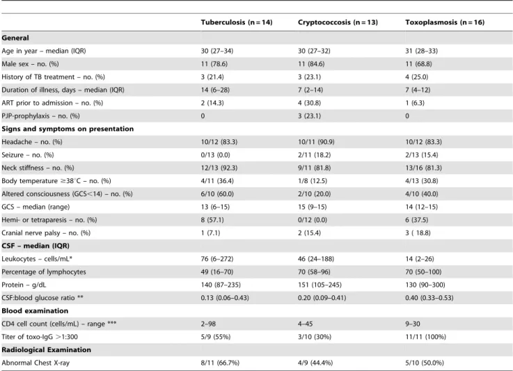
Table 1.Characteristics of patients according to microbiological diagnosis.
Tuberculosis (n = 14) Cryptococcosis (n = 13) Toxoplasmosis (n = 16)
General
Age in year – median (IQR) 30 (27–34) 30 (27–32) 31 (28–33)
Male sex – no. (%) 11 (78.6) 11 (84.6) 11 (68.8)
History of TB treatment – no. (%) 3 (21.4) 3 (23.1) 4 (25.0)
Duration of illness, days – median (IQR) 14 (6–28) 7 (2–14) 7 (4–12)
ART prior to admission – no. (%) 2 (14.3) 4 (30.8) 1 (6.3)
PJP-prophylaxis – no. (%) 0 3 (23.1) 0
Signs and symptoms on presentation
Headache – no. (%) 10/12 (83.3) 10/11 (90.9) 10/12 (83.3)
Seizure – no. (%) 0/13 (0.0) 2/11 (18.2) 2/13 (15.4)
Neck stiffness – no. (%) 12/13 (92.3) 9/11 (81.8) 13/16 (81.3)
Body temperature$38uC – no. (%) 4/11 (36.4) 1/8 (12.5) 4/13 (30.8)
Altered consciousness (GCS,14) – no. (%) 6/10 (60.0) 2/10 (20.0) 4/10 (40.0)
GCS – median (range) 13 (6–15) 15 (9–15) 14 (12–15)
Hemi- or tetraparesis – no. (%) 8 (57.1) 0/12 (0.0) 6 (37.5)
Cranial nerve palsy – no. (%) 1 (7.1) 2 (15.4) 3 ( 18.8)
CSF – median (IQR)
Leukocytes – cells/mL* 76 (6–272) 46 (24–188) 14 (2–26)
Percentage of lymphocytes 49 (16–70) 70 (58–96) 70 (50–100)
Protein – g/dL 140 (87–235) 151 (105–245) 130 (90–300)
CSF:blood glucose ratio ** 0.13 (0.06–0.43) 0.20 (0.09–0.41) 0.40 (0.33–0.53)
Blood examination
CD4 cell count (cells/mL) – range *** 2–98 4–45 9–30
Titer of toxo-IgG.1:300 5/9 (55%) 3/10 (30%) 11/11 (100%)
Radiological Examination
Abnormal Chest X-ray 8/11 (66.7%) 4/9 (44.4%) 5/10 (50.0%)
Patients with combined TB-toxoplasmosis (n = 5), combined TB-Cryptococcus (n = 2), and no definite diagnosis (n = 14) were excluded from the table. Data are presented as no. of patients/no. evaluated (%) unless stated otherwise.
GCS = Glasgow Coma Scale.
*significantly different: TB vs. toxoplasmosis (p = .01); cryptococcosis vs. toxoplasmosis (p = .02). **significantly different: TB vs. toxoplasmosis (p = .03), cryptococcosis vs. toxoplasmosis (p = .02). ***CD4 cell counts were only available for 16 patients with definite diagnoses.
doi:10.1371/journal.pntd.0001994.t001
Figure 1. CSF characteristics according to causative pathogen.CSF cell count (A), protein concentration (B) and CSF:blood glucose ratio (C) for cases with confirmed toxoplasmosis (
N
), crypotoccococis (&), and tuberculosis (m). Toxo = toxoplasmosis; crypto = cryptococcosis; TB = TB meningitis.Table 2.Line list of all patients.
No Age Sex CSF examination* Causative pathogen Diagnosis**
Cell number Protein Glucose ratio ToxoQ TBe Crypton
1 31 M 4 26 0.33 pos neg neg toxoplasmosis
2 30 M 17 100 0.35 pos neg neg toxoplasmosis
3 33 M 18 160 n/a pos neg neg toxoplasmosis
4 18 F 11 150 n/a pos neg neg toxoplasmosis
5 29 F 2 90 0.32 pos neg neg toxoplasmosis
6 24 F 1 300 0.54 pos neg neg toxoplasmosis
7 28 M 13 630 0.4 pos neg neg toxoplasmosis
8 19 F 22 310 0.33 pos neg neg toxoplasmosis
9 30 M 49 130 0.83 pos neg neg toxoplasmosis
10 36 M 1 60 0.58 pos neg neg toxoplasmosis
11 33 M 30 434 0.32 pos neg neg toxoplasmosis
12 35 F 14 95 0.52 pos neg neg toxoplasmosis
13 32 M 26 131 0.5 pos neg neg toxoplasmosis
14 30 M 180 100 0.43 pos neg neg toxoplasmosis
15 34 M 0 50 0.38 pos neg neg toxoplasmosis
16 32 M n/a n/a n/a pos neg neg toxoplasmosis
17 25 F 143 100 0.13 neg pos neg TB
18 31 M 696 39 0.07 neg pos neg TB
19 30 M 7 260 0.06 neg pos neg TB
20 21 F 105 700 0.05 neg pos neg TB
21 52 M 193 190 0.55 neg pos neg TB
22 29 M 1120 n/a 0.03 neg pos neg TB
23 32 M 104 160 0.04 neg pos neg TB
24 30 M 48 40 0.24 neg pos neg TB
25 28 M 507 6980 0.1 neg pos neg TB
26 23 F 0 73 0.4 neg pos neg TB
27 31 M 26 140 n/a neg pos neg TB
28 40 M 4 112 0.47 neg pos neg TB
29 56 M 0 100 0.4 neg pos neg TB
30 29 M 10 210 n/a neg pos neg TB
31 31 M 109 120 0.64 pos pos neg TB-toxo
32 39 M 919 630 0.04 pos pos neg TB-toxo
33 22 M 13 10 0.25 pos pos neg TB-toxo
34 31 M 0 200 0.43 pos pos neg TB-toxo
35 23 F 23 64 0.46 pos pos neg TB-toxo
36 27 M 34 200 0.43 neg neg pos Cryptococcosis
37 30 M 2 22 0.01 neg neg pos Cryptococcosis
38 27 M 419 110 0.06 neg neg pos Cryptococcosis
39 30 M 167 120 0.2 neg neg pos Cryptococcosis
40 38 M 20 230 0.12 neg neg pos Cryptococcosis
41 32 M 209 610 0.01 neg neg pos Cryptococcosis
42 25 F 28 70 0.4 neg neg pos Cryptococcosis
43 32 M 49 100 0.15 neg neg pos Cryptococcosis
44 36 M 46 270 0.51 neg neg pos Cryptococcosis
45 29 M 3 200 0.17 neg neg pos Cryptococcosis
46 22 F 35 151 0.38 neg neg pos Cryptococcosis
47 32 M 248 130 n/a neg neg pos Cryptococcosis
between patients with toxoplasmosis, cryptococcal and tuber-culosis CNS infection.
Patients with confirmed cryptococcosis received amphotericine B, followed by fluconazole; all others received empiric tuberculosis treatment combined with adjunctive corticosteroids [13]. No toxoplasmosis treatment was given, as T. gondii PCR was performed retrospectively and was not available at time of presentation. Eight patients were lost to follow up and were not included in Kaplan Meier analysis. Mortality among those with positive CSF T. gondii PCR was 2.16-fold (95% CI 1.04–4.47) higher compared to those who had a negative PCR result; median survival was 7 days for toxoplasmosis, 7 days for tuberculosis meningitis, 110 days for cryptococcosis, and 32 days for patients with an unknown cause of meningitis (Figure 2).
Discussion
In our cohort of HIV-infected patients presenting with clinical signs and symptoms of CNS infection, CSF T. gondii PCR was positive in 32.8% of patients, sometimes in conjunction with tuberculosis. In the absence of CT or MRI of the brain, toxoplasmosis could not be distinguished from tuberculosis or cryptococcosis. Mortality in this cohort of newly diagnosed and advanced HIV infection was extremely high and associated with a positiveT. gondiiPCR.
Cerebral toxoplasmosis typically causes space occupying lesion(s), leading to subacute or acutely developing confusion, with or without focal neurological deficits [14]. In the absence of CT or MRI of the brain, common findings like headache, fever, hemiparesis and decreased level of consciousness [10] may mimic those of meningitis [4,15–17]. In previous series of cerebral toxoplasmosis [18,19], meningeal signs have been reported in 3 to
16% of the patients, although in many reports neck stiffness is not mentioned [14]. Although rare, cases of spinal cord toxoplasmosis have also been reported [20]. No typical mass lesions were found in 6 patients with an available CT scan. This is not surprising, as this study depended on the availability of CSF samples, that would not have been obtained if typical mass lesions had been found.
We usedT. gondiiPCR for diagnosis of cerebral toxoplasmosis. In previous studies CSFT. gondiiPCR had a sensitivity of 50–60% to confirm cerebral toxoplasmosis in HIV-infected patients [21,22]. The sensitivity is possibly higher among patients with meningoencephalitis compared to those with space-occupying lesions only, but this has not been examined. Specificity ofT. gondii Table 2.Cont.
No Age Sex CSF examination* Causative pathogen Diagnosis**
Cell number Protein Glucose ratio ToxoQ TBe Crypton
49 27 M 0 n/a 0.16 neg pos pos TB-crypto
50 34 M 0 120 0.27 neg pos pos TB-crypto
51 30 F 0 44 0.39 neg neg neg unknown
52 27 M 0 180 0.49 neg neg neg Unknown
53 33 M 259 110 0.57 neg neg neg Unknown
54 32 M 2 130 0.48 neg neg neg Unknown
55 30 M n/a n/a n/a neg neg neg Unknown
56 28 M 143 9850 0.04 neg neg neg Unknown
57 30 M 146 40 0.19 neg neg neg Unknown
58 36 M 17 16 0.51 neg neg neg Unknown
59 24 M 0 30 0.6 neg neg neg Unknown
60 29 M 43 44 0.21 neg neg neg Unknown
61 39 M 18 n/a 0.24 neg neg neg Unknown
62 32 M 184 480 0.08 neg n/a neg Unknown
63 28 M 298 317 0.34 neg neg neg Unknown
64 34 M 166 133 n/a neg n/a neg Unknown
M = male; F = female; pos = positive; neg = negative; n/a = not available.
*cell numbers are in cells/mL; protein in mg/dL; glucose ratio = CSF glucose:blood glucose. **TB-toxo = tuberculosis and toxoplasmosis, TB-crypto = tuberculosis and cryptococcosis. Qtoxoplasmosis, based on toxoplasma PCR;
e
TB meningitis, based on either culture or real time PCR;
ncryptococcosis based on either direct staing, culture, or antigen testing. doi:10.1371/journal.pntd.0001994.t002
Figure 2. Kaplan Meier survival estimates.Patients with available long term follow up data: Toxoplasmosis (n = 14), Cryptococcosis (n = 13), TB meningitis (n = 14), no diagnosis (n = 13).
PCR is high, between 97 and 100% [22–24]. The positivity rate of 32.8% in our study might therefore be an underestimate, especially in the category of patients in whom no other pathogen was isolated despite extensive microbiological testing.
In our cohort, toxoplasmosis could not be distinguished clinically from tuberculosis and cryptococcosis. From our previous series [4], CSF samples were available for the current study for 36/47 HIV-infected patients. Ten out of 17 patients who were diagnosed with ‘probable TB meningitis’ and ‘unknown’ in the previous study were found to have a positiveT. gondiiPCR (and no bacteriological confirmation of tuberculosis) in the current study. Diagnosis of cerebral toxoplasmosis is usually based on clinical findings and CT or MRI of the brain. However, if cerebral imaging is lacking, toxoplasmosis may not be considered. Positive toxoplasma serology, which has a high sensitivity but very poor specificity, is helpful to exclude but not to confirm cerebral toxoplasmosis, although some reports suggest that high toxoplas-ma IgG titers are only found in patients with symptotoxoplas-matic toxoplasmosis [25]. Indeed, in our study, patients with a positive
T. gondiiPCR had a higher IgG titers compared to those who had a negative PCR. An autopsy study from India provides further support for the notion that cerebral toxoplasmosis is not always considered; among 233 HIV patients, toxoplasmosis accounted for 6.8% of deaths, but in none of these cases toxoplasmosis had been suspected clinically [26].
The incidence of cerebral toxoplasmosis varies between countries [14] and is related to the seroprevalence of toxoplasmosis in the general population [19,25]. In the United States, toxoplasma seroprevalence varies from 3% to 30%, whereas in France 73%–90% of the population is infected [10]. Reported seroprevalence rates were varied from 13–31% in the general population, and 45–68% in HIV patients in studies from several developing countries [8,27,28]. In our study, 78% of patients had detectable toxoplasma IgG, but this does not reflect the seroprevalence in the general population or among unselected HIV-infected patients, as only meningitis patients were examined. Mortality in this cohort of patients was very high, higher compared to reported rates in other series [8,9,29]. One explanation is that patients mostly presented with advanced and untreated HIV infection. In addition, no toxoplasmosis treatment was provided, as PCR was done retrospectively on archived samples. In our previous study, HIV infection was associated with a 2.5-fold increased mortality among patients presenting with
meningitis [4]. Data from the current study suggests that this may is at least in part attributable to a high prevalence of (unrecognized and untreated) toxoplasmosis. Future studies should examine the benefit of timely diagnosis and/or empiric treatment of toxoplas-mosis for patients in settings like ours. Empiric treatment for subacute meningitis in HIV-infected patients should probably also include tuberculosis, which is difficult to exclude as culture is slow and microscopy and commercial PCR assays have insufficient sensitivity [30].
Our study suffers from several limitations. Most importantly, no CT or MRI of the brain could be performed. In addition, clinical data, CD4 cell counts and other laboratory parameters were missing in a number of patients. Despite these limitations the data strongly suggest that toxoplasmosis should be included in the differential diagnosis of HIV-infected with clinically suspected subacute meningitis, and that molecular testing or empiric treatment for toxoplasmosis should be considered in these patients, especially if no CT or MRI can be performed. Obviously, timely diagnosis and treatment of HIV will help prevent this severe opportunistic infection.
Supporting Information
Checklist S1 STROBE checklist for the manuscript: ‘‘Cerebral toxoplasmosis mimicking subacute meningi-tis in HIV-infected patients; a cohort study from Indonesia’’.
(DOC)
Acknowledgments
We thank the patients, drs. Regina Loprang, Ela Hayati, and Christian Budiman (field physicians), Lies Ratnasari and Witri Indrasari (laboratory technicians), Kristianto (social worker), and the residents of the Department of Neurology, Hasan Sadikin Hospital, Bandung for their help in conducting this study.
Part of these data has been presented as an oral presentation at the 43rd World Conference on Lung Health of the International Union Against Tuberculosis and Lung Disease 2012 (abstract number: 0P-212-17).
Author Contributions
Conceived and designed the experiments: ARG RW AvdV RvC. Performed the experiments: ARG SD AI LC RW PS WM IP. Analyzed the data: ARG SD LC RW RvC. Wrote the paper: ARG RvC.
References
1. Jarvis JN, Meintjes G, Williams A, Brown Y, Crede T, et al. (2010) Adult meningitis in a setting of high HIV and TB prevalence: findings from 4961 suspected cases. BMC Infect Dis 10: 67.
2. Kongsiriwattanakul S, Suankratay C (2011) Central nervous system infections in HIV-infected patients hospitalized at King Chulalongkorn Memorial Hospital. J Med Assoc Thai 94: 551–558.
3. Hakim JG, Gangaidzo IT, Heyderman RS, Mielke J, Mushangi E, et al. (2000) Impact of HIV infection on meningitis in Harare, Zimbabwe: a prospective study of 406 predominantly adult patients. AIDS 14: 1401–1407.
4. Ganiem AR, Parwati I, Wisaksana R, van der Zanden A, van de Beek D, et al. (2009) The effect of HIV infection on adult meningitis in Indonesia: a prospective cohort study. AIDS 23: 2309–2316.
5. Antinori A, Larussa D, Cingolani A, Lorenzini P, Bossolasco S, et al. (2004) Prevalence, associated factors, and prognostic determinants of AIDS-related toxoplasmic encephalitis in the era of advanced highly active antiretroviral therapy. Clin Infect Dis 39: 1681–1691.
6. Hoffmann C, Ernst M, Meyer P, Wolf E, Rosenkranz T, et al. (2007) Evolving characteristics of toxoplasmosis in patients infected with human immunodefi-ciency virus-1: clinical course and Toxoplasma gondii-specific immune responses. Clin Microbiol Infect 13: 510–515.
7. Naba MR, Kanafani ZA, Awar GN, Kanj SS (2010) Profile of opportunistic infections in HIV-infected patients at a tertiary care center in Lebanon. J Infect Public Health 3: 130–133.
8. Nissapatorn V, Lee C, Quek KF, Leong CL, Mahmud R, et al. (2004) Toxoplasmosis in HIV/AIDS patients: a current situation. Jpn J Infect Dis 57: 160–165.
9. Mayor AM, Fernandez Santos DM, Dworkin MS, Rios-Olivares E, Hunter-Mellado RF (2011) Toxoplasmic encephalitis in an AIDS cohort at Puerto Rico before and after highly active antiretroviral therapy (HAART). Am J Trop Med Hyg 84: 838–841.
10. Skiest DJ (2002) Focal neurological disease in patients with acquired immunodeficiency syndrome. Clin Infect Dis 34: 103–115.
11. Savelkoul PH, Catsburg A, Mulder S, Oostendorp L, Schirm J, et al. (2006) Detection of Mycobacterium tuberculosis complex with Real Time PCR: comparison of different primer-probe sets based on the IS6110 element. J Microbiol Methods 66: 177–180.
12. Lin MH, Chen TC, Kuo TT, Tseng CC, Tseng CP (2000) Real-time PCR for quantitative detection of Toxoplasma gondii. J Clin Microbiol 38: 4121–4125. 13. Thwaites GE, Nguyen DB, Nguyen HD, Hoang TQ, Do TT, et al. (2004)
Dexamethasone for the treatment of tuberculous meningitis in adolescents and adults. N Engl J Med 351: 1741–1751.
14. Montoya JG, Liesenfeld O (2004) Toxoplasmosis. Lancet 363: 1965–1976. 15. Helbok R, Pongpakdee S, Yenjun S, Dent W, Beer R, et al. (2006) Chronic
16. Marais S, Pepper DJ, Schutz C, Wilkinson RJ, Meintjes G (2011) Presentation and outcome of tuberculous meningitis in a high HIV prevalence setting. PLoS One 6: e20077.
17. Thwaites GE, Duc Bang N, Huy Dung N, Thi Quy H, Thi Tuong Oanh D, et al. (2005) The influence of HIV infection on clinical presentation, response to treatment, and outcome in adults with Tuberculous meningitis. J Infect Dis 192: 2134–2141.
18. Porter SB, Sande MA (1992) Toxoplasmosis of the central nervous system in the acquired immunodeficiency syndrome. N Engl J Med 327: 1643–1648. 19. Renold C, Sugar A, Chave JP, Perrin L, Delavelle J, et al. (1992) Toxoplasma
encephalitis in patients with the acquired immunodeficiency syndrome. Medicine (Baltimore) 71: 224–239.
20. Garcia-Gubern C, Fuentes CR, Colon-Rolon L, Masvidal D (2010) Spinal cord toxoplasmosis as an unusual presentation of AIDS: case report and review of the literature. Int J Emerg Med 3: 439–442.
21. Bretagne S (2003) Molecular diagnostics in clinical parasitology and mycology: limits of the current polymerase chain reaction (PCR) assays and interest of the real-time PCR assays. Clin Microbiol Infect 9: 505–511.
22. Remington JS, Thulliez P, Montoya JG (2004) Recent developments for diagnosis of toxoplasmosis. J Clin Microbiol 42: 941–945.
23. Cinque P, Scarpellini P, Vago L, Linde A, Lazzarin A (1997) Diagnosis of central nervous system complications in HIV-infected patients: cerebrospinal fluid analysis by the polymerase chain reaction. AIDS 11: 1–17.
24. Correia CC, Melo HR, Costa VM (2010) Influence of neurotoxoplasmosis characteristics on real-time PCR sensitivity among AIDS patients in Brazil. Trans R Soc Trop Med Hyg 104: 24–28.
25. Barratt JL, Harkness J, Marriott D, Ellis JT, Stark D (2010) Importance of nonenteric protozoan infections in immunocompromised people. Clin Microbiol Rev 23: 795–836.
26. Lanjewar DN (2011) The spectrum of clinical and pathological manifestations of AIDS in a consecutive series of 236 autopsied cases in mumbai, India. Patholog Res Int 2011: 547618.
27. Shin DW, Cha DY, Hua QJ, Cha GH, Lee YH (2009) Seroprevalence of Toxoplasma gondii infection and characteristics of seropositive patients in general hospitals in Daejeon, Korea. Korean J Parasitol 47: 125–130. 28. Osunkalu VO, Akanmu SA, Ofomah NJ, Onyiaorah IV, Adediran AA, et al.
(2011) Seroprevalence of Toxoplasma gondii IgG antibody in HIV-infected patients at the Lagos University Teaching Hospital. HIV AIDS (Auckl) 3: 101– 105.
29. Vidal JE, Hernandez AV, de Oliveira AC, Dauar RF, Barbosa SP, Jr., et al. (2005) Cerebral toxoplasmosis in HIV-positive patients in Brazil: clinical features and predictors of treatment response in the HAART era. AIDS Patient Care STDS 19: 626–634.