Porcine Intestinal Epithelial Cells
Peter Schierack1,2*, Sylvia Kleta1,3, Karsten Tedin1, Julius Tachu Babila4, Sibylle Oswald5, Tobias A. Oelschlaeger5, Rico Hiemann2, Susanne Paetzold6,7, Lothar H. Wieler1
1Institut fu¨r Mikrobiologie und Tierseuchen, Freie Universita¨t Berlin, Berlin, Germany,2Fachbereich Bio-, Chemie- und Verfahrenstechnik, Hochschule Lausitz (FH), Senftenberg, Germany,3Bundesinstitut fu¨r Risikobewertung, Berlin, Germany,4Department of Neuroproteomics, Max Delbrueck Center for Molecular Medicine, Berlin, Germany,5Institut fu¨r Molekulare Infektionsbiologie, Universita¨t Wu¨rzburg, Wu¨rzburg, Germany,6Max Planck Institut fu¨r Infektionsbiologie, Berlin, Germany,7Research Center Borstel, Borstel, Germany
Abstract
Background:The probioticEscherichia colistrain Nissle 1917 (EcN) has been shown to interfere in a humanin vitromodel with the invasion of several bacterial pathogens into epithelial cells, but the underlying molecular mechanisms are not known.
Methodology/Principal Findings:In this study, we investigated the inhibitory effects of EcN onSalmonella Typhimurium
invasion of porcine intestinal epithelial cells, focusing on EcN effects on the various stages ofSalmonellainfection including intracellular and extracellularSalmonellagrowth rates, virulence gene regulation, and adhesion. We show that EcN affects the initialSalmonella invasion steps by modulatingSalmonella virulence gene regulation andSalmonellaSiiE-mediated adhesion, but not extra- and intracellularSalmonellagrowth. However, the inhibitory activity of EcN againstSalmonella
invasion always correlated with EcN adhesion capacities. EcN mutants defective in the expression of F1C fimbriae and flagellae were less adherent and less inhibitory towardSalmonellainvasion. AnotherE. colistrain expressing F1C fimbriae was also adherent to IPEC-J2 cells, and was similarly inhibitory againstSalmonellainvasion like EcN.
Conclusions:We propose that EcN affectsSalmonellaadhesion through secretory components. This mechanism appears to be common to manyE. colistrains, with strong adherence being a prerequisite for an effective reduction of SiiE-mediated
Salmonellaadhesion.
Citation:Schierack P, Kleta S, Tedin K, Babila JT, Oswald S, et al. (2011)E. coliNissle 1917 AffectsSalmonellaAdhesion to Porcine Intestinal Epithelial Cells. PLoS ONE 6(2): e14712. doi:10.1371/journal.pone.0014712
Editor:Leonardo A. Sechi, Universita di Sassari, Italy
ReceivedJuly 1, 2010;AcceptedJanuary 10, 2011;PublishedFebruary 17, 2011
Copyright:ß2011 Schierack et al. This is an open-access article distributed under the terms of the Creative Commons Attribution License, which permits unrestricted use, distribution, and reproduction in any medium, provided the original author and source are credited.
Funding:This study was supported by the German Research Foundation (DFG) through the Collaborative Research Centre 852 (grant no. SFB852/1), by grant FOR 438/1-1 from the DFG and by InnoProfile IP 03 IP 611 funded by the Bundesministerium fu¨r Bildung und Forschung (BMBF, Germany). The funders had no role in study design, data collection and analysis, decision to publish, or preparation of the manuscript.
Competing Interests:The authors have declared that no competing interests exist.
* E-mail: Peter.Schierack@HS-Lausitz.de
Introduction
E. coli Nissle 1917 (EcN; Mutaflor) is a widely employed probiotic strain and several in vivo studies demonstrated its promising probiotic activity in humans and animals [1,2,3,4,5,6]. Proposed probiotic actions of EcN include effects on pathogens, host epithelial cells, host smooth muscle cell activity and the host immune system [7,8,9,10,11,12,13].In vitro, EcN has been shown to prevent invasion of host cells by several pathogens, including Salmonella,Yersinia,Shigella,Legionella,Listeriaand adherent-invasive E. coli[14,15,16]. These studies demonstrated that EcN inhibited invasion in a dose-dependent manner. Interestingly, EcN super-natants were also effective in inhibiting invasion. However, the underlying molecular mechanisms involved in this process are poorly understood to date.
Successful probiotic action of a bacterial strain is often associated with its colonization of the intestine. The colonization of hosts by EcN can be very successful, but is likely specific for each individual [15,17]. EcN has been shown to express type 1 fimbriae and F1C fimbriae (which have usually been associated
with uropathogenic E. coli), but not P and S fimbriae [18]. Recently, it was demonstrated that EcN F1C fimbriae or cellulose production play an important role in EcN biofilm formation, adherence to intestinal epithelial cells in vitro, and intestinal colonization of mice [19,20].
The aim of the present study was to characterize the effects of EcN onSalmonellainvasion of the porcine intestinal epithelial cell line IPEC-J2 [21], with a focus on EcN effects on singleSalmonella infection steps. In addition, EcN adhesion capacities were tested as possible requisites for an EcN specific probiotic activity. We show that adhesion rates always correlate with inhibitory effects against Salmonella invasion, and the initial Salmonella invasion process, especially adhesion, is likely affected.
Materials and Methods
Bacterial strains used in this study
entericaserovar Typhimurium (Salmonella Typhimurium) strain SL1344 was kindly provided by F. Norel (Paris, France). SL1344::kanwas generated by introduction of a kanamycin resistance cassette into the downstream, non-coding region between theavrAandsitDgenes ofSalmonella typhimuriumLT2 as previously described [22]. Following PCR screening for correct insertion and orientation, the kanamycin resistance cassette was introduced into strain SL1344 by bacterio-phage P22 transduction using standard protocols. The non-invasive Salmonella Typhimurium strain VV341 (SL1344 hilA-339::kan) was kindly provided by C.A. Lee (Boston, USA). The non-invasive Salmonella Typhimurium strain SB161 (SL1344 invG) was kindly provided by M. Hensel (Erlangen, Germany) and was transformed with the pEGFP plasmid.b-Galactosidase assays were performed with strains CL87 (SL1344 hilA(iagB)::lacZY), EE635 (SL1344 hilC9::Tn5lacZY), and RL696 (SL1344hilD696::lacZY) provided by C.A. Lee. Strain KT4166 harboring a lacZ fusion to the SPI4-encodedsiiEgene (SL1344icgA1(siiE)::MudJ(kan)) was constructed by P22 transduction of thesiiE::MudJ(kan) fusion into strain SL1344 with kanamycin selection using lysates prepared on strain JS296 (J.M. Slauch, Urbana, USA).
E. coli140815 (IMT13962) was isolated from feces of a clinically healthy pig and found by PCR to be negative for the virulence geneseae,stx2e,faeG,fanA,fasA,fedA,fimF41a,est-1a, eltB-Ip[15]. A
Dfim mutant of EcN was provided by T. O¨ lschla¨ger [14]. Additional DfocA and DfliA mutants of EcN were generated in this study using the protocol of Datsenko and Wanner [22]. Both mutants were complemented with the plasmid pACYC177 harboring sequences encodingfocAorfliAofE. coliCFT073. This was necessary as there were no gene sequences from EcN forfocA andfliAavailable, but the genome of EcN shows high homology to uropathogenicE. coliCFT073 [18].E. coliWS15C1, WS30C1 and WS46C1 were isolated from intestinal contents of wild boars ([23] and this study).
In vitroassays
IPEC-J2 cell culture conditions. The porcine intestinal epithelial cell line IPEC-J2 [21] was grown in Dulbecco’s modified Eagle Medium (DMEM) HAM’S/F-12 (1:1) (Biochrom, Germany), supplemented with 5% fetal calf serum, and maintained in an atmosphere of 5% CO2 at 37uC. Cells
reached confluence after 3–4 days and were used consistently within 8 days from seeding. Cell cultures were tested routinely and found to be free from mycoplasma contamination.
In vitro Salmonella invasion assays
Invasion assays were performed essentially as previously described [24]. E. coliwere grown in LB medium to an optical density at 600 nm (OD600) of approximately 1, washed by
centrifugation, re-suspended in cell culture medium and adjusted by dilution to provide a multiplicity of infection (MOI) of 100:1 or 10:1E. colito host cells in culture wells of a 24-well plate using a conversion of approximately 36108bacteria/ml/OD600.
Conflu-ent monolayers of IPEC-J2 cells were first incubated with the respectiveE. colistrain for 2 or 6 hours at 37uC. Cells were washed three times with PBS to remove non-adherent E. coli. S. Typhimurium was grown in LB medium to an OD600 of
approximately 2, washed by centrifugation, re-suspended in cell culture medium and adjusted by dilution to provide a MOI of 100:1 or 1:1 Salmonella to host cells using a conversion of approximately 36108bacteria/ml/OD600. Confluent monolayers
were infected afterE. colipre-incubation for one hour, followed by an additional hour of incubation in media containing 50mg/ml
gentamicin to kill extracellular bacteria. Infected cells were washed twice with PBS and lysed with 0.1% Triton X-100 in deionized,
distilled water. Dilutions of the resulting cell lysates were plated on LB agar plates for determination of intracellular bacterial counts. For kinetics of intracellular Salmonella growth, IPEC-J2 cells were infected withSalmonella Typhimuriumat a MOI of 1:1 for one hour, with an additional incubation of one hour in media containing 50mg/ml gentamicin to kill extracellular bacteria (time
point ‘‘2 hours’’). IPEC-J2 cells were washed three times with media and incubated with the respectiveE. colistrain for 2 hours with a MOI of 100:1E. colito host cells, followed by incubation in media containing 50mg/ml gentamicin for one hour to kill
extracellular bacteria (time point ‘‘5 hours’’). Finally, cells were incubated over 19 hours in media containing 10mg/ml
gentami-cin (end time point ‘‘24 hours’’).
To determine the effects of EcN in mixedE. colicultures, EcN was mixed with otherE. coliin a ratio of 1:1 (mixture with one otherE. colistrain) or in a ratio of 1:1:1 (mixture with two otherE. colistrains) with a final MOI in the mixture of 100:1E. colito host cells.
To determine the effects of E. coli supernatants, E. coli were grown in DMEM HAM’S/F-12 (1:1) supplemented with 5% fetal calf serum at 37uC to an OD600of 1. Bacteria were centrifuged at
8006g for 5 min. Supernatants were sterile-filtered (pore size 0.22mm) and used in the cell culture assays.
To determine the effects of E. coli supernatants on Salmonella growth rates, Salmonella counts were determined in the culture supernatants during the respective invasion assays. After a one-hour incubation step withSalmonella, the supernatant of each well was removed and plated in serial dilutions on LB agar plates. Invasion and adhesion assays were performed in duplicate.
In vitro Salmonellaadhesion assays
Salmonella adhesion assays with SL1344 hilA-339::kan were performed similarly to the invasion assays.E. coli were grown in LB medium to an OD600 of approximately 1, washed by
centrifugation, re-suspended in cell culture medium and adjusted by dilution to provide a MOI of 100:1E. colito host cells in culture wells of a 24-well plate. Confluent monolayers of IPEC-J2 cells were first incubated with the respective E. colistrain for 2 or 6 hours at 37uC. Cells were washed three times with PBS to remove non-adherent E. coli. SL1344 hilA-339::kan was grown in LB medium to an OD600of approximately 2, washed by
centrifuga-tion, re-suspended in cell culture medium and adjusted by dilution to provide a MOI of 100:1 Salmonella to host cells. Confluent monolayers were infected afterE. colipre-incubation for one hour. After the one hour incubation of IPEC-J2 cells with SL1344hilA -339::kan, cells were washed 3 times with PBS to remove non-adherent Salmonella. IPEC-J2 cells were lysed and lysates were plated on to LB agar plates containing 50mg/ml kanamycin for
determination of adherentSalmonellacounts.
Salmonella adhesion assays with SL1344 pEGFP invG-339::kan were performed similarly to the Salmonella adhesion assays with SL1344 hilA-339::kan until the washing step after the 1 hour incubation with Salmonella. After washing IPEC-J2 cells 3 times with PBS to remove non-adherentSalmonella, cell culture plates were analyzed by the Aklides apparatus (GA Generic Assays GmbH, Germany). This apparatus automatically recognizes fluorescent Salmonella cells, photographs cell monolayers with adherent fluorescent bacteria, and analyses pictures by counting fluorescent cell numbers. IPEC-J2 cell lysis, dilutions of lysates, plating of dilutions and counting of bacterial colonies are omitted.
In vitro E. coliadhesion assays
E. coliwere grown in LB medium to an OD600of approximately
cells in culture wells of a 24-well plate. Confluent monolayers of IPEC-J2 cells were incubated with the respective E. coli strain (EcN, EcN mutants,E. coliWS15C1,E. coliWS30C1 andE. coli WS46C1) for 2 hours at 37uC. Cells were washed three times with PBS to remove non-adherentE. coli. IPEC-J2 cells were lysed and lysates were plated on to LB agar plates for determination of adherentE. colicounts.
b-Galactosidase assays
b-Galactosidase assays were performed as previously described [25,26] withSalmonellastrains grown aerobically to late-log/early stationary phase (OD600,2), unless otherwise noted.
Statistical analysis
Pvalues were calculated using the Student’s t test implemented in the Statistical Package for the Social Sciences (SPSS Statistics; version 17.0).
Results
E. coliNissle 1917 as well asE. coli supernatants inhibit
Salmonella invasion into IPEC-J2 cells
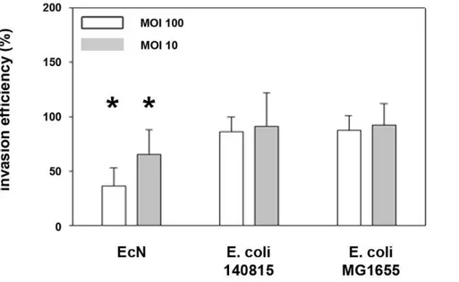
Initially, we verified a probiotic effect ofE. coliNissle 1917 (EcN) onSalmonellainvasion of porcine intestinal epithelial cells (IPEC-J2). We compared the effects of EcN with those of two controlE. colistrains (E. coli140815 andE. coliMG1655), and the effects of EcN in a mix with these two strains, and included two multiplicities of infections (MOI). In general, employing the gentamicin protection assay the invasion efficiency of Salmonella Typhimuriumstrain SL1344 into the porcine intestinal epithelial cell line IPEC-J2 was 27%. A two-hour pre-incubation of IPEC-J2 cells with EcN resulted in a decrease in Salmonella invasion efficiency, while pre-incubation with E. coli 140815 or E. coli MG1655 did not. This effect was stronger using an MOI of 100:1 (E. coli:epithelial cells) compared to 10:1 (Figure 1). The inhibitory
effect of EcN was markedly increased by a pre-incubation period of six hours compared to two hours (Figure 2A).
Inhibition ofSalmonella invasion by EcN was also observed in mixed E. colicultures although the effects on invasion were less effective in mixed E. coli cultures compared to EcN in the monoculture model. Using a mixture of EcN:E. coli140815:E. coli MG1655 (1:1:1), at a total MOI of 10:1 the inhibitory effects of EcN were abolished (Figure 3).
We also tested the effects of cell-free EcN supernatants compared to supernatants of the two control strains. To create a scenario relevant to the initial experiments with bacteria (for which EcN was present in the pre- and co-incubation period), we included pre- and pre-/co-incubation experiments using E. coli culture supernatants. As shown in Figure 4, pre-incubation withE. coli supernatants (E. coli 140815) showed little or no effects on Salmonellainvasion efficiency, while pre-/co-incubation noticeably inhibited Salmonella invasion. We tested the hypothesis that co-incubation of E. colisupernatants with Salmonella was predomi-nantly responsible for the inhibitory effect in subsequent co-incubation experiments. However, co-co-incubation of supernatants of all three tested E. coli strains similarly inhibited Salmonella invasion (Figure 4) as was the case withE. coli pre-incubations. Also supernatants of EcN mutants DfocA (F1C fimbriae), DfimA (type 1 fimbriae) and DfliA inhibited Salmonella invasion compa-rable to EcN wildtype (Figure 4).
Effects ofE. coli supernatants on extracellular growth
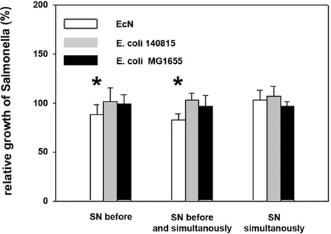
The inhibitory effect of EcN againstSalmonella invasion might have been due to the inhibition of Salmonella growth by E. coli supernatants, which inevitably affects invasion efficiencies. To exclude such effects,Salmonella numbers were determined in cell culture supernatants in parallel to the invasion assays. Superna-tants of all three E. coli did not, or only slightly (EcN), affect extracellular growth ofSalmonella within the one hour Salmonella incubation time (Figure 5).
Effects ofE. colion adhesion ofSalmonella
We determined whether EcN inhibited Salmonella adhesion, which is also a prerequisite for Salmonella invasion. Salmonella adhesion assays were performed with non-invasiveS. Typhimurium SL1344hilA-339::kan. All threeE. colistrains showed no effects on Salmonellaadhesion to IPEC-J2 cells in pre-incubation experiments (Figure 6). Even after a six-hour pre-incubation period withE. coli,
adhesion ofS. TyphimuriumSL1344hilA-339::kanwas not affected (Figure 6).
AdditionalSalmonella adhesion tests were performed with non-invasiveS. TyphimuriumSL1344 pEGFPinvG-339::kan. This mutant adhered twice to IPEC-J2 cells than non-invasiveS. Typhimurium SL1344hilA-339::kan. EcN inhibited SL1344 pEGFPinvG-339::kan adhesion by 40%. In contrast,E. colistrain MG1655 and 140815
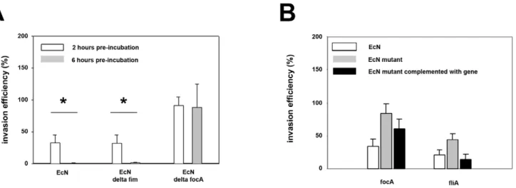
Figure 2. Inhibitory effects ofE. coliNissle 1917 onSalmonella Typhimuriuminvasion is dependent on adhesion.Confluent monolayers of IPEC-J2 cells were pre-incubated withE. coliNissle 1917 (EcN), EcNDfocA, EcNDfimor EcNDfliAusing an MOI of 100:1E. colito host cells. After two or six hours, cells were washed and infected withSalmonella Typhimuriumusing an MOI of 100:1Salmonellato host cells. Invasion levels in percent (%) are expressed as invasion ofSalmonellarelative to invasion without pre-incubation withE. coli(Salmonellamono-infection). The data are the mean6S.E.M. of at least three separate experiments in duplicate wells. * = p,0.01 compared toSalmonellamono-infection. A) Effects of EcNDfocA and EcNDfimmutants onSalmonellainvasion after a 2 or 6 hours pre-incubation period. B) Effects of EcNDfocAand EcNDfliAmutants and their respective strains complemented with the plasmid pACYC 177 containing the relevant gene onSalmonellainvasion after 2 hours pre-incubation. doi:10.1371/journal.pone.0014712.g002
enhanced Salmonella adhesion to IPEC-J2 cells in pre-incubation experiments (Figure 6). The kanamycin resistance of non-invasive Salmonella mutants does not contribute to this effect since strain SL1344::kan harboring a chromosomal kanamycin resistance
cassette in a non-coding, intergenic region not affecting the expression of any genes was similarly invasive and had a similar probiotic effect againstSalmonellainvasion (reduction of invasion by 55.3%) as the wild type strain itself.
Figure 4. Invasion efficiency ofSalmonella Typhimuriuminto IPEC-J2 cells after incubation withE. coliculture supernatants.Confluent monolayers of IPEC-J2 cells were pre-incubated (SN before) and/or co-incubated (SN simultaneously) withE. colisupernatants (SN). Cells were infected withSalmonella Typhimuriumusing an MOI of 100:1Salmonellato host cells. Invasion levels in percent (%) are expressed as invasion of Salmonellarelative to invasion without pre- and/or co-incubation with E. coliSN. The data are the mean 6S.E.M. of at least three separate experiments in duplicate wells. * = p,0.05 compared toSalmonellainfection without influence ofE. coliSN. EcN:E. coliNissle 1917.
doi:10.1371/journal.pone.0014712.g004
Figure 5. Effects ofE. colisupernatants on growth ofSalmonella Typhimurium.Confluent monolayers of IPEC-J2 cells were pre- and/or co-incubated withE. colisupernatants (SN). Cells were infected withSalmonella Typhimuriumusing an MOI of 100:1Salmonellato host cells for one hour. Thereafter, numbers of extracellular, non-adherentSalmonellawere determined. Growth rates are expressed as growth in percent (%) relative to Salmonellagrowth in cell culture medium (Salmonellamono-infection). The data are the mean6S.E.M. of at least three separate experiments in duplicate wells. * = p,0.01 compared toSalmonellamono-infection. EcN:E. coliNissle 1917.
E. coliNissle 1917 does not inhibit intracellular growth of
Salmonella
To test whether EcN might have been able to affectSalmonella post-invasion, EcN and the two control strains were incubated with Salmonella in post-incubation experiments. Here, IPEC-J2 cells were initially infected with Salmonella for one hour, and extracellular Salmonella were inactivated by incubation with gentamicin for another hour. After removal of gentamicin IPEC-J2 cells with intracellularSalmonellawere incubated withE. colifor 2 hours and then incubated for up to 20 hours with medium containing gentamicin to kill extracellular bacteria. Intracellular Salmonella were determined at 2, 5 and 24 hours after the initial Salmonellaincubation period. As shown in Figure 7, all threeE. coli had no effect on intracellularSalmonella growth at 2, 5 as well as 24 hours after the initialSalmonellaincubation.
F1C fimbriae mediates inhibitory effects of EcN on
Salmonella invasion
Adhesion might be a prerequisite for the inhibitory effect of EcN. To test this, we used EcNDfocA(F1C fimbriae),DfimA(type 1 fimbriae) andDfliA(flagellae) mutants. Adhesion by strains EcNDfocA(reduction by 92.7%) and EcNDfliA(reduction by 47.9%) on IPEC-J2 cells was reduced compared to EcN wild type strain. After complementation with the pACYC 177 plasmids, respectively containing thefocAorfliA gene, adhesion was enhanced to 53.4% (focA) and 122.1% (fliA), compared to the EcN wild type strain. Adhesion by strain EcNDfimA on IPEC-J2 cells was not reduced compared to EcN wild type strain. A decrease or increase in adhesion correlated with a decrease or increase of the inhibitory effect onSalmonellainvasion, respectively. Thus EcN
DfocAdid not inhibitSalmonellainvasion compared to EcN wildtype strain, whereas EcNDfliAinhibitedSalmonellainvasion by 50%. EcN
DfimAinhibitedSalmonellainvasion at levels similar to those of EcN wildtype strain (Figure 2). The results were more prominent using a 6-hour EcN pre-incubation period.
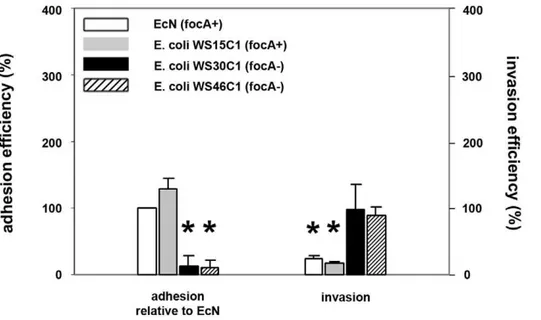
To test whether adhesion via F1C fimbriae is essential for a subsequent inhibitory effect of EcN and the specificity of EcN inhibition, we compared adhesion rates as well as inhibition between EcN and anotherfocAgene-positive (WS15C1), and two otherfocAgene-negative (WS30C1 and WS46C1) E. colistrains. These bacteria all carry the type 1 fimbriae and flagellae and were isolated from the intestine of clinically healthy wild boars [23]. As shown in Figure 8, adhesion rates of thefocAgene-positive strain WS15C1 were higher compared to EcN, butfocA gene-negative strains WS30C1 and WS46C1 adhered much less compared to EcN. Additionally, these high adhesion rates correlated with high inhibitory effects of EcN and WS15C1 on Salmonella invasion (Figure 8).
Effects of EcN on expression ofSalmonella invasion genes
As the previous results indicated thatSalmonellaadhesion - but not extracellular or intracellularSalmonellagrowth - was affected by EcN, we tested the effects of EcN supernatants on the expression of Salmonella invasion gene regulatory and dependent genes. Salmonella strains harboring lacZ fusions to the invasion locus regulatory geneshilC,hilD, hilA, and the SPI4-encodedsiiEgene were incubated with supernatants of EcN,E. coli140815 orE. coli MG1655. As shown in Figure 9, expression of HilC was not affected by any of the threeE. colisupernatants. HilD expression was slightly enhanced by all threeE. colisupernatants and HilA and SiiE expressions were inhibited by all three E. coli supernatants.
Discussion
ProbioticE. coliNissle 1917 (EcN) is being successfully used for the prevention and treatment of various intestinal diseases of humans and animals. However, the basis of its mode of action
Figure 6. Adhesion efficiency ofSalmonella Typhimuriumto IPEC-J2 cells after pre-incubation withE. coli.Confluent monolayers of IPEC-J2 cells were pre-incubated withE. coliNissle 1917 (EcN),E. coli140815 orE. coliMG1655 using an MOI of 100:1E. colito host cells. After two or six hours, cells were washed and infected with non-invasiveSalmonella TyphimuriumSL1344hilA-339::kanor SL1344 pEGFPinvG-339::kanusing an MOI of 100:1Salmonellato host cells. Adhesion levels in percent (%) are expressed as adhesion ofSalmonellarelative to adhesion without pre-incubation withE. coli(Salmonellamono-infection). The data are the mean6S.E.M. of at least three separate experiments in duplicate wells. * = p,0.01 compared toSalmonellamono-infection.
remains largely unanswered. EcN has been shown inin vitrostudies to protect human embryonic intestinal epithelial cells (INT407 cells) against infection by different enteropathogens, including
enteroin-vasive bacteria, but the underlying mechanisms were not clarified [14,16]. In the present study, we initially defined possible probiotic effects of EcN against Salmonella invasion of porcine intestinal
Figure 8. Inhibitory effects offocA-positive andfocA-negativeE. coliisolates onSalmonella Typhimuriuminvasion.Confluent monolayers of IPEC-J2 cells were pre-incubated withE. coliNissle 1917 (EcN,focA-positive strain),E. coliWS15C1 (focA-positive strain),E. coliWS30C1 (focA -negative strain) andE. coliWS46C1 (focA-negative strain) using an MOI of 100:1E. colito host cells. After two hours, cells were washed and adhesion efficiencies ofE. coliisolates were determined (left side); alternatively, cells were washed after two hours and infected withSalmonella Typhimurium using an MOI of 100:1Salmonellato host cells (right side). Adhesion levels in percent (%) were expressed relative to adhesion of EcN. Invasion levels in percent (%) were expressed relative toSalmonellainvasion without pre-incubation with bacteria (Salmonellamono-infection). The data are the mean6S.E.M. of at least three separate experiments in duplicate wells. * = p,0.01 compared to EcN adhesion (left side) orSalmonella mono-infection (right side).
doi:10.1371/journal.pone.0014712.g008
Figure 7. Intracellular growth ofSalmonella Typhimuriumin IPEC-J2 cells after post-incubation with E. coliNissle 1917.Confluent monolayers of IPEC-J2 cells were infected withSalmonella Typhimuriumfor one hour using an MOI of 1:1Salmonellato host cells. After one additional hour of incubation in media containing gentamicin, IPEC-J2 cells were incubated withE. coliNissle 1917 (EcN),E. coli140815 orE. coliMG1655 for two hours using an MOI of 100:1E. colito host cells, followed by incubation in media with gentamicin. IntracellularSalmonellanumbers are presented per well of a 24-well plate. The data are the mean6S.E.M. of at least three separate experiments in duplicate wells.
epithelial cells (IPEC-J2) and subsequently verified such probiotic effects against single stages of theSalmonellainvasion process.
EcN successfully inhibitedSalmonellainfection of IPEC-J2 cells. The specificity of this effect was demonstrated in that two control E. coli strains, 140815 and MG1655, showed no such inhibitory effects. Inhibition ofSalmonella infection could have been due to effects on several infection steps including inhibition of intracel-lular and extracelintracel-lularSalmonellagrowth, inhibition of adhesion, or other factors. The probiotic effect of EcN was found to be highly dependent upon its adherence to IPEC-J2 cells, preferentially through F1C fimbriae (Figure 2).Salmonellaadhesion andSalmonella adhesion gene expressions were affected by EcN and/or EcN supernatants respectively, but not Salmonella extracellular or intracellular growth, supporting a role of adhesion genes in the probiotic effects.
We propose two mechanisms for the probiotic effect of EcN. First, EcN supernatants could be responsible for this effect. This might be mediated through interactions with theSalmonella SiiE-dependent adhesion mechanism, as well as down-regulation of the expression of the adhesion factor SiiE. We suggest thatSalmonella adhesion was reduced via SiiE-mediated adhesion by EcN since adhesion of a non-invasiveinvG Salmonellamutant with a functional SiiE mediation system was affected, whereas adhesion of a non-invasive Salmonella mutant defective in hilA with an SiiE-minus genotype was not, however;E. coli supernatants suppressed both SiiE and HilA expression, as well as suppression of both genes contributed to the probiotic effect. HilA is a transcriptional regulator of the OmpR/ToxR family that is encoded onSalmonella pathogenicity island 1 and plays a key role in the regulation of invasiveness ofSalmonella Typhimurium. HilA has been shown to be
Figure 9.Salmonellainvasion gene regulation byE. colisupernatants.E. coliwere cultivated in cell culture medium (DMEM HAM’S/F-12) until an OD600nm= 1. Supernatants were collected by centrifugation with subsequent sterile filtration. Subsequently, SL1344 fusion strains (SL1344hilC -lacZ, SL1344 hilD-lacZ, SL1344 hilA-lacZ, SL1344 icgA(siiE)-lacZ) were cultivated in supernatants of EcN, E. coli 140815 or E. coli MG1655. B-Galactosidase activity was measured as previously described [25,26]. The results shown are representative of at least two independent experiments. White bar:Salmonellagrown in EcN supernatant, gray bar:Salmonellagrown inE. coli140815 supernatant, black bar:Salmonellagrown inE. coli MG1655 supernatant, patterned bar:Salmonellagrown in pure cell culture medium.
required for the optimal expression of both invasion genes as well as the adhesin SiiE [27,28]. While HilA-dependent regulation has been intensively studied, other HilA-dependent adhesion genes have not been reported so far. HilA expression is also regulated by the regulators HilC and HilD [29]. In our study, HilC expression was not influenced byE. colisupernatants, while HilD expression was slightly enhanced. Thus the regulation of HilA by HilD, which would have up-regulated HilA and SiiE expression, was weaker than the direct effects of E. coli supernatants on HilA. SiiE is secreted fromSalmonellaby a type I secretion system into the cell culture supernatant and can function as an adhesin when in contact with polarized epithelial cells. The IPEC-J2 cell line has also been shown to form a polarized cell monolayer [21,30]. This mechanism might not be EcN-specific since supernatants from other E. coli also similarly inhibited Salmonella invasion, and similarly regulatedSalmonellavirulence gene expression.
A second mechanism is suggested by the strong adherence of EcN on IPEC-J2 cells and the subsequent inhibition ofSalmonellainvasion which may be a major contributing factor in the probiotic effect of EcN. This strong adhesion was EcN-specific compared to the control strains 140815 and MG1655 and was predominantly mediated by F1C fimbriae. However, other F1C fimbriae expressingE. colistrains could also strongly adhere to IPEC-J2 cells and reduce Salmonella invasion, as was the case withE. colistrain WS15C1.
In summary, in the presence ofE. coli, components present in culture supernatants appear to reduce Salmonella SiiE-mediated adhesion by down-regulation of SiiE production.E. colisupernatant compounds might also block SiiE-mediated adhesion by binding to either the SiiE protein itself or to SiiE receptors on the host cell surface. Such effects on the adhesion mediated by the SiiE protein are a subject for future research. In the presence of high concentrations of E. colisupernatants components might bind to the host cell surface. After washing host cells, followed by incubation in fresh cell culture medium (pre-incubation experiments), these bound components might detach and be diluted by added fresh cell culture medium to concentrations that become ineffective in inhibiting Salmonella adhesion. If cells are however incubated at high concentrations of supernatants after washing (pre- plus post-incubation with superna-tants), components of supernatants will still bind to the host cell surface in high concentrations and affectSalmonellaadhesion. This might explain why pre- and co-incubation withE. colisupernatants resulted in a higher reduction of Salmonella invasion than pre-incubation or post-pre-incubation only. Attachment to the cell surface through F1C fimbriae may support intimate EcN contact at the host cell surface and increase the local concentrations of supernatant compounds which decreaseSalmonellaadhesion and invasion.
A supernatant-dependent mechanism seems to be common for differentE. colistrains (this study, [14]) as well as other bacterial species e.g. Lactobacillus acidophilus, Lactobacillus casei, Lactobacillus
plantarum, Pediococcus pentosaceus and Leuconostoc mesenteroides [31,32,33]. In these latter studies, acidification of the medium followed by a bacteriocidal effect, the production of microcins, and other undefined bacteriocidal effects or inhibition of Salmonella growth were found to be responsible for probiotic supernatant-dependent mechanisms. However, in this study,Salmonellagrowth rates were similar in all different E. coli culture supernatants indicating thatE. coli culture supernatants had no bacteriocidal effects against Salmonella. Such a finding does not rule out the possibility of other unknown supernatant factors binding to, and inhibiting SiiE or other molecules. Other authors reported thatL. caseisupernatant inhibitedSalmonellainvasion, but did not modify the viability of Salmonella. These authors supposed that a hypothetical substance of L. casei supernatant directly modified the ability ofSalmonellato invade enterocyte-like cellsin vitroin an acidic environment [33].
The adhesion of EcN to IPEC-J2 cells was a prerequisite for its probiotic effects. As shown in our study, F1C fimbriae as well as flagellae contributed to the adherence but not type 1 fimbriae. This is in accordance with recent observations that F1C fimbriae, not type 1 fimbriae, contributed to the adherence of EcN to the human larynx epithelial cell line Hep-2, and to their persistence in infant mouse colonization and biofilm formation [19]. EcN is therefore able to adhere via F1C fimbriae to different types of cells. The contribution of adherence to the probiotic effect was clearly indicated. For example, the adhesion of the EcN DfliA mutant complemented with a plasmid containing the fliA gene was enhanced to 122.1% compared to the EcN wild type strain, while Salmonellainvasion decreased to 14.9% in the presence of the EcN
DfliA/pACYC177fliAconstruct, compared to 35% in the presence of the EcN wild type strain.
The conclusion of this study is three-fold. First, EcN suppressed Salmonella invasion by suppressing Salmonella adhesion. Second, effects of bacterial supernatants might be also common for otherE. coli. And third, strong adherence is a prerequisite for the probiotic effect of EcN.
Acknowledgments
We thank Matthias Filter (Bundesinstitut fu¨r Risikobewertung, Berlin, Germany) for helping with the statistical analysis and Bryon Nicholson (Department of Veterinary Microbiology and Preventive Medicine, Ames, USA) for helpful comments on the manuscript.
Author Contributions
Conceived and designed the experiments: PS. Performed the experiments: PS SK RH. Analyzed the data: PS SK KT JTB. Contributed reagents/ materials/analysis tools: PS SO TAO SP LHW. Wrote the paper: PS SK KT JTB LHW.
References
1. Kruis W, Fric P, Pokrotnieks J, Lukas M, Fixa B, et al. (2004) Maintaining remission of ulcerative colitis with the probiotic Escherichia coli Nissle 1917 is as effective as with standard mesalazine. Gut 53: 1617–1623.
2. Schultz M, Strauch UG, Linde HJ, Watzl S, Obermeier F, et al. (2004) Preventive effects of Escherichia coli strain Nissle 1917 on acute and chronic intestinal inflammation in two different murine models of colitis. Clin Diagn Lab Immunol 11: 372–378.
3. Schroeder B, Duncker S, Barth S, Bauerfeind R, Gruber AD, et al. (2006) Preventive effects of the probiotic Escherichia coli strain Nissle 1917 on acute secretory diarrhea in a pig model of intestinal infection. Dig Dis Sci 51: 724–731. 4. Henker J, Laass MW, Blokhin BM, Maydannik VG, Bolbot YK, et al. (2008) Probiotic Escherichia coli Nissle 1917 versus placebo for treating diarrhea of greater than 4 days duration in infants and toddlers. Pediatr Infect Dis J 27: 494–499. 5. von Buenau R, Jaekel L, Schubotz E, Schwarz S, Stroff T, et al. (2005)
Escherichia coli strain Nissle 1917: significant reduction of neonatal calf diarrhea. J Dairy Sci 88: 317–323.
6. Krammer HJ, Kamper H, von Bunau R, Zieseniss E, Stange C, et al. (2006) [Probiotic drug therapy with E. coli strain Nissle 1917 (EcN): results of a prospective study of the records of 3,807 patients]. Z Gastroenterol 44: 651–656. 7. Reissbrodt R, Hammes WP, dal Bello F, Prager R, Fruth A, et al. (2009) Inhibition of growth of Shiga toxin-producing Escherichia coli by nonpathogenic Escherichia coli. FEMS Microbiol Lett 290: 62–69.
8. Bar F, Von Koschitzky H, Roblick U, Bruch HP, Schulze L, et al. (2009) Cell-free supernatants of Escherichia coli Nissle 1917 modulate human colonic motility: evidence from an in vitro organ bath study. Neurogastroenterol Motil 21: 559–566, e516-557.
9. Ukena SN, Singh A, Dringenberg U, Engelhardt R, Seidler U, et al. (2007) Probiotic Escherichia coli Nissle 1917 inhibits leaky gut by enhancing mucosal integrity. PLoS One 2: e1308.
11. Bickert T, Trujillo-Vargas CM, Duechs M, Wohlleben G, Polte T, et al. (2009) Probiotic Escherichia coli Nissle 1917 suppresses allergen-induced Th2 responses in the airways. Int Arch Allergy Immunol 149: 219–230.
12. Zyrek AA, Cichon C, Helms S, Enders C, Sonnenborn U, et al. (2007) Molecular mechanisms underlying the probiotic effects of Escherichia coli Nissle 1917 involve ZO-2 and PKCzeta redistribution resulting in tight junction and epithelial barrier repair. Cell Microbiol 9: 804–816.
13. Helwig U, Lammers KM, Rizzello F, Brigidi P, Rohleder V, et al. (2006) Lactobacilli, bifidobacteria and E. coli nissle induce pro- and anti-inflammatory cytokines in peripheral blood mononuclear cells. World J Gastroenterol 12: 5978–5986.
14. Altenhoefer A, Oswald S, Sonnenborn U, Enders C, Schulze J, et al. (2004) The probiotic Escherichia coli strain Nissle 1917 interferes with invasion of human intestinal epithelial cells by different enteroinvasive bacterial pathogens. FEMS Immunol Med Microbiol 40: 223–229.
15. Kleta S, Steinruck H, Breves G, Duncker S, Laturnus C, et al. (2006) Detection and distribution of probiotic Escherichia coli Nissle 1917 clones in swine herds in Germany. J Appl Microbiol 101: 1357–1366.
16. Boudeau J, Glasser AL, Julien S, Colombel JF, Darfeuille-Michaud A (2003) Inhibitory effect of probiotic Escherichia coli strain Nissle 1917 on adhesion to and invasion of intestinal epithelial cells by adherent-invasive E. coli strains isolated from patients with Crohn’s disease. Aliment Pharmacol Ther 18: 45–56. 17. Prilassnig M, Wenisch C, Daxboeck F, Feierl G (2007) Are probiotics detectable in human feces after oral uptake by healthy volunteers? Wien Klin Wochenschr 119: 456–462.
18. Grozdanov L, Raasch C, Schulze J, Sonnenborn U, Gottschalk G, et al. (2004) Analysis of the genome structure of the nonpathogenic probiotic Escherichia coli strain Nissle 1917. J Bacteriol 186: 5432–5441.
19. Lasaro MA, Salinger N, Zhang J, Wang Y, Zhong Z, et al. (2009) F1C fimbriae play an important role in biofilm formation and intestinal colonization by the Escherichia coli commensal strain Nissle 1917. Appl Environ Microbiol 75: 246–251.
20. Monteiro C, Saxena I, Wang X, Kader A, Bokranz W, et al. (2009) Characterization of cellulose production in Escherichia coli Nissle 1917 and its biological consequences. Environ Microbiol 11: 1105–1116.
21. Schierack P, Nordhoff M, Pollmann M, Weyrauch KD, Amasheh S, et al. (2006) Characterization of a porcine intestinal epithelial cell line for in vitro studies of microbial pathogenesis in swine. Histochem Cell Biol 125: 293–305.
22. Datsenko KA, Wanner BL (2000) One-step inactivation of chromosomal genes in Escherichia coli K-12 using PCR products. Proc Natl Acad Sci U S A 97: 6640–6645.
23. Schierack P, Romer A, Jores J, Kaspar H, Guenther S, et al. (2009) Isolation and characterization of intestinal Escherichia coli clones from wild boars in Germany. Appl Environ Microbiol 75: 695–702.
24. Lee CA, Falkow S (1990) The ability of Salmonella to enter mammalian cells is affected by bacterial growth state. Proc Natl Acad Sci U S A 87: 4304–4308. 25. Thompson A, Rolfe MD, Lucchini S, Schwerk P, Hinton JC, et al. (2006) The
bacterial signal molecule, ppGpp, mediates the environmental regulation of both the invasion and intracellular virulence gene programs of Salmonella. J Biol Chem 281: 30112–30121.
26. Hernandez VJ, Bremer H (1990) Guanosine tetraphosphate (ppGpp) depen-dence of the growth rate control of rrnB P1 promoter activity in Escherichia coli. J Biol Chem 265: 11605–11614.
27. Bajaj V, Hwang C, Lee CA (1995) hilA is a novel ompR/toxR family member that activates the expression of Salmonella typhimurium invasion genes. Mol Microbiol 18: 715–727.
28. Main-Hester KL, Colpitts KM, Thomas GA, Fang FC, Libby SJ (2008) Coordinate regulation of Salmonella pathogenicity island 1 (SPI1) and SPI4 in Salmonella enterica serovar Typhimurium. Infect Immun 76: 1024–1035. 29. Ellermeier CD, Ellermeier JR, Slauch JM (2005) HilD, HilC and RtsA constitute
a feed forward loop that controls expression of the SPI1 type three secretion system regulator hilA in Salmonella enterica serovar Typhimurium. Mol Microbiol 57: 691–705.
30. Gerlach RG, Jackel D, Geymeier N, Hensel M (2007) Salmonella pathogenicity island 4-mediated adhesion is coregulated with invasion genes in Salmonella enterica. Infect Immun 75: 4697–4709.
31. Bernet-Camard MF, Lievin V, Brassart D, Neeser JR, Servin AL, et al. (1997) The human Lactobacillus acidophilus strain LA1 secretes a nonbacteriocin antibacterial substance(s) active in vitro and in vivo. Appl Environ Microbiol 63: 2747–2753.
32. Chiu HH, Tsai CC, Hsih HY, Tsen HY (2008) Screening from pickled vegetables the potential probiotic strains of lactic acid bacteria able to inhibit the Salmonella invasion in mice. J Appl Microbiol 104: 605–612.