b
ig-h3 Promotes Human Osteosarcoma Cells Metastasis
by Interacting with Integrin
a
2
b
1 and Activating PI3K
Signaling Pathway
Yun-Shan Guo1,2., Rui Zhao1., Jie Ma3., Wei Cui4., Zhen Sun1
, Bo Gao1, Shu He1, Yue-Hu Han1, Jing Fan1, Liu Yang1*, Juan Tang2*, Zhuo-Jing Luo1*
1Department of Osteology, Xijing Hospital, Fourth Military Medical University, Xi’an, China,2Cell Engineering Research Centre & Department of Cell Biology, State Key Laboratory of Cancer Biology, State Key Discipline of Cell Biology, Fourth Military Medical University, Xi’an, China,3Department of Neurosurgery, Tangdu Hospital, Fourth Military Medical University, Xi’an, China,4Department of Endocrinology and Metabolism, Xijing Hospital, Fourth Military Medical University, Xi’an, China
Abstract
Osteosarcoma, the most common primary bone tumor in children and young adolescents, is characterized by local invasion and distant metastasis. But the detailed mechanisms of osteosarcoma metastasis are not well known. In the present study, we found thatbig-h3 promotes metastatic potential of human osteosarcoma cells in vitro and in vivo. Furthermore,big-h3 co-localized with integrina2b1 in osteosarcoma cells. Butbig-h3 did not change integrina2b1 expression in Saos-2 cells. Interaction ofbig-h3 with integrina2b1 mediates metastasis of human osteosarcoma cells. The second FAS1 domain ofb ig-h3 but not the first FAS1 domain, the third FAS1 domain or the fourth FAS1 domain mediates human osteosarcoma cells metastasis, which is thea2b1 integrin-interacting domain. We further demonstrated that PI3K/AKT signaling pathway is involved inbig-h3-induced human osteosarcoma cells metastasis process. Together, these results revealbig-h3 enhances the metastasis potentials of human osteosarcoma cells via integrina2b1-mediated PI3K/AKT signal pathways. The discovery of big-h3-mediated pathway helps us to understand the mechanism of human osteosarcoma metastasis and provides evidence for the possibility thatbig-h3 can be a potential therapeutic target for osteosarcoma treatment.
Citation:Guo Y-S, Zhao R, Ma J, Cui W, Sun Z, et al. (2014)big-h3 Promotes Human Osteosarcoma Cells Metastasis by Interacting with Integrina2b1 and Activating PI3K Signaling Pathway. PLoS ONE 9(3): e90220. doi:10.1371/journal.pone.0090220
Editor:Fre´de´ric Andre´, Aix-Marseille University, France
ReceivedNovember 11, 2013;AcceptedJanuary 27, 2014;PublishedMarch 4, 2014
Copyright:ß2014 Guo et al. This is an open-access article distributed under the terms of the Creative Commons Attribution License, which permits unrestricted use, distribution, and reproduction in any medium, provided the original author and source are credited.
Funding:This work is supported by the National Basic Research Program of China (2011CB964703) and the National Natural Science Foundation of China (31101005). The funders had no role in study design, data collection and analysis, decision to publish, or preparation of the manuscript.
Competing Interests:The authors have declared that no competing interests exist.
* E-mail: [email protected] (LY); [email protected] (JT); [email protected] (ZJL)
.These authors contributed equally to this work.
Introduction
Osteosarcoma is a high-grade malignant bone neoplasm that occurs primarily in children and young adolescents. It occurs with an incidence of approximately three cases per million people per year [1]. The principles of treatment of osteosarcoma have undergone dramatic improves in the past 20 years. Multi-agent chemotherapy increased the 5-year overall survival of patients with localized disease to between 60% and 78% [2]. The survival of patients with metastatic osteosarcoma, however, remains poor with survival rates ranging from 11% to 20% [3,4]. This outcome suggested that 80% of the patients had metastasis at the time of presentation. Hence, a novel strategy that would efficiently inhibit osteosarcoma metastasis is highly desirable.
Tumor metastasis consists of a trail of complex procedures, all of which must be successfully completed to result in clinically detectable metastatic tumors at distal tissues [5,6]. To complete the process, primary cancer cells have to attach to extracellular matrix (ECM) components, invade through the basement mem-brane, intravasate into the circulation, and extravasate to distal tissues [7,8]. The entire process regulated by interactions between cancer cells and ECM. As a major component of the tumor microenvironment, ECM proteins potentially affect the metastasis
process [9]. Thus, molecular alterations of the ECM proteins in the tumor microenvironment have a considerable impact on the metastatic process during tumorigenesis.
not clear yet whether big-h3 is involved in osteosarcoma metastasis.
This study sought to examine whetherbig-h3 expression could influence osteosarcoma cells metastasis and to determine the molecular mechanism by which this occurred, in an effort to elucidate the role of big-h3 in the regulation of osteosarcoma metastasis. In the present study, we showed thatbig-h3 promotes adhesion, invasion and migration of human osteosarcoma cells. big-h3 mediates human osteosarcoma cells metastasis through interacting with integrin a2b1, and then activates downstream PI3K/AKT signaling pathway. Furthermore, we identified that only the second FAS1domain of big-h3 was involved in osteosarcoma cells metastasis.
Results
Downregulation ofbig-h3 decreases adhesion, invasion and migration of human osteosarcoma cells in vitro
As an ECM protein, big-h3 is involved in cell proliferation, migration, invasion, apoptosis and tumorigenesis [18–21]. To test the role ofbig-h3 in human osteosarcoma cells, small interfering RNAs against big-h3 (big-h3 siRNA) were transfected into the human osteosarcoma cell lines, Saos-2 cells and MG63 cells, for 48 hours to knockdown big-h3 mRNA and protein expression. Silencer negative control siRNAs (control siRNA) were also used as a negative control. As compared with control siRNA treated cells, thebig-h3 siRNA could effectively decrease the mRNA and protein expression of big-h3 in Saos-2 cells and MG63 cells (P,0.05, Figure 1A and 1B). Cell function assays demonstrated that the amounts of cell adhesion were significantly decreased after big-h3 siRNA treatment in Saos-2 cells and MG63 cells (57.6%611.9% and 52.3%69.4%, respectively) (P,0.05, Figure 1C). In addition, the abilities of cells to invade through Transwell chambers were decreased after transfected withbig-h3 siRNA in Saos-2 cells and MG63 cells. (37.1%618.5% and 31.2%612.8%, respectively) (P,0.05, Figure 1D). Similarly, treatment withbig-h3 siRNA also decreased the amounts of cell migration in Saos-2 cells and MG63 cells (39.2%615.3% and 45.4%610.7%, respectively) (P,0.05, Figure 1E). This finding suggests that big-h3 may enhance adhesion, invasion and migration potential of human osteosarcoma cells.
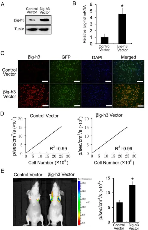
big-h3 promotes metastasis of human osteosarcoma cells in vivo
To extend these studies in vivo, Saos-2 cells stably expressing GFP were established by lentiviral infection. Then the big-h3 vector and a control vector were stably transfected into Saos-2 cells which were stably expressing GFP. As compared with control vector-treated cells, the big-h3 vector could stably increase the mRNA and protein expression ofbig-h3 in Saos-2 cells (P,0.05, Figure 2A and 2B). Moreover, we observed stable expression of GFP and a strong correlation between GFP fluorescence signals and cell number in both control vector-treated cells and big-h3 vector-treated cells (Figure 2C and Figure 2D). Furthermore, Saos-2 cells expressing the GFP were injected into immunodeficient mice through the tail vein. GFP fluorescence imaging was used to monitor the presence of Saos-2 cells. Due to size restrictions imposed by mouse capillaries, human tumor cells are rarely able to pass from the venous to the arterial system by way of the lung. Cells that failed to metastasize were not able to survive. Detectable GFP fluorescence signals indicated that cells had succeeded in metastasizing [23–24]. We found that the GFP signal in the group ofbig-h3 vector-transfected cells was significantly higher than the GFP signal in the group of control vector-transfected cells in the
lung (P,0.05, Figure 2E). Therefore, the result indicated thatb ig-h3 significantly promotes metastasis of human osteosarcoma cells in vivo.
big-h3 immunoprecipitates witha2b1 integrin in human osteosarcoma cells
Integrins are cell surface adhesive receptors composed ofaand b chain heterocomplexes, which play an essential role in osteosarcoma metastasis [25–28]. It has been reported that both a2 andb1 subunits are expressed in human osteosarcoma cells and they serve as bidirectional transducers of extracellular and intracellular signals in tumor metastasis processes [29–31]. Accordingly, we hypothesized that big-h3 might interact with integrina2b1 to affect the metastasis ability of osteosarcoma cells. Immunofluorescent double staining was performed to examine cellular distribution of integrina2b1 andbig-h3 in Saos-2 cells. The results showed co-localizations ofbig-h3 with integrina2 and integrinb1 subunits on the cell membrane (Figure 3A). To further confirm this result, co-immunoprecipitation assay were performed to detect the interaction ofbig-h3 with integrina2 and integrinb1 subunits. integrina2 and integrinb1 subunits were found to co-immunoprecipitate with endogenousbig-h3 in Saos-2 cells lysates, (Figure 3B–3D), indicating thatbig-h3 and integrina2b1 interact in their native conformations. To elucidate the effects ofbig-h3 on integrin a2b1 expression, we tested the protein expressions of integrina2 and integrinb1 subunits inbig-h3 siRNA transfected Saos-2 cells. We found that there were no significant expression modifications of integrin a2 and integrin b1 in big-h3 siRNA transfected cells compared with control cells (Figure 3E). This finding suggested that big-h3 did not change integrin a2b1 expression in Saos-2 cells.
Interaction ofbig-h3 with integrina2b1 mediates metastasis of human osteosarcoma cells
To identify whether integrin a2b1 is involved in big-h3 mediated human osteosarcoma metastasis, the function blocking antibodies, mouse anti-human integrina2 mAb (P1E6) and mouse anti-human integrinb1 mAb (6S6) were used. We found thatb ig-h3 siRNA markedly reduced the amounts of cell adhesion in blank control group (p,0.05, Figure 4A). However, the addition of P1E6 and 6S6, alone or combination reduced the amounts of cell adhesion in control siRNA transfected cells to levels comparable with that inbig-h3 siRNA transfected cells.big-h3 siRNA did not further reduce the amounts of cell adhesion after blocking integrin a2b1 (p.0.05, Figure 4A). The result indicated that integrina2b1 is involved inbig-h3 mediated cell adhesion. In addition, invasion and migration assay were performed. We found thatbig-h3 siRNA markedly reduced the amounts of cell invasion and migration in blank control groups (p,0.05, Figure 4B and 4C). However, the addition of P1E6 and 6S6, alone or combination reduced the amounts of cell invasion and migration in control siRNA transfected cells to levels comparable with that inbig-h3 siRNA transfected cells.big-h3 siRNA did not further reduce the amounts of cell invasion and migration after blocking integrin a2b1 (p.0.05, Figure 4B and 4C). The above results indicate that integrina2b1 is required forbig-h3 mediates metastasis of human osteosarcoma cells.
The second FAS1 domain ofbig-h3 promotes human osteosarcoma cells metastasis
function via interactions with various integrins [15–17]. To identify which FAS1domain mediated human osteosarcoma cells metastasis, we cloned the total gene ofbig-h3 (WT) and its four segments of highly conserved sequence, the first FAS1domain (D-I), the second FAS1domain (D-I(D-I), the third FAS1domain (D-III) and the fourth FAS1domain (D-IV) and then we transfected them into Saos-2 cells. We found that mRNA of the four FAS1domains ofbig-h3 were overexpressed in Saos-2 cells respectively (P,0.05, Figure 5B). Cell adhesion, invasion and migration assay demon-strated that overexpression of the second FAS1domain promoted the amounts of cell adhesion, invasion and migration to levels comparable with that in the total gene of big-h3 overexpressed cells (P,0.05, Figure 5C–5E). However, overexpression of the first FAS1 domain, the third FAS1 domain and the fourth FAS1do-main were not able to promote cell adhesion, invasion and migration in Saos-2 cells (P.0.05, Figure 5C–5E). These results suggested that only the second FAS1domain, but not the first FAS1domain, the third FAS1domain or the fourth FAS1domain ofbig-h3 was involved in osteosarcoma cells metastasis
big-h3 induces human osteosarcoma cells metastasis by activating PI3K signaling pathway
Currently, the identities of integrin a2b1-associated signaling molecules that are responsible for mediating human osteosarcoma cells metastasis in response tobig-h3 are unclear. To determine the signaling pathways that contribute to human osteosarcoma cells metastasis induced bybig-h3, an examination was conducted into the effects ofbig-h3 on the phosphorylation status of AKT. Knockdown ofbig-h3 was found to decrease phosphorylation of AKT in Saos-2 cells (Figure 6A). To further test whether PI3K is involved inbig-h3 mediated Saos-2 cells metastasis, LY294002, a reversible inhibitor of PI3K was employed. big-h3 siRNA markedly reduced phosphorylation of AKT in control group. However,big-h3 siRNA did not further reduce phosphorylation of AKT following LY294002 treatment (Figure 6B). The result suggested that activity of PI3K is required for big-h3 induced phosphorylation of AKT. Moreover, the addition of P1E6 and 6S6, alone or combination reduced the levels of phosphorylation of AKT in control siRNA transfected cells to levels comparable with that inbig-h3 siRNA transfected cells (Figure 6C). The result indicated that integrin a2b1 is involved in big-h3 induced phosphorylation of AKT. In addition, adhesion, invasion and
Figure 1. Downregulation ofbig-h3 decreases adhesion, invasion and migration of human osteosarcoma cells in vitro.(A) Western blot was performed to examine thebig-h3 protein levels in Saos-2 cells and MG63 cells which were transfected with control siRNA orbig-h3 siRNA. (B) Real Time PCR was performed to examine thebig-h3 mRNA levels in Saos-2 cells and MG63 cells which were transfected with control siRNA orbig-h3 siRNA. (C) The amounts of cell adhesion was tested in Saos-2 cells and MG63 cells which were transfected with control siRNA orbig-h3 siRNA. (D) The amounts of cell invasion was tested in Saos-2 cells and MG63 cells which were transfected with control siRNA orbig-h3 siRNA. (E) The amounts of cell migration was tested in Saos-2 cells and MG63 cells which were transfected with control siRNA orbig-h3 siRNA. Control siRNA were used as a negative control. Scale = 100mm. The adhension assay, invasion assay, and migration assay were adopted as described in Materials and Methods.
Values are the means6SE from six independent experiments. *P,0.05 by Student’s t test. doi:10.1371/journal.pone.0090220.g001
migration assay were performed. We found that big-h3 siRNA markedly reduced the amounts of cell adhesion, invasion and migration of Saos-2 cells in control groups (p,0.05, Figure 6D– 6F). However, the addition of LY294002 reduced the amounts of cell adhesion, invasion and migration in control siRNA transfected
cells to levels comparable with that inbig-h3 siRNA transfected cells. big-h3 siRNA did not further reduce the amounts of cell adhesion, invasion and migration following LY294002 treatment (p.0.05, Figure 6D–6F). The above results indicated that PI3K/
Figure 2.big-h3 promotes metastasis of human osteosarcoma cells in vivo.(A) Western blot was performed to examine thebig-h3 protein levels in Saos-2 cells which were transfected with control vector orbig-h3 vector. (B) Real Time PCR was performed to examine thebig-h3 mRNA levels in Saos-2 cells which were transfected with control vector orbig-h3 vector. (C) Immunofluorescence was performed to examine the expression levels of GFP andbig-h3 in Saos-2 cells which were transfected with control vector orbig-h3 vector. Scale = 100mm. (D) A strong correlation exists
between the cell number and GFP fluorescence intensity (control vector treated cells, R2 = 0.99; big-h3 vector treated cells, R2 = 0.99). (E) Representative fluorescence imaging of mice injected with Saos-2 cells stably expressing control vector or big-h3 vector. Quantification of fluorescence imaging data is shown at the right. Bars represent the mean of triplicate samples; error bars represent standard deviation. Data are representative of three independent experiments. *P,0.05 by Student’s t test.
AKT signaling pathway is involved in big-h3 induced human osteosarcoma cells metastasis.
Discussion
Osteosarcoma is the most common primary bone tumor in children and young adolescents. Although significant improve-ments in the treatment of patients with osteosarcoma recently, patients with metastatic osteosarcoma still have very poor prognosis [1,2]. Compounding the problem is that the molecular basis underlying metastatic osteosarcoma is poorly understood. Progression of osteosarcoma is thought to owing to cells attaching to ECM, invading through the basement membrane and migrating to distant tissues [3,4]. Thus, selectively blocking these metastatic abilities, through targeted therapy of key molecules should be an attractive strategy to inhibit osteosarcoma metastasis. big-h3, an ECM protein mainly induced by TGF-b, was first identified in the human lung adenocarcinoma cell line A549 [13]. It is expressed in many tumor cells and tissues including the liver,
lung, prostate and kidney [10–12]. Although its roles are largely unknown, it has been suggested that it is involved in the regulation of many aspects of tumor cell processes, including cell adhesion, spreading, invasion, proliferation and apoptosis [18–21]. In the present study, the effects ofbig-h3 on cell adhesion, invasion and migration were determined in osteosarcoma cells. The cell adhesion assay revealed that knockdown ofbig-h3 counteracted the adhesion of osteosarcoma cells to matrigel. Moreover, knockdown of big-h3 effectively inhibited the cell invasion and migration of osteosarcoma cells using transwell chamber and wound healing assay. We further discovered that big-h3 signifi-cantly promoted metastasis of human osteosarcoma cells in vivo using lung metastasis experiment. These results indicated thatb ig-h3 acts as a major contributor to metastatic potential of osteosarcoma.
It has been suggested that biochemical signals ofbig-h3 can be transmitted across the plasma membrane through integrins to regulate various cellular functions, including adhesion, invasion, migration, survival, growth and differentiation [15–17]. Integrins,
Figure 3.big-h3 immunoprecipitates witha2b1 integrin in human osteosarcoma cells.(A) Localization ofbig-h3 and integrina2b1 in Saos-2 cells. Saos-Saos-2 cells were double-stained forbig-h3 (green) and integrina2 and integrinb1 (red). Scale = 2mm. (B)big-h3 immunoprecipitation with
integrina2b1. Lysates of Saos-2 cells were subjected to immunoprecipitation with anti-big-h3 antibody pre-bound coupling gel, integrina2 and integrinb1 in the immune complexes were detected by western blot analysis. (C) Integrina2 immunoprecipitation withbig-h3. Lysates of Saos-2 cells were subjected to immunoprecipitation with anti-integrina2 antibody pre-bound coupling gel,big-h3 in the immune complexes was detected by Western blot analysis. (D) Integrinb1 immunoprecipitation withbig-h3. Lysates of Saos-2 cells were subjected to immunoprecipitation with anti-integrinb1 antibody pre-bound coupling gel,big-h3 in the immune complexes was detected by Western blot analysis. Immunoprecipitated with anti-IgG antibody was used as the negative control. (E) Western blot was performed to examine the integrina2 and integrinb1 protein levels in Saos-2 cells which were transfected with control siRNA orbig-h3 siRNA.
doi:10.1371/journal.pone.0090220.g003
a large family of cell matrix adhesion receptors, have been demonstrated to play important roles in many types of tumor cells. Through the interaction with the basement membrane, integrins can mediate cell adhesion and invasion [25–28]. The overexpres-sion of integrina2b1 has been reported to be associated with poor overall survival in patients with osteosarcoma [29–31]. In this study,big-h3 was found to colocalize and co-immunoprecipitate with integrin a2b1 in osteosarcoma cells. These results demon-strate thatbig-h3 and integrina2b1 at least are in proximity, if not directly associated in osteosarcoma cells. Even though the interaction ofbig-h3 and integrins has been largely described in many other cellular lines [15–17], the exact mechanisms that link big-h3 to integrina2b1 have not been reported yet. Our results further show that blocking the functions of integrin a2b1 with antibodies specific for integrina2 andb1 reduces cell adhesion, invasion and migration in control siRNA transfected cells. However, no significant inhibitory effect is obtained in big-h3 siRNA transfected cells. These results indicate that the enhancing effect ofbig-h3 on cell metastasis potential is mediated through integrina2b1. We also demonstrated that the expression levels of integrina2 and integrinb1 are not influenced by the expression levels ofbig-h3 in osteosarcoma cells. That means the enhancing effect of big-h3 is not mediated through the overexpression of integrina2b1. It is known that cells can change the conformation of their integrins in response to cellular stimulation in a process often termed ‘‘integrin activation’’. This conformational change
mediates events such as cell migration, platelet aggregation, and assembly of ECM [32,33]. From the above results, we speculate that the positive effect of big-h3 is mediated through the up-regulation of integrina2b1 activity.
big-h3 contains four repetitive highly conserved FAS1 domains and a C-terminal arginyl-glycyl-aspartic acid (RGD) motif [14]. Previous studies reported that big-h3 mediates cell functions through these motifs that interact with different integrins on various cell types. The second and the fourth FAS1 domains of big-h3 mediate corneal epithelial cell adhesion by interacting with integrina3b1. However, all four FAS1 domains ofbig-h3 mediate fibroblastic cell adhesion by interacting with the integrin avb5 [15–17]. To gain insight into the molecular mechanisms, we assessed the effect of four FAS1 domain proteins on b ig-h3-induced metastasis of osteosarcoma cells. The results showed that only the second FAS1 domain displayed comparable cell metastasis potential to the full lengthbig-h3 protein, indicating the existence of an integrina2b1 -interacting motif in the second FAS1 domain ofbig-h3 in osteosarcoma cells.
Integrins influence cell behavior not only by providing a docking site for ECM proteins at the cell surface, but also by acting to active signaling pathway regarding cell growth, survival and migration [34,35]. To gain insight into integrin signaling mechanisms by which big-h3 promotes metastasis potential of osteosarcoma cells, we investigated the activation of integrin downstream molecules. Various studies have suggested that
Figure 4. Interaction ofbig-h3 with integrina2b1 mediates metastasis of human osteosarcoma cells.The amounts of cell adhesion (A), invasion (B) and migration (C) were tested in control siRNA orbig-h3 siRNA transfected Saos-2 cells which were incubated with P1E6 and 6S6, alone or combination. Scale = 100mm. Bars represent the mean of triplicate samples; error bars represent standard deviation. Data are representative of three
integrins and their ligands collaborate closely with growth factors in transducing signals through the phosphoinositide 3-kinase (PI3K)-AKT pathway [36–38]. In our study, knockdown ofbig-h3
caused reduction of AKT phosphorylation. At the same time, inhibition of PI3K resulted in inhibition ofbig-h3-mediated AKT phosphorylation. These results indicated thatbig-h3 may act via
Figure 5. The second FAS1 domain ofbig-h3 promotes human osteosarcoma cells metastasis.(A) Schematic representation of the total gene ofbig-h3 (WT), the first FAS1domain (D-I),the second FAS1domain (D-II), the third FAS1domain (D-III) and the fourth FAS1domain (D-IV). (B) The total gene ofbig-h3 (WT) and its four segments of highly conserved sequence (D-I, D-II, D-III and D-IV) were cloned and transfected into Saos-2 cells. Real Time PCR was used to test the mRNA expression levels of D-I, D-II, D-III and D-IV in Saos-2 cells respectively.big-h3 (WT) transfected cells were used as positive control. The amounts of cell adhesion (C), invasion (D) and migration (E) were tested in big-h3 (WT), D-I, D-II, D-III and D-IV transfected Saos-2 cells. Scale = 100mm. Bars represent the mean of triplicate samples; error bars represent standard deviation. Data are
representative of three independent experiments. * p,0.05 by one-way ANOVA analysis. doi:10.1371/journal.pone.0090220.g005
PI3K-dependent pathways to activate AKT. In addition, inhibi-tion of PI3K resulted in inhibiinhibi-tion of big-h3-mediated cell adhesion, invasion and migration in osteosarcoma cells. These data highlight the involvement of PI3K/AKT signaling pathway as an intracellular pathway of big-h3-mediated effects on metastasis of human osteosarcoma. Multiple intracellular path-ways lead to PI3K/AKT signaling pathway activation. Ligand binding to integrins causes FAK phosphorylation, which in turn activates the PI3K/AKT signaling pathway and activation of PI3K/AKT signaling pathway by integrins has been described in other tumor cell types [39–41]. Our study presents that big-h3 interacted with integrina2b1. And blocking of integrina2b1 with specific antibodies dismissed phosphorylating effect ofbig-h3 on AKT. These results indicated that integrina2b1 is involved inb ig-h3 induced PI3K/AKT pathway activation in osteosarcoma cells. PI3K/AKT signaling is a promising therapeutic target for metastatic osteosarcoma [42,43]. Therefore, this study revealed that downregulation of big-h3 might contribute to the
anti-metastatic therapy of human osteosarcoma through inhibiting PI3K/AKT signaling.
It is well recognized that the prognosis of patients with osteosarcoma are very poor due to the incurable nature of distant metastasis. Thus, preventing human osteosarcoma metastasis is an all-important issue nowadays. The results of this study identified that big-h3 increases the adhesion, invasion and migration of human osteosarcoma cells via interacting with integrina2b1 and activating PI3K/AKT signaling pathway. The discovery ofb ig-h3-mediated pathway helps us to understand the mechanism of human osteosarcoma metastasis and provides evidence for the possibility thatbig-h3 can be a potential therapeutic target for osteosarcoma treatment.
Materials and Methods
Cell culture
The human osteosarcoma cell lines, Saos-2 and MG-63, were purchased from the American Type Culture Collection. Cells were
Figure 6. big-h3 induces human osteosarcoma cells metastasis by activating PI3K signaling pathway. (A) Expression levels of phosphorylated AKT (p-AKT) and AKT were analyzed in control siRNA orbig-h3 siRNA transfected Saos-2 cells. (B) Expression levels p-AKT and AKT were analyzed in control siRNA orbig-h3 siRNA transfected Saos-2 cells which were incubated with PI3K inhibitor LY294002. (C) Expression levels of p-AKT and p-AKT were analyzed in control siRNA orbig-h3 siRNA transfected Saos-2 cells which were incubated with P1E6 and 6S6, alone or combination. The amounts of cell adhesion (D), invasion (E) and migration (F) were tested in control siRNA orbig-h3 siRNA transfected Saos-2 cells which were incubated with LY294002. Scale = 100mm. Bars represent the mean of triplicate samples; error bars represent standard deviation. Data are
cultured in RPMI 1640 medium (Gibco, Grand Island, NE, USA), supplemented with 10% FBS, 1% penicillin/streptomycin and 2%L-glutamine at 37uC in a humidified atmosphere of 5% CO2.
Gene silencing
The sense sequence forbig-h3 small interfering RNAs (siRNAs) was 59- CCUUUACGAGACCCUGGGATT-39 and the anti-sense sequence was 59-UCCCAGGGUCUCGUAAAGGTT-39
(Ambion, Austin, TX, USA). big-h3 siRNA was suspended in serum-free DMEM with LipofectAMINE 2000 reagent (Invitro-gen, Carlsbad, CA, USA) for 20 min. The mixture was then aliquoted into 6-well plates containing pre-plated Saos-2 or MG63 cells to a final concentration of 40 nM of siRNA per well. Forty-eight hours later, cells were lysed for Western blot analysis. Silencer negative control siRNA (control siRNA) was used as a negative control under similar conditions (Ambion, Austin, TX, USA).
Western blot analysis
Protein expression was analyzed by Western blotting as previously described [39]. In brief, cells were lysed in 1% n-octyl-p-D-glucopyrano-side (OG) buffer (20 mM Tris-HCl pH 8.0, 150 mM NaCl, 1% OG, 1 mM EDTA, 10 g/ml leupeptin, 2 g/ml aprotinin,1 mM PMSF). BCA Protein Assay Kit (Pierce Biotechnology, Rockford, IL, USA) was employed to determine the total protein density and equal amounts of proteins were separated by 10% SDS-polyacrylamide gel electrophoresis (SDS-PAGE), and then electransferred to polyvinylidene fluoride (PVDF) microporous membrane (Millipore, Boston, MA, USA). After blocking with 5% non-fat milk, the membrane was incubated for 2 h at room temperature with the designated antibody. Immunodetection was performed by using the Western-Light chemiluminescent detection system (Applied Biosystems, Foster, CA, USA).
Real-time quantitative PCR
Total RNA was extracted from the cells with TRIzol reagents (Invitrogen, Carlsbad, CA, USA) and reverse transcribed into cDNA with a ReverTra Ace-a kit (Toyobo, Shanghai, China). All primers and probes were synthesized by Shanghai Sangon Co., and their sequences were as follows:big-h3: forward primer, 59 -CATTGAGAACAGCTGCATCG-39 and reverse primer, 59 -AGTCTGCTCCGTTCTCTTGG-39; the first FAS1 domain (D-I),forward primer, 59-AGGCCTGAGATGGAGGG-39 and reverse primer, 59-GTTGGTGATGGTGGAGA-39; the second FAS1 domain (D-II), forward primer, 59- TCCACCATCAC-CAACAAC-39and reverse primer, 59 -GATGAGCTACTCATC-39; the third FAS1 domain (D-III), forward primer, 59 -GAT-GAGCTACTCATC-39 and reverse primer, 59 -CATGA-CAGTCCCCAT-39; and the fourth FAS1domain (D-IV), forward primer, 59- CTGACCCCCCCAATG-39and reverse primer, 59 -GTTGGCTGGAGGCTG-39; b-actin (control): forward primer, 59-CCCAGCCATGTACGTTGCTA-39 and reverse primer, 59 -TCACCGGAGTCCATCACGAT-39. Real-time quantitative PCR was performed with SYBR green PCR reagent (Takara, Japan) and a Stratagene M63005P Multiplex Quantitative PCR System (Agilent Technologies, Germany).
Cell adhesion assay
The wells of a 96-well culture plate were coated with Matrigel (BD Biosciences, San Jose, CA, USA) at a concentration of 5 mg/ ml and incubated at 4uC overnight. The coated wells were blocked with PBS containing 2% BSA for 30 min and then washed with
PBS. Cells suspended in serum-free medium containing 0.1% BSA were added to the wells (26104/well) and incubated at 37uC, 5% CO2 for 30–60 min with or without antibodies. After removing
medium and non-attached cells, 0.2% crystal violet was added for 10 min. The plate was gently washed with tap water and dried in air for 24 h. Then, 0.1 ml 5% SDS/50% ethanol was added for 20 min and then the plates were read at 540 nm.
Cell invasion assay
The chemotactic cell invasion assay was performed using 24-well trans24-well units with an 8-mm pore size polycarbonate filter (Millipore, MA, USA). Each lower compartment of the transwell contained 600 ml of 0.5% FBS as chemo-attractant or 0.5% BSA as negative control in RPMI 1640. The upper side of a polycarbonate filter was coated with Matrigel (5 mg/ml in cold medium) to form a continuous thin layer. Prior to addition of the cell suspension, the dried layer of matrigel matrix was rehydrated with medium without FBS for 2 h at room temperature. Cells (16105) pre-incubated with antibodies were suspended in 0.1 ml RPMI 1640 containing 0.1% BSA and added into the upper compartment of the transwell unit and incubated for 24 h or 18 h at 37uC in a humidified atmosphere containing 5% CO2. Cells
remaining in the upper compartment were completely removed with gentle swabbing. The number of cells that had invaded through the filter into the lower compartment was determined using a colorimetric crystal violet assay.
Cell migration assay
Cells (26106) were plated in six-well plates and cultured to approximately 90% confluence. The cells were scraped with a pipette tip, washed several times in serum-free medium, and then examined under a phase contrast microscope (Olympus, Tokyo, Japan). The cells were re-fed with 10% FBS medium for 24 h or 18 h, and images were obtained.
Immunofluorescence assay
Cells were allowed to attach for 8 h to glass coverslips. They were then fixed in 3.7% formaldehyde in PBS, permeabilized with 0.5% Triton X-100 and blocked with 1% BSA (Fraction V) in PBS for 1 h. Coverslips were incubated with the indicated antibodies at a 1:500 dilution, or with rhodamine-phalloidin (Molecular Probes, Eugene, OR, USA) at a 1:40 dilution in PBS for 20 min. Antibody-treated cells were washed in PBS and incubated with FITC or Texas red-conjugated secondary antibodies (Pierce Biotechnology, Rockford, IL, USA) at a 1:500 dilution in PBS for 1 h. Cell nuclei were stained with DAPI (Vector Labs, Burlingame, CA). Finally, the cells were mounted using glycerol. Cells probed with rhodamine-phalloidin were washed and immediately mounted and observed by FV1000 laser scanning confocal microscopy (Olympus, Tokyo, Japan).
Co-Immunoprecipitation (Co-IP) assay
itation buffer. Immune complexes were eluted from coupling gel with Elution buffer and resolved by 10% SDS-PAGE. Integrina2, Integrinb1 orbig-h3 in the immune complexes was detected by Western blot using, respectively, anti-integrina2 anti-integrin b1 or anti-big-h3 antibodies (BD Biosciences, San Jose, CA, USA). anti-IgG were used as the negative control.
Plasmid construction and transfection
Full-lengthbig-h3 (GenBank, NM_000358) and the four FAS1 domains were PCR-amplified, and the primers were designed as follows: Full-length big-h3 (WT), forward primer, 59 -TTTTCTCGAGAGGCCTGAGATGGAGGG-39(XhoI) and re-verse primer, 59- TAAATTCGAAATGATTVATCCTCTC-TAA-39(HindIII); the first FAS1 domain (D-I),forward primer, 59-TTTTCTCGAG AGGCCTGAGATGGAGGG -39(XhoI) and reverse primer, 59-TAAATTCGAA GTTGGTGATGGTG-GAGA -39(HindIII); the second FAS1 domain (D-II), forward primer, 59 TTTTCTCGAG TCCACCATCACCAACAAC -39(XhoI) and reverse primer, 59-TAAATTCGAA GATGAGC-TACTCATC -39(HindIII); the third FAS1 domain (D-III), forward primer, 59-TTTTCTCGAG GATGAGCTACTCATC -39(XhoI) and reverse primer, 59-TAAATTCGAA CATGA-CAGTCCCCAT -39(HindIII); and the fourth FAS1domain (D-IV), forward primer, 59- 2TTTTCTCGAG CTGACCCCCC-CAATG -39(XhoI) and reverse primer, 59- TAAATTCGAA GTTGGCTGGAGGCTG -39(HindIII). The products of full-lengthbig-h3 (WT) and the four FAS1 domains were confirmed by sequencing (Shanghai Sangon, Shanghai, China) and then cloned into the pcDNA3.1 vector (Promega, Madison, WI, USA), respectively. After cells were grown to 60–80% confluency, transfection was performed using the LipofectAMINE 2000 reagent (Invitrogen, Carlsbad, CA, USA) according to the manufacturer’s instructions. The pcDNA3.1 empty vector was used as a negative control under similar conditions.
Cells stably expressing GFP
The cells were infected with a reconstructed pWPT–GFP–puro lentiviral vector, which contains a puromycin selection marker as
previously reported [44]. For selection, 2mg/ml puromycin was added to the medium, and after 14 days, the medium was changed to medium without puromycin. In this manner, the cells stably expressing GFP were established.
Lung metastasis experiment
NOD/SCID immunodeficient mice were used for experimental lung metastasis experiment. Saos-2 human osteosarcoma cells expressing GFP were trypsinized and washed with PBS. Subse-quently, 16106 cells in 0.2 ml PBS were injected into the lateral tail vein. After 7 days, GFP fluorescence imaging was performed using the Xenogenin vivo Imaging System (IVIS 200, Xenogen, Alameda, CA, USA). GFP fluorescence images were analyzed with Igor image analysis software (Wavemetrics, Lake Oswego, OR, USA). The regions of interest were drawn over the signals, and the GFP fluorescence images were quantified in units of maximum photons per second per centimeter squared per steradian (p/s/ cm2/s).
Statistical analysis
Statistical analysis was performed using SPSS 13.0 statistical software. The results were expressed as mean values6SD. And the Student’s t-test or one-way ANOVA were used to evaluate the statistical significance in the groups. The differences were considered significant whenP,0.05.
Acknowledgments
The authors gratefully acknowledge professor Zhi-Nan Chen and professor Jian-Li Jiang for excellent assistance.
Author Contributions
Conceived and designed the experiments: ZJL JT LY. Performed the experiments: YSG RZ JM WC. Analyzed the data: ZS BG SH YHH JF. Contributed reagents/materials/analysis tools: ZJL JT LY. Wrote the paper: YSG RZ JM WC.
References
1. Messerschmitt PJ, Garcia RM, Abdul-Karim FW, Greenfield EM, Getty PJ (2009) Osteosarcoma. J Am Acad Orthop Surg 17(8):515–527.
2. Kim HJ, Chalmers PN, Morris CD (2010) Pediatric osteogenic sarcoma. Curr Opin Pediatr 22(1):61–66.
3. Harris MB, Gieser P, Goorin AM, Ayala A, Shochat SJ, et al. (1998) Treatment of metastatic osteosarcoma at diagnosis: a Pediatric Oncology Group Study. J Clin Oncol 16:3641–3648.
4. Meyers PS, Heller G, Healey G, Huvos A, Applewhite A, et al. (1993) Osteogenic sarcoma with clinically detectable metastasis at initial presentation. J Clin Oncol 11:449–453.
5. Woodhouse EC, Chuaqui RF, Liotta LA (1997) General mechanisms of metastasis. Cancer 80 (Suppl. 8): 1529–1537.
6. Hanahan D, Weinberg RA (2000) The hallmarks of cancer. Cell 100:57–70. 7. Chambers AF, Groom AC, MacDonald IC (2002) Dissemination and growth of
cancer cells in metastatic sites. Nat Rev Cancer 2:563–572.
8. Bierie B, Moses HL (2006) Tumour microenvironment: TGF-b: The molecular Jekyll and Hyde of cancer. Nat Rev Cancer 6:506–520.
9. Bernstein LR, Liotta LA (1994) Molecular mediators of interactions with extracellular matrix components in metastasis and angiogenesis. Curr Opin Oncol 6:106–113.
10. Ween MP, Lokman NA, Hoffmann P, Rodgers RJ, Ricciardelli C, et al. (2011) Transforming growth factor-beta-induced protein secreted by peritoneal cells increases the metastatic potential of ovarian cancer cells. Int J Cancer 128(7):1570–84.
11. Tang J, Wu YM, Zhao P, Jiang JL, Chen ZN (2009)big-h3 interacts witha3b1 integrin to promote adhesion and migration of human hepatoma cells. Exp Biol Med 234:35–39.
12. Shah JN, Shao G, Hei TK, Zhao Y (2008) Methylation screening of the TGFBI promoter in human lung and prostate cancer by methylation-specific PCR. BMC Cancer 8:284.
13. Skonier J, Bennett K, Rothwell V, Kosowski S, Plowman G, et al. (1994)big-h3, a transforming growth factor-bresponsive gene encoding a secreted protein that inhibits cell attachment in vitro and suppresses the growth of CHO cells in nude mice. DNA Cell Biol 13:571–84.
14. Kawamoto T, Noshiro M, Shen M, Nakamasu K, Hashimoto K, et al. (1998) Structural and phylogenetic analysis of RGD-CAP/beta-ig-h3, a fasciclin like-adhesion protein expressed in chick chondrocytes. Biochim Biophys Acta 1395:288–92.
15. Kim JE, Jeong HW, Nam JO, Lee BH, Choi JY, et al. (2002) Identification of motifs in the fasciclin domains of the transforming growth factor-beta-induced matrix protein betaig-h3 that interact with the alphavbeta5 integrin. J Biol Chem 277:46159–46165.
16. Kim JE, Kim SJ, Lee BH, Park RW, Kim KS, et al. (2000) Identification of motifs for cell adhesion within the repeated domains of transforming growth factor-beta-induced gene, betaig-h3, J Biol Chem 275:30907–30915. 17. Park SW, Bae JS, Kim KS, Park SH, Lee BH, et al. (2004) Beta igh3 promotes
renal proximal tubular epithelial cell adhesion, migration and proliferation through the interaction with alpha3beta1 integrin. Exp Mol Med 36:211–9. 18. Bae JS, Lee SH, Kim JE, Choi JY, Park RW, et al. (2002) Betaigh3supports
keratinocyte adhesion, migration, and proliferation through alpha3beta1 integrin. Biochem Biophys Res Commun 294:940–8.
19. Ma C, Rong Y, Radiloff DR, Datto MB, Centeno B, et al. (2008) Extracellular matrix protein big-h3/TGFBI promotes metastasis of colon cancer by enhancing cell extravasation. Genes Dev 22:308–321.
20. Ma J, Cui W, He SM, DuanYH, Heng LJ, et al. (2012) Human U87 astrocytoma cell invasion induced by interaction ofbig-h3 with integrina5b1 involves calpain-2. Plos One 7(5):e37297.
anti-angiogenic and anti-tumor effects. Biochimica et Biophysica Acta 1833:2378– 2388.
22. Zamilpa R, Rupaimoole R, Phelix CF, Somaraki-Cormier M, Haskins W, et al. (2009) C-terminal fragment of transforming growth factor beta-induced protein (TGFBIp) is required for apoptosis in human osteosarcoma cells. Matrix Biology 28:347–353.
23. Minn AJ, Gupta GP, Siegel PM, Bos PD, Shu W, et al. (2005). Genes that mediate breast cancer metastasis to lung. Nature 436:518–524.
24. Minn AJ, Kang Y, Serganova I, Gupta GP, Giri DD, et al. (2005). Distinct organ-specific metastatic potential of individual breast cancer cells and primary tumors. J Clin Invest 115:44–55.
25. Kimura H, Tome Y, Momiyama M, Hayashi K, Tsuchiya H, et al. (2012) Imaging the inhibition by anti-b1 integrin antibody of lung seeding of single osteosarcoma cells in live mice. Int J Cancer 131(9):2027–33.
26. Uehara F, Tome Y, Yano S, Miwa S, Mii S, et al. (2013) A color-coded imaging model of the interaction ofav integrin-GFP expressed in osteosarcoma cells and RFP expressing blood vessels in Gelfoam vascularized in vivo. Anticancer Res 33(4):1361–6.
27. Tome Y, Sugimoto N, Yano S, Momiyama M, Mii S, et al. (2013) Real-time imaging ofav integrin molecular dynamics in osteosarcoma cells in vitro and in vivo. Anticancer Res 33(8):3021–5.
28. Tome Y, Kimura H, Maehara H, Sugimoto N, Bouvet M, et al. (2013) High lung-metastatic variant of human osteosarcoma cells, selected by passage of lung metastasis in nude mice, is associated with increased expression ofa(v)b(3) integrin. Anticancer Res 33(9):3623–7.
29. Nissinen L, Westermarck J, Koivisto L, Ka¨ha¨ri VM, Heino J (1998) Transcription of alpha2 integrin gene in osteosarcoma cells is enhanced by tumor promoters. Exp Cell Res 243(1):1–10.
30. Scotlandi K, Serra M, Manara M, Nanni P, Nicoletti G, et al. (1993) Human osteosarcoma cells, tumorigenic in nude-mice, express beta(1)-integrins and low-levels of alkaline-phosphatase activity. Int J Oncol 3(5):963–9.
31. Vihinen P, Riikonen T, Laine A, Heino J (1996) Integrin alpha 2 beta 1 in tumorigenic human osteosarcoma cell lines regulates cell adhesion, migration, and invasion by interaction with type I collagen. Cell Growth Differ 7(4):439–47. 32. Tadokoro S, Shattil SJ, Eto K, Tai V, Liddington RC, et al. (2003) Talin binding to integrin beta tails: A final common step in integrin activation. Science 302:103–106.
33. Tanentzapf G, Brown NH (2006) An interaction between integrin and the talin FERM domain mediates integrin activation but no linkage to the cytoskeleton. Nat Cell Biol 8:601–606.
34. Guo W, Giancotti FG (2004) Integrin signalling during tumour progression. Nat Rev Mol Cell Biol 5:816–826.
35. Calderwood DA, Zent R, Grant R, Rees DJ, Hynes RO, et al. (1999) The Talin head domain binds to integrin beta subunit cytoplasmic tails and regulates integrin activation. J Biol Chem 274:28071–28074.
36. Shaw LM (2001) Identification of insulin receptor substrate 1 (IRS-1) and IRS-2 as signaling intermediates in the alpha6beta4 integrin-dependent activation of phosphoinositide 3-OH kinase and promotion of invasion. Mol Cell Biol 21:5082–5093.
37. Izmailyan R, Hsao JC, Chung CS, Chen CH, Hsu PW, et al. (2012) Integrinb1 mediates vaccinia virus entry through activation of PI3K/Akt signaling. J Virol 86(12):6677–87.
38. Krishnamurthy M, Li J, Fellows GF, Rosenberg L, Goodyer CG, et al. (2011) Integrin {alpha}3, but not {beta}1, regulates islet cell survival and function via PI3K/Akt signaling pathways. Endocrinology 152(2):424–35.
39. Tang J, Wu YM, Zhao P, Yang XM, Jiang JL, et al. (2008) Overexpression of HAb18G/CD147 promotes invasion and metastasis viaa3b1 integrin mediated FAK-paxillin and FAK-PI3K-Ca2+
pathways. Cell Mol Life Sci 65: 2933–2942. 40. Murillo CA, Rychahou PG, Evers BM (2004) Inhibition of alpha5 integrin decreases PI3K activation and cell adhesion of human colon cancers. Surgery 136, 143–149.
41. Qin J, Tang J, Jiao L, Ji J, Chen WD, et al. (2013) A diterpenoid compound, excisanin A, inhibits the invasive behavior of breast cancer cells by modulating the integrinb1/FAK/PI3K/AKT/b-catenin signaling. Life Sci 93(18–19):655– 63.
42. Jin S, Pang RP, Shen JN, Huang G, Wang J, et al. (2007) Grifolin induces apoptosis via inhibition of PI3K/AKT signalling pathway in human osteosar-coma cells. Apoptosis 12(7):1317–26.
43. Tsubaki M, Satou T, Itoh T, Imano M, Ogaki M, et al. (2012) Reduction of metastasis, cell invasion, and adhesion in mouse osteosarcoma by YM529/ ONO-5920-induced blockade of the Ras/MEK/ERK and Ras/PI3K/Akt pathway. Toxicol Appl Pharmacol 259(3):402–10.
44. Tang J, Guo YS, Zhang Y, Chen ZN, Jiang JL, et al. (2012) CD147 induces UPR to inhibit apoptosis and chemosensitivity by increasing the transcription of Bip in hepatocellular carcinoma. Cell Death Differ 19(11):1779–90.