All ceramic restorations, tooth-colored inlays, onlays, veneers and crowns based on silica con-tent can be bonded to tooth substrate by adhesive
resin cements.1-5 Basically, two techniques can
be used to achieve this purpose: the conventional technique based on etching and rinse protocol, and the self-etching technique.
The conventional technique is a very sensitive method, requiring several sequential steps in
or-AbstrAct
Ceramics have been widely used for esthetic and functional improvements. The resin cement is the material of choice for bonding ceramics to dental substrate and it can also dictate the final esthetic appearance and strength of the restoration. The correct use of the wide spectrum of resin luting agents available depends on the dental tooth substrate. This article presents three-year clini-cal results of a 41 years old female patient B.H.C complaining about her unattractive smile. Two all-ceramic crowns and two laminates veneers were placed in the maxillary incisors and cemented with a self-adhesive resin luting cement and conventional resin luting cement, respectively. After a three-year follow-up, the restorations and cement/teeth interface were clinically perfect with no chipping, fractures or discoloration. Proper use of different resin luting cements shows clinical ap-propriate behavior after a three-year follow-up. Self-adhesive resin luting cement may be used for cementing all-ceramic crowns with high predictability of success, mainly if there is a large dentin surface available for bonding and no enamel at the finish line. Otherwise, conventional resin luting agent should be used for achieving an adequate bonding strength to enamel. (Eur J Dent 2011;5:478-485)
Key words: Conventional resin luting agent; Self-adhesive resin luting cement; Dental ceramic.
Rodolfo Bruniera Anchietaa
Eduardo Passos Rochaa
Erika Oliveira de Almeidaa
Amilcar Chagas Freitas Juniora
Ana Paula Martinia
Bonding All-Ceramic Restorations with
Two Resins Cement Techniques: A Clinical
Report of Three-Year Follow-Up
a Sao Paulo State University-UNESP, Faculty of Dentistry of Araçatuba, Department of Dental Materials and Prosthodontics, Sao Paulo, Brazil.
Corresponding author: Dr. Rodolfo Bruniera Anchieta Universidade Estadual Paulista Julio de Mesquita Filho-UNESP, Faculdade de Odontologia de Araçatuba. Departamento de Materiais Odontológicos e Prótese Rua José Bonifacio, 1193, Vila Mendonça,
CEP: 16015-050
Phone/fax: 55 18 3636 3290
E-mail: [email protected]
der to achieve adequate bond strength between the ceramics and dental substrate, mainly for dentin. Failure or limitations in any step might im-pair the dentin hybridization and compromise the success rate of the restoration.6,7
In order to avoid such risks and the dental sen-sitivity regarding the conventional technique, the self-adhesive resin cements have been introduced for a one-step technique, avoiding the limitations and risks of each step of the conventional cemen-tation. The dentin hybridization has been achieved
and considered adequate.6
However, the differences in these resin luting cements, such as differences in chemical compo-sition, filler rate, particle size, and initiation sys-tem can influence their bond strength to ceramic
and tooth.4 Besides, the behavior of these luting
agents under enamel and dentin is different. The bond strength on enamel has been considered in-adequate in comparison to that one achieved by
conventional technique.8
Thus, the objective of this study is to present a three-year follow-up of a case report in which the two cementation techniques were used for bond-ing 2 all-ceramic crowns and 2 laminate veneers in maxillary incisors.
cAsE rEPort
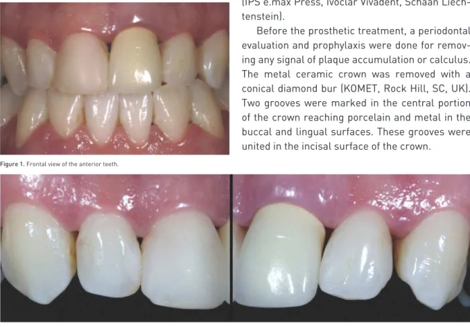
A 41-year-old female, B.H.C, Caucasian, pre -sented at the Sao Paulo State University, Araça-tuba Dental School, complaining about her unat-tractive smile (Figure 1).
The clinical exam revealed an unsatisfactory metal ceramic crown in the left central incisor re-garding marginal fit, color and anatomy, associ-ated to grayish color in gingival region (metallic margin) and gingival inflammation. The right cen-tral and lateral incisors and the left lateral incisor presented Class III and IV resin composite resto-rations with inadequate anatomy and pigmenta-tions at the tooth/restoration interface (Figure 2).
After clinical exam, impressions of maxillary and mandible arches were taken with polyvinyl-siloxane (Express, 3M/ESPE, St. Paul, Minnesota USA) to obtain preliminary casts for diagnostic waxing from right to left lateral incisors and
fabri-cation of 4 provisional crowns in acrylic resin (Vipi -Cor, Sao Paulo, Brazil) (Figure 3).
The treatment planning was established ac-cording to the clinical exam and diagnostic waxing including 2 all-ceramic crowns (left central e lat-eral incisors) and 2 laminate veneers (right cen-tral and lateral incisors) based on ceramic system (IPS e.max Press, Ivoclar Vivadent, Schaan Liech-tenstein).
Before the prosthetic treatment, a periodontal evaluation and prophylaxis were done for remov-ing any signal of plaque accumulation or calculus. The metal ceramic crown was removed with a conical diamond bur (KOMET, Rock Hill, SC, UK). Two grooves were marked in the central portion of the crown reaching porcelain and metal in the buccal and lingual surfaces. These grooves were united in the incisal surface of the crown.
Figure 1.Frontal view of the anterior teeth.
After crown removal, a cast metal post and core was replaced by a glass fiber post (Refor-post, Angelus, Brazil). The esthetic core was built up with composite resin (Z350, 3M/ESPE, St. Paul, Minnesota USA) and cemented with self-adhesive
resin luting cement RelyX Unicem (3M/ESPE,
Seefeld, Germany) (Figure 4).
Dental reduction was guided by a silicone index based on the diagnostic waxing cast.
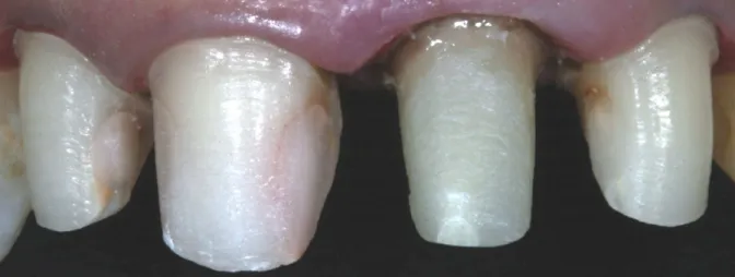
After dental reduction (Figure 5), simultaneous
Figure 3.Aspect of 4 provisional crowns pressed in acrylic resin.
Figure 5.Finished dental reductions. The tooth’s reductions were guided by a silicone index based on the diagnostic wax cast.
Figure 4. A. Testing of the insertion of the glass fiber post; B. Customized glass fiber post with composite resin and cementing with self-adhesive resin luting ce-ment; C. Customized glass fiber post cemented; D. Dental reductions.
impression technique with retraction cord (double retraction cord technique) was taken with poly-ether (Impregum, 3M/ESPE, St. Paul, Minnesota USA). The polyether was manipulated with the automatic mixing machine Pentamix 3 (3M/ESPE, St. Paul, Minnesota USA) and used due to its ad-equate hydrophilic behavior (Figure 6).
Then the provisional restorations were adjust-ed and cementadjust-ed with temporary cement (RelyX
Temp, 3M/ESPE, St. Paul, Minnesota USA). The color selection was digitally recorded to be sent to dental laboratory.
After the restorations are ready (Figure 7) and clinical proof, the ceramic restorations were pre-pared for cementation. The internal surfaces of the ceramic restorations were etched according to the following steps: 1st – conditioning with 10% hydrofluoric acid for 20 seconds; 2nd – water rinse
Figure 8.Etching of internal surface of the laminate veneers. A. Etching with a 10% hydrofluoric acid for 20 seconds; B. Application of silane for 1 minute; C. Application of bond resin.
Figure 9.Self-adhesive resin cementation of total-ceramic crowns (left central and lateral incisors).
and air drying, 3rd – application of silane coupling
agent for 1 minute; 4th – hot air drying; 5th – appli -cation of adhesive system and drying for removal of material excess (only for conventional technique); 6th – light polymerization of adhesive agent (only for conventional technique) (Figure 8).
The cementation procedures were initiated after gently cleaning all teeth preparations with pumice stone and water. The self-adhesive resin luting
ce-ment (RelyX Unicem, 3M/ ESPE, Seefeld, Germany)
was used for full-coverage crowns (left cental and lateral incisor) while the conventional resin cement
(RelyX ARC, 3M/ ESPE, Seefeld, Germany) was
used for laminate veneers in which there was more enamel surface available.
The self-adhesive resin luting cement was ma-nipulated according to the manufacturer’s instruc-tion and inserted in the full-coverage crown indi-vidually (Figure 9 A,B). The excess was removed
followed by polymerization during 40 seconds with
a halogen light device (QHL75 Lite, Dentsply Inter-national, York, Pa.).
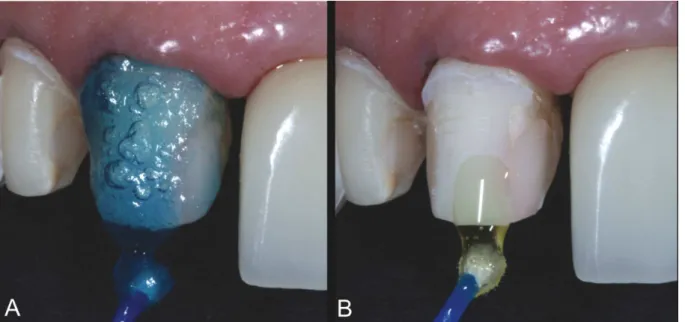
For laminate veneers, the dental substrate was etched with phosphoric acid for 15 seconds, which was carried out in each right central and lateral in-cisor (Figure 10A).9,10 After water rinse and drying,
the hybridization was achieved by using two step etch and rinse adhesive system (Figure 10B) (Adper
Single Bond 2, 3M/ ESPE, Seefeld, Germany). The
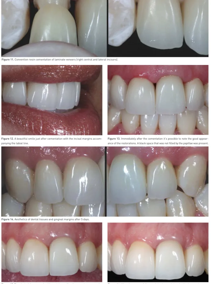
adhesive layer was polymerized during 15 seconds. The conventional resin luting cement (RelyX ARC, 3M/ ESPE, St. Paul, USA) was manipulated according to the manufacturer´s instruction and dispensed on the inner surface of the laminates for cementation. The laminates were individually placed in position (Figure 11).
It was noted the good appearance of restora-tions (Figure 12). After removal of material excess and retraction cord, the final adjustments of the restorations were taken as shown in the Figure 13, although a black space between the central inci-sors was present.
Five days after cementation, the health of gin-gival tissues surrounding restorations were ob-served, enhancing the integration between ceram-ics, gingival margin and lips. The esthetics results
were very satisfactory (Figure 14). The quality of
the restorations and the appearance of the gingival margin were maintained after two-year and three-year follow-up. Note that black space between the
central incisors was totally filled by papilla, contrib-uting further to the aesthetic and harmony (Figures 15 and 16).
dIscussIon
The spectrum of materials for indirect restora-tions has been raised during recent decades. Ce-ramics have been used without any metallic sup-port, mainly because of their superior esthetical properties. Silica-based ceramics require adhesive bond to tooth structures, which can help the me-chanics of the ceramics.6,11-12
The integration between porcelain and cement has been reported to reinforce both substrates re-ducing the microleakage at the tooth-restoration interface.13,14
In the present report, two different techniques for bonding porcelain to the dental substrate were used: the conventional resin cementation and the self-adhesive resin cementation. Although there are many differences between both techniques, one aspect that might drive the choice is the dental substrate. Enamel substrate requires conventional technique for cementation once the bond strength of the self-adhesive resin cements on enamel is still problematic.15
It is important to highlight that the preparation of the right central and lateral incisors was kept on enamel while the dentin was reached for the left central and lateral incisors. According to these characteristics, the self-adhesive resin cement was indicated for the left central and lateral inci-sors while the conventional cementation was used for the right teeth.
The self-adhesive properties are claimed to be based upon functional phosphoric-acid methacry-lates that demineralize the dentin, reacting with in-organic fillers (72 wt %), and infiltrate the tooth sub-strate to create the hybrid layer. This characterizes also the micromechanical retention. Secondary reactions have been suggested to provide chemi-cal adhesion to hydroxyapatite.6 The basic inorganic
fillers are able to undergo a cement reaction with the phosphoric-acid methacrylates. The dominant setting reaction starts with free radical polymeriza-tion, which can be initiated either by light or by a
redox system (dual-curing composite materials).6
Figure 11.Convention resin cementation of laminate veneers (right central and lateral incisors).
Figure 12.A beautiful smile just after cementation with the incisal margins accom-panying the labial line.
Figure 13.Immediately after the cementation it’s possible to note the good appear-ance of the restorations. A black space that was not filled by the papillae was present.
Figure 14.Aesthetics of dental tissues and gingival margins after 5 days.
Figure 15.The gingival tissues are healthy and the quality of restorations was main-tained after two-year follow-up.
Others authors claimed that without any con-ditioning, the self-adhesive cement RelyX Unicem
(3M/ ESPE, Seefeld, Germany) showed improved
sealing of dentin at the cervical margin when
com-pared to a conventional resin cement.8 The RelyX
Unicem showed bond strength to dentin not statis-tically different from the other resin based luting materials.6,15
The conditioning of inner surfaces of the ce-ramic restorations was performed in the present study with 10% hydrofluoric acid for 10 seconds, followed by silane application, to improve bond re-sults.17,18
The conditionings of the ceramics were carried out since literature states that the lack of ceramic conditioning previously to cementation results in low bond strength between cement and
ceram-ic.4,19 This is in agreement with other authors that
demonstrated proper bonding between these sub-strates as a result of porcelain conditioning and silanization.20-22
In this context, the application of a silane cou-pling agent to the pretreated ceramic surface
pro-vides a chemical covalent and hydrogen bond23
and it is a major factor for a sufficient resin bond
to silica-based ceramics.24 Silanes are
bifunction-al molecules that bond silicone dioxide with the OH groups on the ceramic surface. They also have a degradable functional group that copolymerizes
with the organic matrix of the resin.23
For the teeth with preparation on enamel, the laminates were cemented with the conventional technique using RelyX ARC (3M/ ESPE, Seefeld,
Germany).
In addition, the proper enamel hybridization resulted from conditioning with phosphoric acid for 15 seconds9,10,25 followed by application of
one-bottle adhesive system (Adper Single Bond 2, 3M/
ESPE, Seefeld, Germany) and light polymerization
during 15 seconds. Then, the porcelain
restora-tions were cemented.21
The conventional technique was used in this case since the self-adhesive cements (Unicem) may not be the ideal material for luting inlays and partial crowns, where a considerable enamel
sur-face area is present.6 So, many authors6 showed
that the bond strength of Unicem to enamel was statistically lower than all other luting resins and it was also observed greater leakage at the enam-el interface with this cement without any previous
conditioning.8 These results suggest an
insuf-ficient etching ability of the cement to the smear layer covering enamel and, therefore, the lack of development of adequate micromechanical reten-tion.
According to this, conventional adhesion pro-cedures are indicated for enamel using adhesive systems for bonding between tooth and cement due to proper bond strength and marginal seal-ing.26,27
So, the procedures based on protocols estab-lished by literature and clinical experience pro-vided patient’s satisfaction regarding the imme-diate result of the restorations maintained up to three-year follow-up. No sign of misfit, leakage or staining in tooth/cement/ceramic interface was clinically observed after three years. In addition, health of gingival tissue surrounding restorations was noticed and the restorations were harmoni-cally integrated with soft tissues.28
concLusIons
Conventional and self-adhesive resin cements are appropriate for cementation of all-ceramic restorations. However, the proper indication de-pends on the dental substrate available after preparation. The use of both cements in the same clinical case has been shown to be adequate to achieve satisfactory esthetic and functional re-sults after three year follow up.
rEFErEncEs
1. Manhart J, Scheibenbogen-Füchsbrunner A, Chen HY, Hickel R. A 2-year clinical study of composite and ceramic inlays. Clin Oral Investig 2000;4:192-198.
2. Mitchell CA, Abbariki M, Orr JF. The influence of luting ce-ment on the probabilities of survival rate and modes of
fail-ure of cast full-coverage crowns. Dent Mater
2000;16:198-206.
3. Peumans M, Van Meerbeek B, Lambrechts P, Vanherle
G. Porcelain veneers: review of the literature. J Dent
2000;28:163-177.
4. Peumans M, Hikita K, De Munck J, Van Landuyt K, Poitevin
A, Lambrechts P & Van Meerbeek B. Bond durability of composite luting agents to ceramic when exposed to
long-term thermocycling. Oper Dent 2007;32:372-379.
5. Chen Y-W, Raigrodski AJ. A conservative approach for treating young adult patients with porcelain laminate
6. Abo-Hamar SE, Hiller KA, Jung H, Federlin M, Friedl KH &
Schmalz G. Bond strength of a new universal self-adhesive
resin luting cement to dentin and enamel. Clin Oral Investig
2005;9:161-167.
7. Pospiech P. All-ceramic crowns: bonding or cementing?
Clin Oral Investig 2002;6:189-197.
8. Ibarra G, Johnson GH, Geurtsen W, Vargas MA. Microle -akage of porcelain veneer restorations bonded to enamel and dentin with a new self-adhesive resin-based dental
ce-ment. Dent Mater 2007;23:218-225.
9. Yazici AR, Çelik Ç, Özgünaltay G, Dayangaç B. Bond
strength of different adhesive systems to dental hard
tis-sues. Oper Dent 2007;32:166-172.
10. Pivetta MR, Moura SK, Barroso LP, Lascala AC, Reis A,
Loguercio AD, Grande RH. Bond strength and etching
pattern of adhesive systems to enamel: effects of
condi-tioning time and enamel preparation. J Esthet Restor Dent
2008;20:322-336.
11. Burke FJ. The effect of variations in bonding procedure on fracture resistance of dentin-bonded all-ceramic crowns.
Quintessence Int 1995; 26:293-300.
12. Burke FJ, Fleming GJ, Nathanson D, Marquis PM. Are ad -hesive technologies needed to support ceramics? An
as-sessment of the current evidence. J Adhes Dent
2002;4:7-22.
13. Dietschi D, Maeder M, Meyer JM, Holz J. In vitro resistance to fracture of porcelain inlays bonded to tooth. Quintessence
Int 1990;21:823-831.
14. Burke FJ, Watts DC. Fracture resistance of teeth restored
with dentin-bonded crowns. Quintessence Int
1994;25:335-340.
15. De Munck J, Vargas M, Van Landuyt K, Hikita K, Lambrechts P, Van Meerbeek B.. Bonding of an auto-adhesive luting
material to enamel and dentin. Dent Mater
2004;20:963-991.
16. Behr M, Rosentritt M, Regnet T, Lang R, Handel G. Mar -ginal adaptation in dentin of a self-adhesive universal
res-in cement compared with well-tried systems. Dent Mater
2004;20:191-197.
17. Chen JH, Matsumura H, Atsuta M. Effect of etchant, etch-ing period, and silane primetch-ing on bond strength to
porce-lain of composite resin. Oper Dent 1998;23:250-257.
18. Sorensen JA, Engelman MJ, Torres TJ, Avera SP. Shear
bond strength of composite resin to porcelain. Int J
Prosth-odontics 1991;4:17-23.
19. Kumbuloglu O, Lassila LV, User A, Toksavul S, Vallittu PK. Shear bond strength of composite resin cements to lithium disilicate ceramics. J Oral Rehabil 2005;32:128-133.
20. Piwowarczyk A, Lauer HC, Sorensen JA. In vitro shear bond strength of cementing agents to fixed prosthodontic
restorative materials. J Prosthetic Dent 2004;92:265-273.
21. Reich SM, Wichmann M, Frankenberger R, Zajc D. Effect of surface treatment on the shear bond strength of three
res-in cements to a machres-inable feldspatic ceramic. J Biomed
Mater Res Part B Applied Biomater 2005;74:740-746.
22. Soares CJ, Soares PV, Pereira JC, Fonseca RB. Surface treatment protocols in the cementation process of ceramic and laboratory-processed composite restorations: a
lit-erature review. J Esthet Restor Dent 2005;17:224-235.
23. Blatz MB, Sadan A, Kern M. Resin-ceramic bonding: a re-view of the literature. J Prosthetic Dent 2003;89:268-274. 24. Frankenberger R, Kramer N, Sindel J. Repair strength of
etched vs silicacoated metal-ceramic and all-ceramic
res-torations. Oper Dent 2000;25:209-215.
25. Swift EJ, Perdigao J, Heymann HO. Bonding to enamel and dentin: a brief history and state of the art. Quintessence Int
1995;26:95-110.
26. Deliperi S, Bardwell DN, Wegley C. Restoration interface microleakage using one total-etch and three self-etch
ad-hesives. Oper Dent 2007;32:179-184.
27. Hikita K, Van Meerbeek B, De Munck J, Ikeda T, Van Lan-duyt K, Maida T, Lambrechts P & Peumans M. Bonding ef-fectiveness of adhesive luting agents to enamel and dentin.
Dent Mater 2007;23:71-80.
28. Chu SJ, Tan J H-P, Stappert CFJ, Tarnow DP. Gingival ze -nith positions and levels of the maxillary anterior dentition.