Bonding of Y-TZP to Dentin: Effects
of Y-TZP Surface Conditioning,
Resin Cement Type, and Aging
MA Bottino
C Bergoli
EG Lima
SMS Marocho
RO Souza
LF Valandro
Clinical Relevance
The application of the low-fusing glaze porcelain followed by hydrofluoric acid etching and silanization and tribochemical silicatization generates strong bonds between resin cement and zirconia. Panavia generates stronger bonds than does Clearfil.
SUMMARY
Purpose: To evaluate the effects of two sur-face treatments, aging, and two resin cements on shear bond strength between dentin and yttrium-stabilized tetragonal zirconia poly-crystal ceramic (Y-TZP).
Materials and Methods: Eighty human molars were embedded in acrylic resin and sectioned 3 mm below the occlusal plane. These teeth and 80 cylindrical Y-TZP specimens (height, 4 mm; diameter, 3.4 mm) were divided into eight groups (n=10) using the following factors: Y-TZP surface treatment (Vi: low-fusing porce-lain [vitrification] + hydrofluoric acid etching + silanization or Si: tribochemical silicatiza-tion); cementation strategies (PF: Panavia or CC: Clearfil); and storage (nonaging or aging). Bonding surfaces of 40 Y-TZP specimens re-ceived Vi treatment, and the rest rere-ceived Si treatment. Half of the ceramic-tooth assem-blies were cemented with Panavia, the rest with Clearfil. Shear tests were executed using 0.4-mm–thick wire at 0.5 mm/min. Data were analyzed by three-way analysis of variance and Tukey test (a=0.05). Fractures were
ana-lyzed. Marco Antonio Bottino, DDS, PhD, chair and professor,
MScD-PhD Graduate Program in Restorative Dentistry, Prosthodontic Unit, Science and Technology Institute, Den-tal School, Sa˜o Paulo State University (UNESP), Sa˜o Jose´ dos Campos/SP, Brazil
Ce´sar Dalmolin Bergoli, DDS, MScD, PhD student in Prosthodontics, Graduate Program in Restorative Dentistry, Science and Technology Institute, Dental School, Sa˜o Paulo State University (UNESP), Sa˜o Jose´ dos Campos/SP, Brazil.
Elen Guerra Lima, DDS, MScD student in Prosthodontics, Graduate Program in Restorative Dentistry, Science and Technology Institute, Dental School, Sa˜o Paulo State Uni-versity (UNESP), Sa˜o Jose´ dos Campos/SP, Brazil.
Susana Marı´a Salazar Marocho, DDS, MScD, PhD, Science and Technology Institute, Dental School,Sa˜o Paulo State University (UNESP), Sa˜o Jose´ dos Campos/SP, Brazil.
Rodrigo Othavio Assunc¸a˜o Souza, DDS, MScD, PhD, adjunct professor, Federal University of Paraı´ba (UFPB), Depart-ment of Restorative Dentistry, Division of Prosthodontics, Joa˜o Pessoa/Paraı´ba, Brazil.
*Luiz Felipe Valandro, DDS, MScD, PhD, associate professor, Federal University of Santa Maria (UFSM), Head of MSciD-PhD Graduate Program in Oral Science, Prosthodontic Unit, Faculty of Odontology, Santa Maria, RS, Brazil.
*Corresponding author: R. Floriano Peixoto 1184, Santa Maria, RS 97015-372, Brazil; e-mail: lfvalandro@hotmail. com
Results: Y-TZP surface treatments did not affect bond strength (p=0.762, Vi = Si), while resin cements (p,0.001, Panavia . Clearfil) and aging (p=0.006, nonaging.aging) showed a significant effect. Most failures were in adhesive at dentin-cement interfaces; no fail-ure occurred between zirconia and cement.
Conclusion: When Y-TZP ceramic is bonded to dentin, the weakest interface is that be-tween dentin and resin cement. The resin cement/Y-TZP interface was less susceptible to failures, owing to Y-TZP surface treatments.
INTRODUCTION
Zirconia-based dental ceramics have better mechan-ical properties than do other commercially available ceramic materials.1However, their bond strength to resin cement has been reported2to be weak because zirconia-based ceramics have a large crystalline phase, rendering conventional hydrofluoric acid etching treatment impossible. Therefore, in the last few years, many researchers2-6 have studied alter-native methods of improving the adhesion between zirconia and resin cements.
Among the possible treatments being investigated to improve the adhesion, air-particle abrasion proto-cols have attracted much interest because of their simplicity. These methods are called tribochemical silicatization and involve air abrasion of the ceramic surface with alumina or with silica-modified alumi-na particles (30lm), followed by the application of a silane coupling agent.1,7-9 The alumina- or silica-modified alumina particles produce microroughness on the zirconia surface, while the silane coupling agent acts as a link between the sandblasted surface and the cement matrix.8-13
In addition to the air-abrasion protocols, resin cements containing phosphate ester monomer 10-methacryloyloxydecyl dihydrogen phosphate (MDP) have been shown to form strong bonds with zirconia. These cements react with oxides, creating a good interaction with the zirconia surface.8,10-13 Howev-er, a study10 has showed that the use of cements with MDP alone is not sufficient for creating a stable union between the resin cement and zirconia. Therefore, air-abrasion protocols14are also required to be a part of the bonding technique. On the other hand, airborne particle abrasion procedures can structurally damage the zirconia surface, decreas-ing the mechanical properties of this ceramic15; hence, the use of these procedures remains contro-versial.
As a new alternative to the surface treatment of zirconia, some researchers4,16-18have evaluated the application of a low-fusing porcelain material (glaze) on the yttrium-doped tetragonal zirconia (Y-TZP) intaglio surface. This technique aims to create a surface that can be etched by hydrofluoric acid, similar to feldspathic ceramics. Although this is a promising technique, it has been introduced very recently, and data on resin bond strength and bond durability as a result of using this method are scarce.
In fixed dental prostheses (FDPs), in which mechanical retention plays an important role, the mechanism responsible for adhesion with dentin appears to be less important.19On the other hand, in bonded FDPs for which mechanical retention is not the primary retention mechanism the bond strength between the resin cement and dentin is essential to the success of the treatment.20,21Hence, evaluation of the adhesion between Y-TZP, resin cements, and dentin is important.5,22
Chai and others5 reported that studies that evaluate simply the bond strength between resin cements and restorative materials are limited in scope from a clinical perspective, as FDP restora-tions are normally cemented to an enamel/dentin substrate. When zirconia specimens are cemented onto a dental substrate, it is possible to assess both interfaces, such as the ceramic and cement-dentin interfaces, which leads to better evaluation of adhesion, from the clinical point of view.
Therefore, the aim of the present study is to evaluate two cementation strategies (involving the use of the resin cements Panavia F and Clearfil SA Cement, respectively), two Y-TZP surface treatment techniques (involving the application of a low-fusing porcelain and silicatization, respectively), and the effect of thermo-cycling aging on the shear bond strength between zirconia and dentin. The following hypotheses were tested: 1) the application of a low-fusing glaze porcelain application and silanization as Y-TZP sur-face treatments will not influence the bond strength; 2) the cementation strategies will not influence the bond strength; and 3) aging will decrease the bond strength.
MATERIALS AND METHODS
The product names, manufacturers, chemical com-positions, and batch numbers of the materials used in the study are listed in Table 1.
Selection of Teeth
(48C) until needed. With the assistance of a cylindri-cal metallic mold (diameter, 20 mm; height, 15 mm), each tooth was embedded 2 mm apical to the cemento-enamel junction with a self-curing acrylic resin (JET, Artigos Odontolo´gicos Classico Ltda, Sao Paulo, Brazil).
Then, each tooth was sectioned 3 mm below the occlusal surface using a low-speed diamond cutting saw (Labcut 1010, Extec, Enfield, CT, USA) with extensive water cooling in order to expose the superficial coronal dentin surface. These surfaces were then wet-ground with 600-grit silicon paper for 60 seconds using a polishing machine (PSK-2V, Skill TEC, Sao Paulo, Brazil).
Before the cementation procedures, the teeth were numbered from 1 to 80, and eight random sequences consisting of 10 numbers each were generated using the computer program Random Allocator (developed by M Saghaei, Dept of Anesthesia, Isfahan Univer-sity of Medical Sciences, Isfahan, Iran). This proce-dure was performed to homogenize the groups and
randomize the allocation of the specimens to the eight groups.23Next, each of the eight groups, which comprised 10 samples each, was assigned to one of the following categories: Y-TZP surface treatment (Vi: application of the low-fusing glaze porcelain [vitrification]þhydrofluoric acid etchingþ
silaniza-tion or Si: tribochemical silicatizasilaniza-tion), cementasilaniza-tion strategies (PF: Panavia or CC: Clearfil), and storage condition (nonaging or aging): Si þ PF; Si þ PF þ
aging; SiþCC; SiþCC þaging; ViþPF; ViþPFþ
aging; Vi þCC; and Vi þCC þaging.
Preparation of the Y-TZP Specimens
First, blocks of Vita In Ceram YZ 2000 (Vita Zahnfabrik, Bad Sa¨ckingen, Germany) were sec-tioned using a diamond saw (Labcut 1010, Extec) to produce smaller cubes (5315320 mm3). A drill-type trephine was used to perforate these cubes perpen-dicular to the surface (with the aid of a preparation device) to produce presintered zirconia cylinders (diameter, 4.5 mm; height, 5 mm) that were then
Table 1: Material, Manufacturer, Chemical Composition, and Batch Number of the Products Used in the Study
Material Manufacturer Chemical Composition Batch No.
Rocatec Plus 3M ESPE, Seefeld, Germany Silicatized aluminum oxide particles (30lm) 1036301855
Condac 37 FGM, Joinvile, SC, Brazil 37% Phosphoric acid 140111 Porcelain Conditioner Dentstply, Petro´polis, RJ, Brazil 10% Hydrofluoric acid 229431B
Vita Akzent Glaze Spray Vita Zahnfabrick, Germany Not available 21790
Clearfill SA Cement Kuraray Medical Inc, Japan Bis-GMA, TEGDMA, MDP, hydrophobic aromatic dimethacrylate, silanated barium glass filler, silanated colloidal silica, di-camphorquinone, benzoyl peroxide, initiator, hydrophobic aliphatic dimethacrylate, silanated, surface-treated sodium fluoride, accelerators, pigments, 45vol% is inorganic fillers
023AAB
Monobond S Ivoclar Vivadent, Schaan, Liechtenstein
Alcohol solution of silane metacrylate 532888
Panavia F–Adhesive Primer A
Kuraray Medical Inc, Japan 2-Hydroxyethyl methacrylate,
10-methacryloyloxydecyl dihydrogen phosphate, N-methacryloyl-5-aminosalicylic acid, water, accelerators
00282A
Panavia F–Adhesive Primer B
Kuraray Medical Inc, Japan N-methacryloyl-5-aminosalicylic acid, water, catalysts, accelerators
00157A
Panavia F–Cement Paste A Kuraray Medical Inc, Japan 10-Methacryloyloxydecyl dihydrogen phosphate, hydrophobic aromatic dimethacrylate, hydrophobic aliphatic methacrylate, hydrophilic aliphatic dimethacrylate, silanated silica filler, silanated colloidal silica,DL-camphorquinone, catalysts, initiators, others
00251B
Panavia F–Cement Paste B Kuraray Medical Inc, Japan Sodium fluoride, hydrophobic aromatic
dimethacrylate, hydrophobic aliphatic methacrylate, hydrophilic aliphatic dimethacrylate, silanated barium glass filler, catalysts, accelerators, pigments, others
00029A
Vita In Ceram YZ Vita Zahnfabrick, Germany 91% Zirconium oxide (ZrO2), 5% yttrium oxide (Y2O3), 3% hafnium oxide (HfO
2), small amounts (,1%) of
aluminum oxide (Al2O3) and silicon oxide (SiO2)
28070
sintered, as recommended by the manufacturer, in an oven (Vita Zyrcomat, Vita Zahnfabrik). The final dimensions of the ceramic cylinders were 3.4 mm in diameter and 4 mm in height.
The surface of each cylinder that had to undergo cementation was polished with 800-, 1000-, and 1200-grit silicon carbide paper, under water cooling, for 60 seconds each using a polishing machine (PSK-2V, Skill TEC). After polishing, the cylinders were cleaned ultrasonically for five minutes in isopropyl alcohol.
Conditioning of the Y-TZP Surfaces
For 50% of the zirconia cylinders (N=40), a low-fusing porcelain glaze (Vita Akzent Glaze Spray, Vita Zahnfabrik) was applied for one to two seconds on the cementation surface at a 10-mm distance. The conditioned specimen was then sintered (VACUMAT 40T, Vita Zahnfabrik) according to the manufactur-er’s instructions. Then the glaze-coated surfaces were treated with 10% hydrofluoric acid gel (Porce-lain Conditioner, Dentsply, Petropolis, RJ, Brazil) for 60 seconds, washed for 15 seconds, dried, and silanized with a methacryloxypropyltrimethoxysi-lane (MPS)–based simethacryloxypropyltrimethoxysi-lane coupling (Monobond S, Ivoclar Vivadent, Schaan, Liechtenstein). The silan-ized samples were kept aside for 60 seconds to let the solvent evaporate.
The remaining zirconia cylinders (N=40) were treated using the tribochemical silicatization meth-od. First, the surfaces of the cylinders were air-abraded using 30lm silica-coated alumina particles (Rocatec Soft, 3M ESPE, Seefeld, Germany) from a distance of 10 mm and with a pressure of 2.8 bar. Subsequently, the MPS-based silane coupling agent (Monobond S, Ivoclar Vivadent) was applied in the manner described above.
Cementation Procedures
The cementation surface of the specimens was defined by an adhesive tape (Scotch, 3M, Ribeira˜o Preto, Brazil) with a 3.4-mm–diameter hole, aiming to standardize the cementation area and prevent the overflow of the resin cement.
For the PF samples, the dentinal surface treat-ment was performed as follows. Equal amounts of Primers A and B were mixed, and this mixture was applied on the dentinal surface with a microbrush. This was followed by spraying the surface gently with air and letting it stand for one minute to allow the reaction to take place. For CC samples, dentinal
surface treatment was not required, as the Clearfil SA Cement is self-adhesive.
The two resin cements were manipulated as recommended by the manufacturer and were applied on the conditioned surfaces of the zirconia cylinders. The cylinders were placed on top of the area bounded by the adhesive tape, and a load of 750g was applied on the cylinders for 60 seconds. Any excess cement was removed, and all of the surfaces (vestibular, mesial, distal, and lingual) were photoactivated using an LED (1200 mW/cm2) (Radii Cal, SDI, Australia) for 20 seconds.
Storage Conditions
All the specimens were stored for 24 hours in distilled water at 378C. Half of the specimens were submitted to the shear bond strength test and the other half were aged before testing using a thermo-cycling protocol that involved 5000 cycles of alter-nate immersion in baths at 58C and 558C for 30 seconds each with intervals of two seconds between the immersions.
Shear Bond Strength Test
The test was conducted using a universal testing machine (EMIC DL 1000, Emic, Sa˜o Jose´ dos Pinhais, PR, Brazil) with a crosshead speed of 0.5 mm/min and a steel wire with a thickness of 0.4 mm. The test cylinder was aligned with the load cell, and the wire loop was positioned as close as possible to the ceramic/dentin interface and parallel to the direction of the load cell (50 Kgf). The steel wire was then pulled using the universal machine in order to perform the shear bond strength test.
The bond strength was calculated using the formula R = F/A, where R is the bond strength (MPa), F is the load required for rupture of the specimen (N), and A is the bonded cross-sectional area of the specimen (mm2). The bonded cross-sectional area was calculated using the formula for the area of a circle, which is given by A = p3r2, wherep=3.14 andr=1.7 mm (half of the diameter of the cylinder). Using this formula, the bonded cross-sectional area was found to be 9.07 mm2.
Fracture Analysis
Fracture analysis was performed for several speci-mens in order to identify the fracture origin and mode of fracture. Stereomicroscope examinations were performed using various lighting configura-tions to identify the fracture pattern. The identified
fractures were classified on the basis of the following scheme: score A=a detachment of the resin cement from the dentin; score B = a detachment of the ceramic from the resin cement; score C=a fracture of ceramic without an adhesive failure; score D =a
fracture of dentin, without an adhesive failure; and score E = an area of resin cement fracture bigger than an area of adhesive failure.
Measurement of Glaze Thickness
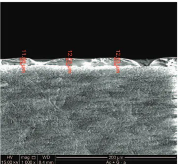
A pilot study was conducted to measure the thickness of the glaze obtained by the vitrification process. To measure the thickness of the glaze layer, three zirconia beams (103232 mm) were sprayed with the glaze, sintered, and broken into two pieces so that the glaze thickness could be measured by SEM at a magnification of 10003. As can be noted in Figure 1, the applied glaze is homogeneously
distributed on the zirconia surface. In addition, the thickness of the glaze layer was also found to be of an acceptable level, with a mean value of 12 6 0.3lm (Figure 2).
Data Analysis
The bond strength data were analyzed using three-way analysis of variance (ANOVA) and Tukey test (a=0.05) with the software Minitab 16.1.0. The specimens with pretest failures (during the aging process) were included in the statistical analysis and were conferred a bond strength value of 0 MPa.
RESULTS
The factors resin cement (p,0.001; Panavia. Clear-fil) and storage condition (p=0.006; nonaging. aging) statistically influenced the bond strength values, while the factor surface treatment (p=0.762; Vi = Si) had no effect. According to the result of the Tukey test, the groups SiþCC þaging
(4.0 6 3.4 MPa) and Vi þ CC þ aging (3.9 6 3.8
MPa) exhibited the lowest bond strength values, while the Vi þPF group (17.26 10.1 MPa) showed
the highest values of bond strength but was not statistically different than ViþPFþTC (14.766.0
MPa), SiþPF (14.668.4 MPa), SiþPFþTC (11.6 6 6.2 MPa), and Si þ CC (10.8 6 3.8 MPa). The
group ViþCC (7.063.4 MPa) showed intermediate
values. When subjected to the Si surface treatment, the zirconia cylinders cemented with Clearfil cement showed a significant decrease in bond strength values after aging.
Fracture analysis revealed that fractures occurred predominantly in the adhesive at the resin cement/
Figure 2. Low-fusing porcelain glaze thickness measured on zirconia surface under SEM.
Table 2: Shear bond strength (MPa) and incidence of failure type data.
Surface Treatment Cement Thermo-cycling Group Abbreviations
Meana(SD) Scores for Failure Typeb
Score A Score B Score C Score D Score E
Tribosilicatization Panavia F No SiþPF 14.6 (8.4)AB 6 0 0 0 4 Yes SiþPFþTC 11.6 (6.2)ABC 4 0 0 0 6 Clearfill SE No SiþCC 10.8 (3.8)ABC 6 0 0 0 4 Yes SiþCCþTC 4.0 (3.4)C 7 0 0 0 3 Glassy application Panavia F No ViþPF 17.3 (10.1)A 6 1 0 0 3 Yes ViþPFþTC 14.7 (6.0)AB 8 0 0 0 2 Clearfill SE No ViþCC 7.0 (3.4)BC 8 0 0 0 2 Yes ViþCCþTC 3.9 (3.8)C 7 1 0 0 2
52 (65%) 2 (2.5%) 0 0 26 (32.5%) aMeans and standard deviations (SDs) of shear bond data (MPa) and Tukey test (a
=0.05). Letters denote statistically similar treatment groups.
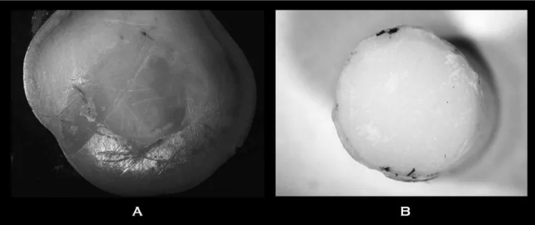
dentin interface (65% of the failures were scored type A), followed by cohesive failures in the resin cement (32.5% were scored type E) and in the adhesive at the resin cement and ceramic interface (2.5% were scored type B) (Table 2). Representative images of the different failure modes are shown in Figures 3 and 4. For the groups SiþCC þaging and ViþCC þ aging, three specimens each failed during aging;
for the group Si þ PF þaging, one specimen failed
during aging; and in the group Vi þ PF þ aging,
there were no specimen failures during the aging process. All of the premature failures that occurred during aging were at the resin cement and dentin interface (scored type A).
DISCUSSION
Different surface treatments have been tested to improve the bond strength between resin cement and zirconia. Nowadays, special attention has been given to the treatment of the zirconia intaglio
Figure 3. (A) Representative image of a cohesive failure. (B) Detailed view of the small black square in (A). Fracture surface topography revealed the configuration of the glaze film on the zirconia surface, which is associated with exposure of zirconium oxides.
surface with low-fusing porcelain glazes (produced by vitrification), which would create an etchable layer on the acid-resistant material, creating a scenario similar to that found in silica-based ceram-ics. For studies on zirconia to have clinical relevance, it is important to consider the dentin surface as well.5
The three-way ANOVA showed that the zirconia surface treatments resulted in similar bond strengths, confirming the first hypothesis of the study and representing a good pattern of condition-ing promoted by Y-TZP surface conditioncondition-ing ap-proaches. The silicatization after air-abrasion by silica-modified aluminum oxide particles is consid-ered a prerequisite for achieving good adhesion between the resin cement and zirconia surfaces10 and leads to the generation of bond strength values that are higher than those obtained from other surface treatments.1,5,7,10 This method creates hy-droxyl groups and enhances the micromechanical retention of the resins on the zirconia surface. In addition, the application of a silane coupling agent after the silica coating generates a siloxane network that improves the bond between the resin cement and zirconia.10 Chai and others5 employed an experimental design similar to that used in our study and observed that the tribochemical silica coating of the zirconia surface generated statistically higher values for the bond strength between the zirconia cylinders and the dentin surface. The vitrification method creates an etchable glassy thin film on the acid-resistant zirconia surface. This surface can then be etched by hydrofluoric acid and silanized using an MPS-based primer. In addition, only two specimens subjected to the vitrification process exhibited adhesive failure at the resin cement/zirconia interface. This result corroborates the findings of Cura and others.4On the other hand,
in contrast to the results obtained by us, Everson and others,16Valentino and others,17and Ntala and others18observed statistically higher values of bond strength for vitrification techniques than for the tribochemical silica-coating approach. This may be because these studies did not use dentin as an adhesion substrate. We utilized dentin as an adhe-sion substrate for the zirconia samples, and the majority of the failures occurred at the dentin/resin cement interface, preventing a real evaluation of the adhesion of the resin cement to the treated zirconia surface (comparison between the Y-TZP surface conditioning).
The second hypothesis of the study was rejected, as the bond strength values are statistically affected
by the resin cements (Panavia F: 14.5 MPa; Clearfil SE: 6.4 MPa). In this study, the resin cement Panavia F showed statistically higher values of shear bond strength than did the self-adhesive resin cement Clearfil SA Cement (Table 2).
According to previously reported studies,24-26 the lower values of bond strength obtained for Clearfil SA Cement can be explained by the inability of this cement to remove the smear layer on the dentin surface, as is the case with other self-adhesive resin cements. This leads to the formation of a poor hybridization layer between the resin cement and dentin. Despite the poor interaction of the self-adhesive resin cement with the dentin, it is impor-tant to note that this cement showed a good interaction with the zirconia surfaces that had been subjected to either of the two surface treatments. This can be confirmed by the absence of adhesive failures between the self-adhesive resin cement and the ceramic. However, this finding should be further investigated by an experiment designed to evaluate the adhesion between this self-adhesive resin ce-ment and zirconia alone without involving the dentin substrate.
Panavia F showed higher values of shear bond strength, and these could be related to a good dentin hybridization generated by the self-etching adhesive. This adhesive is considered to be a ‘‘mild’’ self-etchant and can remove the smear layer and expose the dentinal tubules. In addition, this adhesive contains the phosphate-based functional monomer 10-MDP, which interacts with collagen and hydroxy-apatite components in dentin, resulting in the formation of a strong and stable bond between the resin cement and coronal dentin.27-29
Regardless of the resin cements and surface treatments used in the study, the aging process decreased the bond strength. This could be due to the use of the dentin substrate in the study, as it appears to be more susceptible to the hydrolysis. In contrast, even after the thermo-cycling aging process, the interfaces between the resin cements and the zirconia surfaces showed fewer adhesive failures. This is evidence that this interface exhibited stable bonding even after the aging process.
applying the porcelain glaze and zirconia surface treatments before glaze application should also be investigated further. The fracture analysis provided important information about the system behavior. The cement/dentin interface was weaker than the cement/zirconia interface for all experimental groups. Chai and others5 cemented zirconia cylin-ders that had undergone different surface treat-ments onto a dentin surface and observed a large number of adhesive failures between the cement and dentin. Their results, which are in accordance with the results obtained in this study, highlight the importance of including the dentin substrate in studies on bond strength so that they are more relevant clinically.
CONCLUSION
Considering the experimental design and the results obtained, we can draw the following conclusions:
1. The bond strength between the resin cements and the zirconia substrate was stronger than that between dentin and the cements, as a function of the efficient conditioning methods performed on the Y-TZP surface, even after lengthy storage. 2. The conventional resin cement containing MDP
monomers showed better bond performance than did the self-adhesive resin cement.
3. The dentin/cement interface appears to be the more critical zone in this system.
4. The application of a thin film of low-fusing glaze porcelain on the zirconia surface followed by hydrofluoric acid etching and silanization appears to offer a promising surface treatment method with which to improve the adhesion between zirconia and resin cement. However, more studies should be performed to confirm this.
Conflict of Interest
The authors have no proprietary, financial, or other personal interest of any nature or kind in any product, service, and/or company that is presented in this article.
(Accepted 2 October 2012)
REFERENCES
1. Bottino MA, Valandro LF, Buso L, & Scotti R (2005) Effect of surface treatments on the resin bond to zirconium-based ceramic International Journal of Pros-thodontics18(1)60-65.
2. Atsu SS, Kilicarslan MA, Kucukesmen HC, & Aka PS (2006) Effect of zirconium-oxide ceramic surface treat-ments on the bond strength to adhesive resinJournal of Prosthetic Dentistry95(6)430-436.
3. Ozcan M, Kerkdijk S, & Valandro LF (2008) Comparison of resin cement adhesion to Y-TZP ceramic following manufactures’ instructions of the cements only Clinical Oral Investigation12(3)279-282.
4. Cura C, Ozcan M, Isik G, & Saracoglu A (2011) Comparison of alternative adhesive cementation concepts for zirconia ceramic: Glaze layer VS zirconia primer
Journal of Adhesive Dentistry13(1)1-8.
5. Chai J, Chu FCS, & Chow TW (2011) Effect of surface treatment on shear bond strength of zirconia to human dentinJournal of Prosthodontics20(3)173-179.
6. Kim MJ, Kim YK, Kim KH, & Kwon TY (2011) Shear bond strengths of various luting cements to zirconia ceramic: Surface chemical aspects Journal of Dentistry
39(11)795-803.
7. Valandro LF, Ozcan M, Bottino MC, Scotti R, Bottino MA, & Della Bona A (2006) Bond strength of a resin cement to high-alumina and zirconia-reinforced ceramics: The effect of surface conditioningJournal of Adhesive Dentistry8(3)
175-181.
8. Ozcan M, Alkumru HN, & Gemalmaz D (2001) The effect of surface treatment on the shear bond strength of luting cement to a glass-infiltrated alumina ceramic Interna-tional Journal of Prosthodontics14(4)335-339.
9. Della Bona A, Donassollo TA, Demarco FF, Barret AA, & Mecholsky JJ Jr (2007) Characterization and surface treatment effects on topography of a glass-infiltrated alumina/zirconia-reinforced ceramic Dental Materials
23(6)769-775.
10. Ozcan M, Cura C, & Valandro LF (2011) Early bond strength of two resin cements to Y-TZP ceramic using MPS or MPS/4-META silanesOdontology99(1)62-67.
11. Kern M, & Wegner SM (1998) Bonding to zirconia ceramic: Adhesion methods and their durabilityDental Materials14(1)64-71.
12. Ozcan M, & Valittu PK (2003) Effect of surface condi-tioning methods on the bond strength of luting cement to ceramicsDental Materials19(8)725-731.
13. Friederich R, & Kern M (2002) Resin bond strength to densely sintered alumina ceramicInternational Journal of Prosthodontics15(4)333-338.
14. Aboushelib MN, Kleverlaan CJ, & Feilzer AJ (2007) Selective infiltration-etching technique for a strong and durable bond of resin cements to zirconia-based materials
Journal of Prosthetic Dentistry98(5)379-388.
15. Zhang Y, Lawn BR, Rekow ED, & Thompson VP (2004) Effect of sandblasting on the long-term performance of dental ceramics Journal of Biomedical Material Re-searches Part B71(2)381-386.
16. Everson P, Addison O, Palin WM, & Burke FJT (2012) Improved bonding of zirconia substructures to resin using a ‘‘glaze-on’’ techniqueJournal of Dentistry40(4)347-351.
17. Valentino TA, Borges GA, Borges LH, Platt JA, & Sobrinho-Correr L (2012) Influence of glazed zirconia on dual-cure luting agent bond strengthOperative Dentistry
37(2)181-187.
bonding to zirconia substrates Journal of Dentistry
38(10)773-781.
19. Edelhoff D, & Ozcan M (2007) To what extent does the longevity of fixed dental prostheses depend on the function of cement? Clinical Oral Implants Researches
18(Supplement 3)193-204.
20. Sailer I, Feher A, Filser F, Luthy H, Gauckler LJ, Scharer P, & Franz Ha¨mmerle CH (2006) Prospective clinical study of zirconia posterior fixed partial dentures: 3-year follow-upQuintessence International37(9)685-693.
21. Peutzfeldt A, Sahafi A, & Flury S (2011) Bonding of restorative materials to dentin with various luting agents
Operative Dentistry36(3)266-273.
22. Moon JE, Kim SH, Lee JB, Ha SR, & Choi YS (2011) The effect of preparation order on the crystal structure of yttria-stabilized tetragonal zirconia polycrystal and the shear bond strength of dental resin cements Dental Materials27(7)651-663.
23. Montenegro R, Needleman L, Moles D, & Tonetti M (2002) Quality of RCTs in periodontology—A systematic reviewJournal of Dental Research81(12)866-870. 24. Bitter K, Paris S, Pfuertner C, Neumann K, & Kielbassa
AM (2009) Morphological and bond strength evaluation of different resin cements to root dentinEuropean Journal of Oral Science117(3)326-333.
25. De Munck J, Vargas M, Van Landuyt K, Hikita K, Lambrechts P, & VanMeerbeek B (2004) Bonding of an
auto-adhesive luting material to enamel and dentin
Dental Materials20(10)963-971.
26. Saskalauskaite E, Tam LE, & McComb D (2008) Flexural strength, elastic modulus, and pH profile of self-etch resin luting cements Journal of Prosthodontics 17(4)
262-268.
27. Zicari F, Coutinho E, De Munck J, Poitevin A, Scotti R, Naert I, & Van Meerbeek B (2008) Bonding effectiveness and sealing ability of fiber-post bondingDental Materials
24(7)967-977.
28. Fukegawa D, Hayakawa S, Yoshida Y, Suzuki K, Osaka A, & Van Meerbeek B (2006) Chemical interaction of phosphoric acid ester with hydroxyapatite Journal of Dental Research85(10)941-944.
29. Inoue S, Koshiro K, Yoshida Y, De Munck J, Nagakane K, & Suzuki K (2005) Hydrolytic stability of self-etch adhesives bonded to dentin Journal of Dental Research
84(12)1160-1164.
30. Yeo IS, Yang JH, & Lee JB (2003) In vitro marginal fit of three all-ceramic crown systems Journal of Prosthetic Dentistry90(5)459-464.