Nepenthesin Protease Activity Indicates
Digestive Fluid Dynamics in Carnivorous
Nepenthes
Plants
Franziska Buch1, Wendy E. Kaman2,3, Floris J. Bikker3, Ayufu Yilamujiang1, Axel Mithöfer1*
1Department of Bioorganic Chemistry, Max Planck Institute for Chemical Ecology, Hans Knöll Straße 8, D-07745, Jena, Germany,2Department of Medical Microbiology and Infectious Diseases, Erasmus MC, `s-Gravendijkwal 230, 3015 CE, Rotterdam, The Netherlands,3Department of Oral Biochemistry, Academic Centre for Dentistry Amsterdam, University of Amsterdam and VU University Amsterdam, Gustav Mahlerlaan 3004, 1081 LA, Amsterdam, The Netherlands
Abstract
Carnivorous plants use different morphological features to attract, trap and digest prey, mainly insects. Plants from the genusNepenthespossess specialized leaves called pitch-ers that function as pitfall-traps. These pitchpitch-ers are filled with a digestive fluid that is gener-ated by the plants themselves. In order to digest caught prey in their pitchers,Nepenthes plants produce various hydrolytic enzymes including aspartic proteases, nepenthesins (Nep). Knowledge about the generation and induction of these proteases is limited. Here, by employing a FRET (fluorescent resonance energy transfer)-based technique that uses a synthetic fluorescent substrate an easy and rapid detection of protease activities in the di-gestive fluids of variousNepenthesspecies was feasible. Biochemical studies and the het-erologously expressed Nep II fromNepenthes mirabilisproved that the proteolytic activity relied on aspartic proteases, however an acid-mediated auto-activation mechanism was necessary. Employing the FRET-based approach, the induction and dynamics of nepenthe-sin in the digestive pitcher fluid of variousNepenthesplants could be studied directly with in-sect (Drosophila melanogaster) prey or plant material. Moreover, we observed that
proteolytic activity was induced by the phytohormone jasmonic acid but not by salicylic acid suggesting that jasmonate-dependent signaling pathways are involved in plant carnivory.
Introduction
Charles Darwin’s work“Insectivorous Plants”published in 1875 [1] still contains much of what we know about those specialized plants. However, Darwin never worked with the genus
Nepenthes, which is distributed primarily in Southeast Asia.Nepenthespitcher plants have so-called pitfall-traps that are divided intoi)an upper part representing the attraction zone,ii)a part in the middle representing the slippery zone, andiii)a lower part, the digestion zone.
OPEN ACCESS
Citation:Buch F, Kaman WE, Bikker FJ, Yilamujiang A, Mithöfer A (2015) Nepenthesin Protease Activity Indicates Digestive Fluid Dynamics in Carnivorous
NepenthesPlants. PLoS ONE 10(3): e0118853. doi:10.1371/journal.pone.0118853
Academic Editor:Dawn Sywassink Luthe, Pennsylvania State University, UNITED STATES
Received:October 29, 2014
Accepted:January 16, 2015
Published:March 9, 2015
Copyright:© 2015 Buch et al. This is an open access article distributed under the terms of the Creative Commons Attribution License, which permits unrestricted use, distribution, and reproduction in any medium, provided the original author and source are credited.
Data Availability Statement:All relevant data are within the paper and its Supporting Information files.
Funding:The authors received no specific funding for this work.
Pitchers are filled with a digestive fluid, or enzyme cocktail, to digest caught prey [2,3]. Even closed pitchers have such a fluid, which is both plant-derived and sterile [4]. Since Darwin, sci-entists have known that hydrolytic activity—in particular, proteolytic activity—is present in in-sectivorous plants. In addition to proteases, the digestive fluid ofNepenthesspp. is known to contain various esterases, phosphatases, ribonucleases and different chitinases (e.g.
[2,3,5,6,7,8]). Proteases in digestive fluid from several species ofNepentheshave also been de-scribed early [9], purified and characterized (e.g. [10,11,12]). However, only An et al.[13] cloned nepenthesins from the pitcher tissue ofN.alata. Two years later nepenthesin I and II fromN.distillatoriawere purified and characterized [14]. After the nepenthesin cDNAs were cloned fromN.gracilispitchers [14], these proteases were identified as members of a new sub-family of aspartic proteases [14,15,16]. In addition, Stephenson and Hogan [17] reported a cys-teine protease inN.ventricosa.
Otherwise, little is known about the regulation and induction of hydrolytic enzymes in-volved in the digestive process in carnivorous plants. In recent years, gene induction analyses were carried out on the tissue of the pitcher to search for hydrolytic enzymes [5,6,18]. This also holds true for the very prominent aspartic proteases inNepenthes[13], although the proteolytic activity in the pitcher fluid represents an ideal target to follow and study dynamic processes during carnivory inNepenthespitfalls. But up to now, the low amounts of enzymes in the pitchers have made it impossible to analyze changes in the digestive fluid depending on devel-opmental stages of the pitcher or in response to prey capture.
Here, we report on the introduction of a new technique, the highly sensitive FRET (fluores-cent resonance energy transfer), for the direct, easy and rapid detection and characterization of protease activity in the digestive fluids ofNepenthes. Using a synthetic fluorogenic substrate, i.e. FRET peptide, we investigated the dynamics of protease activity in response to various sti-muli. In addition, we cloned and expressed the proteases involved in the enzymatic reaction, nepenthesin I and II.
Material and Methods
Organisms and culture conditions
Nepenthesplants(N.mirabilis,N.alata)were grown in the greenhouse of the Max Planck Insti-tute for Chemical Ecology in Jena under controlled conditions. The plants were cultivated in a growth chamber with a photoperiod of 15 h light/9 h dark, day/night temperature of 18–20° C/16–18°C and humidity about 55%. Every day, plants were sprayed and every second day they were watered with rain water.
Both tissue from the lower part of the pitchers and pitcher fluid fromN.mirabilisandN.
alatawere used for this study. As well, the pitcher fluid of otherNepenthes species (N. rein-wardtiana,N.distillatoria,N.wittei,N.hookariana,N.boschiana,N.maxima,N.eymaeand the hybridN.alata x N.ventricosa), which were grown in the greenhouses of the Botanical Gar-dens in Jena and Munich, were used for fluorescence intensity measurements. The pitcher fluid samples were collected from closed pitchers using a sterile syringe, from open pitchers by pour-ing fluid directly into 15 ml sterile Falcon tubes.
Measuring protease activity with fluorescent substrate and FRET
Using a small and highly specific FRET peptide substrate (FITC(Ahx)-Val-Val-LysDbc), en-coded as PFU-093 by Kaman et al. [19,20], we measured the proteolytic activity of the pitcher fluid. PFU-093, one of many substrates developed to study the presence of bacteriain situ (sali-va, sputa, serum), was designed with fluorescein isothiocyanate (FITC) operating as a fluoro-phore and LysDbc acting as its quencher. When both molecules are physically close, the connection made by the two-amino acid bridge quenches the fluorescence and no activity can be detected (Fig. Aa inS1 File). However, when proteolytic activity separates the fluorophore and quencher, fluorescence intensity can be measured using a microplate reader (Tecan infinite M200, Männedorf, Switzerland) (Fig. Ab inS1 File). We mixed 50μl ofNepenthespitcher
fluid, 49μl pure water (Gibco) for dilution and 1μl of 80μM PFU-093 in black 96-well
microti-ter plates (Greiner Bio-one GmbH, Frickenhausen, Germany), and measurements were taken for up to 11 h. The fluorescence activity was measured at 42°C and an excitation/ emission wavelength of 485 nm/530 nm.
Biochemical studies
The impact of pH on the FRET method was tested by incubating 500μlN.alatapitcher fluid
mixed with 245μl pure water and 5μl of 80μM PFU-093. After 10 h at 42°C, 50μl each of this
mixture was transferred into 10 different wells and mixed with another 50μl of various 30 mM
buffer solutions (pH 2, pH 3, pH 4, up to pH 10) with H2O as a control. Then fluorescence was
measured and the following categories were used: for an acidic range, citrate buffer from pH 2 to 4.9; for an acid-base balance, phosphate buffer from pH 4.9 to pH 8.5; and for a basic pH range, glycine buffer from pH 8.5–10.
To verify the stability of the reaction, digestive fluid was pre-incubated with 30 mM citrate buffer pH 4.0 for 10 h at 42°C; as a control, pre-incubation was done with water. For fluores-cence measuring, 50μl of this mixture was either mixed with 30 mM phosphate buffer pH 8 or
with 30 mM citrate buffer pH 4.
Inhibitor tests were performed by mixing 50μl of pitcher fluid, 1μl of 80μM PFU-093 and
various inhibitors at different concentrations: including 100μM phenanthroline, 100μM
AEBSF, 20μM E-64 and 100μM pepstatin A. Samples were mixed with pure water to 100μl
final volume and measured as described earlier, including the inhibitors solvents (DMSO, MeOH, H2O) and pure pitcher fluid for control.
Determination of substrate cleavage site
To determine the cleavage site of PFU-093 substrate cleaved byNepenthesaspartic proteases, Nep I/II, HPLC-MS was performed. For this measurement, 495μl pitcher fluid of bothN. mi-rabilisandN.alata, as well as 5μl of 80μM PFU-093 substrate, was added to 1.5 ml safe-lock
tubes (Eppendorf AG, Hamburg, Germany) and incubated for 11 h at 42°C. This mixture was concentrated by drying and subsequently resolved in 60μl H2O. Of the concentrated probe,
20μl was injected into a Dionex UltiMate 3000 HPLC system, equipped with a Kinetex C18
column and connected to a Thermo Fisher LTQ for MS. ESI-MS in positive ion mode was used for searching three different fragment masses;m/z397.47,m/z496.28,m/z619.8 (Fig. Ac inS1 File).
RNA preparation and cDNA synthesis
of oneN.mirabilispitcher was isolated using the InviTrap Spin Plant RNA Mini Kit (Stratec Molecular, Berlin, Germany) following the manufacturer’s protocol. For RNA cleanup and concentration RNeasy MinElute Cleanup Kit (Qiagen, Hilden, Germany) was used. First-strand cDNA was synthesized using SuperScript III First-Strand Synthesis SuperMix (Invitro-gen) as well as up to 5μg total RNA according to the specified protocol.
Rapid amplification of cDNA ends (RACE), cloning and sequencing
Previously sequencedN.gracilisandN.alatanepenthesin mRNA transcripts (GenBank acces-sions:NgNepI, AB114914;NgNepII, AB114915;NaNepI AB266803) were aligned using the CLUSTAL-W multiple sequence alignment program (available from:http://www.genome.jp/ tools/clustalw/) and assessed for percent sequence identity. Specific primers for both nepenthe-sin I and II were designed in regions where 100% sequence identity was found (Table A inS1 File, primer sequences 1–4). These primers were used to amplify fragments of cDNA sequences usingN.mirabiliscDNA as a template for the PCR reactions. Subsequently, the amplified products were cloned into a pCR 2.1-TOPO vector following the described TOPO TA cloning protocol (Invitrogen, Darmstadt, Germany) and sent for sequencing (Eurofins MWG Operon, Ebersberg, Germany). The resulting partial sequences were used to designN.mirabilisNep I and II RACE primers via version 4.0.0. of Primer3Web software [22] (Table A inS1 File, RACE primer sequences 5–8). For generating 5’- and 3’- RACE-Ready cDNA, the manual of the SMARTer RACE cDNA Amplification Kit (Clontech, Mountain View, CA, Canada/ USA) was followed. The generation of 5’and 3’cDNA fragments of Nep I and II was described as 5’ -RACE and 3’- RACE PCR reactions in the manufacturer’s manual. To clean and concentrate DNA, DNA Clean & Concentrator-5 (Zymo Research, Irvine, CA, USA) was used. The result-ing amplified products were cloned into a pCR 2.1-TOPO vector followresult-ing the described pro-cedure (see above). The resulting plasmids were sequenced by Eurofins MWG Operon. The complete Nep I and II cDNA sequences were identified by using the DNASTAR Lasergene Software SeqMan Pro (Madison, WI, USA). Subsequently, known Nep I and II protein se-quences fromN.gracilis (Ng)andN.alata (Na)were compared with the sequences ofN. mira-bilis(Nm). Protein sequences from NCBI Genbank were aligned by using“Jalview-Open SourceBioinformatics”- Software [23] and“MegAlign (DNASTAR)”- software version 10.1.2.
Expression in insect cells and Western blot
For functional identification, cDNA was amplified with primer sequences 9–10 for Nep I and primer sequences 11–12 for Nep. II (Table A inS1 File) to obtain an open reading frame (ORF) that lacks the predicted signal peptide (SP). The cDNA from Nep I and II was subcloned in pMIB/V5-His vector A (Invitrogen, Darmstadt, Germany) for transfection into Sf9 cells using lipid-mediated transfection. Cells were transfected in 60-mm diameter Petri dishes with 4μg of plasmid DNA using Insect Gene Juice (Novagen, Nottingham, UK) as a transfection
re-agent. After 48 h, cells were split 1:5 in a 60-mm diameter Petri dish and selected with 80μg/ ml
blasticidin until they reached confluency. Expression was analyzed by Western blot using the anti-V5 horseradish peroxidase antibody (Invitrogen).
Auto-activation of
Nepenthes mirabilis
nepenthesin II
Recombinant nepenthesin II (NmNepII/Sf9) fromN.mirabilis, expressed in Sf9 culture medi-um, was dialyzed against pure H2O (Slide-A-Lyzer Dialysis Cassettes; 10.000 MWCO, Thermo
Fisher Scientific, Bonn, Germany) for 24 h; the same was done with Sf9 culture medium (Sf9cm) only for a control. Next, 500μl of the dialyzedNmNepII/Sf9 and 500μl of Sf9cm were
auto-cleavage of the protease and to eliminate the pro-peptide (in Nep II protein sequence at position 73) by acidification. After 24 h of incubation at pH 4, 100μl of 100 mM Tris- HCl
buffer pH 8.5 was added to both mixtures to achieve a more basic pH range to measure fluores-cence. Furthermore, 500μl ofNmNepII/SF9 and SF9cm was measured without pre-incubation
in pH 4 buffer. Fluorescence intensity measurements of each mixture were taken every 5 min for 6 h in a microplate reader in a black 96-well plate with 5 technical replicates.
Protease activities in
Nepenthe
s pitcher fluids induced by different
treatments
To induce protease activity, the pitcher fluids of twoNepenthesspecies,N.alataandN. mirabi-lis, were supplemented withi)fruit flies,Drosophila melanogaster, as insects represent the natu-ral food, andii)a piece ofNepenthesleaf as plant-derived food material. In addition, the phytohormonesiii)jasmonic acid (JA), andiiii)salicylic acid (SA) were also tested for their ability to induce protease activity. In both cases the phytohormones were injected directly into the pitcher fluid.
Fluorescence activity was measured first for control values before feeding/ treating Nepen-thesplants (time point 0 h) and subsequently at different time points (from 24 h to 240 h) samples were taken and analyzed for proteolytic activity using the PFU-093 substrate. Ten
D.melanogasterflies, two pieces of 1.5 x 1.5 cm damagedNepenthesleaf, and approximately 200μM each of JA and of SA were added to the pitcher fluid. Usually, three replicates were
done; duplicates for feeding withNepenthesleaf material, and four repeats in the case of SA. Experiments were performed under semi-sterile conditions by covering the pitchers with gauze before plants opened their lids (Fig. 1).
As an additional experiment, the pH regulation in the digestive fluid was observedin vivo: 5 pitchers were each challenged with 40D.melanogasterflies. The pH of digestive fluids was measured before (0 h) and at different time points (between 2 and 192 h) after treatment by im-mersing test strips directly into the pitcher fluid. Measurements included two control pitchers without fruit flies. In addition, 200μl samples taken at various time points (0, 96, 144, 192 h)
were tested for protease activity by incubating 50μl pitcher fluid mixed with 39μl H2O and
1μl of 80μM PFU-093 substrate per reaction for 5 h at room temperature. Afterwards, probes
were mixed with 10μl of 100 mM Tris- HCl buffer, pH 8.5, to a final volume of 100μl and
pH 8 in each sample, and subsequently, samples were measured in a microplate reader. The re-moved volume was replaced with 25 mM KCl.
Results and Discussion
Proteolytic activity in
Nepenthes
pitchers
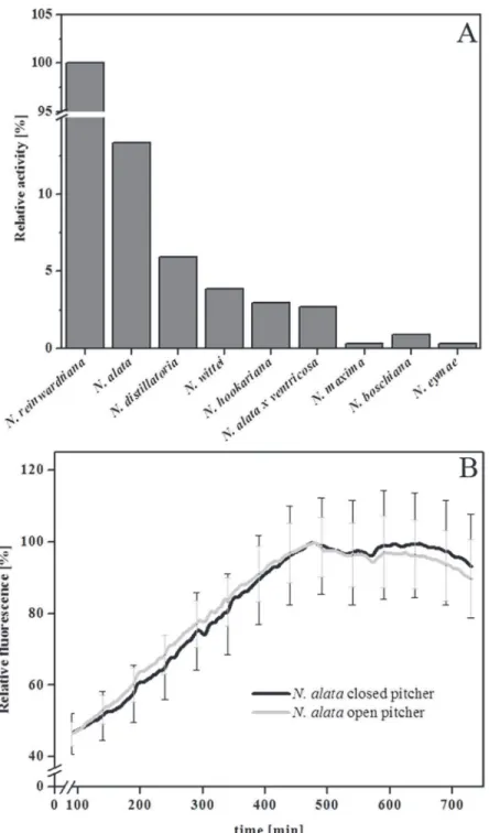
Employing the FRET technique with PFU-093 as substrate, proteolytic activity was detected in the pitcher fluid of all tenNepenthesspecies tested although at different intensities (Fig. 2A). Because fluids from closed pitchers were found to be sterile and contain only plant-derived components [4,24,25], the digestive fluids from closed and open pitchers ofN.alatawere also compared. InFig. 2B, proteolytic activity was detectable in both samples, indicating that the ac-tivity in the closed pitcher originated exclusively from the plant itself. Based on that, sterile di-gestive fluids from closed or newly opened pitchers (kept semi-sterile with gauze,Fig. 1B) were used for further measurements.
adjusted with buffers of high molarity before the final fluorescence was measured. In the con-trol, only water was added. The result (Fig. Ba inS1 File) suggests that the PFU-093 intrinsic fluorescence was quenched under acidic conditions and depended on decreasing pH levels, whereas at neutral and basic pH levels it was detectable. This forced us to keep or adjust the pH value between 7 and 8 in all fluorescence measurements.
In order to analyze whether the proteolytic reaction takes place not only at neutral pH but also under acidic conditions, a subsequent incubation experiment at pH 4 was undertaken. Re-sults revealed that it was possible to restore measurable fluorescence simply by adding a strong buffer and adjusting the pH of the sample to 8 directly after incubation (Fig. Bb inS1 File). This result also demonstrates that the proteases, which cleave the artificial substrate, are stable and active at acidic as well as slightly basic pH ranges, suggesting that these enzymes act like the aspartic proteinases nepenthesin I and II, purified and characterized from several Nepen-thesspecies [9,10,11,12,13,14,15,16,17,26].
Since the most prominent proteases inNepenthespitcher fluid are nepenthesins, it was con-ceivable that the activity we measured was due to those aspartic proteases. To challenge this idea, various inhibitors with specificities against different types of proteases were used in com-bination with the proteolysis assay. Inhibitors for metalloproteases (phenanthroline), serine-proteases (AEBSF), and cysteine-serine-proteases (E-64) showed no or minor effect (Fig. 3). In contrast, the proteolytic activity in the pitcher fluid is strongly inhibited (4.0 to 4.5% remaining activity) by the aspartic protease inhibitor pepstatin A. This result is in agreement with results obtained for pepstatin-inhibition of nepenthesin activity inN.alata[13] andN.distillatoria[14].
A detailed analysis of the cleavage reaction determined the cleavage site in the substrate (Figs. Ab and Ac inS1 File) shows the putative cleavage sites and the resulting products). An
Fig 1.Nepenthes alatapitchers. AWithout andBcovered with gauze. Pitchers/ pitcher fluid ofNepenthes
species can be kept under semi-sterile conditions by using gauze.
HPLC-MS analysis of the PFU-093 cleavage products after treatment withNepenthesproteases proved that, as expected, the substrate was cleaved between the two valines (Fig. 4). Although the molecular weight of the substrate was 1098 g/mol (Fig. 4B), a fragment ion withm/z620, corresponding to the FITC(Ahx)-Val fragment (Fig. 4C), was detected after flies were digested
Fig 2. Proteolytic activity inNepenthes. ASamples of the pitcher fluid of ten differentNepenthesspecies were investigated for their PFU-093 cleaving activity.BKinetics of proteolytic PFU-093 cleaving activity in pitcher fluid from open (grey line) and closed (black line) pitchers ofNepenthes alata. Relative fluorescence was measured over 12 h at 42°C.
in pitcher fluid from bothN.mirabilisandN.alata(Fig. 4A). Searching for a fragmentm/z= 397–398, we found a smaller peak in the chromatogram (Fig. 4B, green line) at Rt17.1 min.
This peak likely represents the“LysDbc”cleavage fragment and indicates that the second valine was also cleaved off. No fragment representing“Val-LysDbc”(m/ z= 497–498) was detected.
Cloning and heterologous expression of nepenthesin
To confirm that nepenthesin is the active protease involved in PFU-093 degradation, the cDNAs of nepenthesin I and II were cloned. First RNA was isolated fromN.mirabilispitchers.
Fig 3. Inhibitor experiments.Different protease inhibitors—phenanthroline, AEBSF, E-64 and pepstatin A—were tested for their ability to inhibit protease activity responsible for PFU-093 cleavage. Individual working concentrations are indicated in brackets. Pitcher fluid fromN.mirabiliswithout any inhibitor was used as a control (from left, first bar). Additional controls were carried out with the particular solvents of the in-hibitors (DMSO, MeOH, H2O). Statistics was done using one-way ANOVA, All Pairwise Multiple Comparison
Procedures (Student-Newman-Keuls Method), P<0.05; different letters indicate significant differences.
doi:10.1371/journal.pone.0118853.g003
Fig 4. Determination of protease cleavage site.A sample of PFU-093 after digestion with pitcher fluid proteases (N.mirabilisandN.alata) was analyzed by HPLC-MS.ASingle ion chromatograms for mass rangesm/z= 620–621 amu (black line),m/z= 497–498 amu (red line) andm/ z= 398–399 amu (green line). BMass spectrum of PFU-093 (FITC(Ahx)-Val-Val-LysDbc).CMass spectrum of the FITC(Ahx)-Val fragment after cleavage of PFU-093, FITC(Ahx)-Val-Val-LysDbc (MS was done with ESI in the positive ion mode).
After synthesizing cDNA, RACE PCR was further used to isolate the missing 5’and 3’regions. The resulting complete cDNA sequences of both Nep I and II, which include 5’- and 3’- un-translated regions, had 1570 and 1610 base pairs, respectively. The ORFs for Nep I and II both had 1314 base pairs, encoding 437 amino acid sequences (GenBank accessions AFV26024 (Nep I), AFV26025 (Nep II). Both Nep I and II possess a signal peptide for secretion which is predicted to have 24 amino acids (prediction was made using SignalP 4.1 Server [27]. The pre-dicted molecular weights of Nep I and II without signal peptides were calculated with 43.7 and 43.5 kDa, respectively. In addition, Nep I possesses seven predicted N-glycosylation sites used NetNGlyc 1.0 Server [28], whereas Nep II possesses only two. These observations support the fact that the glycosylation of nepenthesin proteins has been previously observed inNepenthes distillatoria[14].
Percent identity betweenN.mirabilis(NmNepI/II),N.alata(NaNepI/II) andN.gracilis
(NgNepI/II) amino acid sequences, was determined by alignment by using the CLUSTAL-W software program (see above) (Fig. C inS1 File). We found that the Nep I and Nep II sequences had rather high percent identity: 94.1% (NmNepI:NgNepI), 99.1% (NmNepI:NaNepI) and 96.1% (NmNepII:NgNepII). However, the maximum percent identity is about 66% when se-quences ofNmNepI are compared to those ofNmNepII. In Fig. C inS1 Filethe predicted signal peptide (SP) cleavage sites,predicted by SignalP- software [27] are shown to be located between amino acid positions 24 and 25: THS/TS. Thus, the active proteins start with the amino acids
“NGPS”(Nep I) and“QSSS”(Nep II), respectively. Between the SPs and the active protein, propeptide sequences [14] were detected (Fig. C inS1 File). TheN.mirabilisproteases were found to be typical nepenthesin-aspartic proteases: on one hand they lack the PSI (plant-specif-ic insertion), wh(plant-specif-ich is typ(plant-specif-ical for vacuolar APs [29], and on the other hand, they possess a special insertion assigned to residues 148–169 and known as‘the nepenthesin- type AP (NAP)-specific insertion’[14]. This insertion contains four additional cysteine residues (arranged pair-wise to form disulphide bonds), shown in Fig. C inS1 Fileas yellow, dark green, light green and orange; these residues contribute to the primary structure [14,15] and precede the tyrosine resi-due that is shown as a small green box above the sequence. The other two cysteine resiresi-dues are red and light red. The two catalytic aspartic acid residues are shown as small black boxes.
For heterologous expression in insect Sf9 cells, primers (see above) were used to amplify the ORFs of Nep I and II excluding their native signal peptide and stop codon. These fragments were cloned in frames with the sequence corresponding to the signal peptide from the bee mel-litin at the 5’-end and with a V5-(His)6tag at the 3’-end. After 72 h, culture medium was
har-vested and tested by Western blotting, using an anti-V5 horseradish peroxidase antibody that showed the expression ofNmNepII/Sf9 (Fig. D inS1 File) but not ofNmNepI/Sf9. However,
NmNepII/Sf9 did not show proteolytic activity.
The proteolytic activity ofNmNepII/Sf9 was activated by an acid-mediated cleavage of the expressed propeptide as described for nepenthesin-1 fromN.gracilisexpressed inE.coli[30]. After being activated,NmNepII/Sf9 was incubated for 24h at pH 4; then its protease activity could be measured (Fig. 5) at different protein concentrations (lines 1–3). The control without the presence of PFU-093 substrate (line 4) and theNmNepII/Sf9 control lacking auto-activa-tion (line 5) show no detectable activity. The activity ofNmNepII/Sf9 confirms our initial as-sumption that the aspartic proteases in the pitcher fluid, i.e. nepenthesins, are responsible for cleaving the fluorescent substrate.
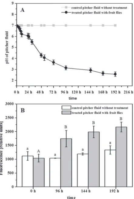
auto-activated when the surrounding medium is acidified (Fig. 6) [18], so protease activity increases when pitcher fluid is acidified by the addition ofD.melanogaster. InFig. 6B, a significant in-crease of protease activity is shown 96 h after the addition of fruit flies, confirming the role of nepenthesins as active proteases.
Induction of nepenthesins
FRET is a tool to directly analyze proteolytic, namely nepenthesin, levels in the digestive fluids ofNepenthes. Compared with others (e.g. [13]), this method is faster, more convenient, and specific for aspartic proteases. Moreover, kinetics can be easily measured with the same sample. The direct analysis of nepenthesins as one of the most prominent enzymes in the digestive fluid ofNepenthespitchers can be seen as the leading-activity for changes in the pitcher fluid activities that occur, for example, after the plants capture prey. As recently shown for the car-nivorous plantDionaea muscipula[31,32], besides nepenthesins the presence of more prote-ases can be expected. However, inNepenthes, only inN.ventricosaa cysteine proteinase activity was described in the pitcher fluid and a cDNA encoding a putative cysteine protease was cloned from pitcher tissue [17].
Up to now, the induction of hydrolytic enzymes in the pitcher was investigated only indi-rectly by gene induction analyses of the pitcher tissue [5,6,13,33]. Now, we are able to follow such dynamic processes simply by determining the Nep protease activity. Such analyses require pitchers ofN.mirabilisandN.alatato first be treated by supplementing them with prey such asD.melanogaster. This treatment resulted in a significant increase in proteolytic activity after 48 h (Fig. 7A). With some delay, the same effect was visible after supplementing the pitchers withNepenthesleaf material (Fig. 7B). Although the latter finding might suggest a kind of‘ can-nibalism’inNepenthes, this was not surprising because it is known that carnivorous plants
Fig 5. Proteolytic activity of recombinantN.mirabilisNepenthesin II (NmNepII/Sf9).PFU-093 fluorescence was measured over 6 h every 5 min after pre-incubation at pH 4 for 24 h for auto-activation. Different concentrations ofNmNepII/Sf9 were tested with constant concentrations of PFU-093 substrate. Line 1: 99μl ofNmNepII/Sf9; line 2: 49μlNmNepII/Sf9; line 3: 24.5μlNmNepII/Sf9. All were mixed with pure H20
and 1μl of 80μM fluorescent substrate to a total volume of 100μl per well. Controls: line 4: 99μlNmNepII/Sf9
and H2O without the addition of fluorescent substrate; line 5: 99μlNmNepII/Sf9 and 1μl of 80μM PFU-093
without pre-incubation/ auto-activation in pH 4 glycine-buffer.
actually take whatever they get for their nutrition. For example,N.ampullariais specialized to capture leaf litter from the canopy above [34]. With any type of organism that falls in the trap all sources of nutrients become available for the plant and justify the induction of various hydrolyzing enzymes.
Recently, it has been shown for different carnivorous plant genera that defense-related phy-tohormones are involved in the trapping process, e.g. forDrosera capensis[35,36] as well as in the digestion process, e.g. inD.muscipula[37,38]. In order to follow up those studies, we inves-tigated the effects of phytohormones inNepentheson protease activity. Interestingly, when
Fig 6. Influence ofDrosophila melanogasteron pitcher fluid- pH and nepenthesin levels.Pitchers were supplemented with 40 fruit flies each.AThe pitcher fluid pH was continuously determined at different time points until 192 h after treatment (black line); control without treatment (gray line).BProtease activity was measured before (0 h) and after the addition of fruit flies (96, 144, 192 h; gray bars) by using PFU-093 substrate for each sample. Measurement included 2 control pitchers (white bars). ForBOne-way ANOVA, P<0.05, Post Doc Test_SNK, was performed.
phytohormones were added to the pitcher, only the addition of jasmonic acid (JA), not salicylic acid (SA), significantly increased the Nep- activities (Fig. 7C and 7D). These results support the hypothesis that carnivory in plants might have evolved from defensive reactions against pathogens or herbivores [7,36].
Conclusion
Carnivorous plants ofNepenthes, unlike other plant carnivores, offer the possibility of working with sterile digestive fluids as long as the pitchers are closed or the open pitchers kept some-what sterile by means of gauze. Here, a novel FRET-based method regardingNepenthesplants was established, optimized and successfully applied by using the fluorogenic PFU-093 substrate for easy and rapid detection of protease activities in digestive fluids ofNepenthesspecies. The specificity of the substrate for aspartic proteases provides a means of unravelling the processes involved in prey digestion in carnivorous pitcher plants and possibly in other carnivorous plants. The ability to measure induced protease activity in pitcher fluids is much more reflect-ing the digestive process as quantifyreflect-ing transcripts of the correspondreflect-ing genes.
Supporting Information
S1 File. Fig. A in S1 File.FRET-peptide,-method and putative cleavage sites.aSimplified structure of the artificial substrate PFU-093, according to Kaman et al. [19]. The substrate con-tains a fluorescein isothiocyanate (FITC) as fluorophore (F) and Lysin-Dabcyl (LysDbc) as its quencher (Q) connected by a two valine (Val) bridge. The close vicinity of Q to F quenches the fluorescence.bPutative proteolytic cleavage sites of PFU-093 substrate either between the two Val or between the last Val and the LysDbc, resulting in cleavage products: 1) FITC-Val, 2) Val-LysDbc, 3) LysDbc;cpredicted molecular masses of these products: 1) 619.68 g/mol, 2)
Fig 7. Induction of nepenthesin protease activity inNepenthes mirabilispitcher fluid.Proteolytic activity was measured with PFU-093 substrate at different time points after various treatments:A
supplementation withD.melanogaster(n= 3);Bsupplementation withNepenthesleaf (n= 2);Cinjection of jasmonic acid (200μM end concentration) (n= 3)Dinjection of salicylic acid (end concentration 200μM) (n= 4). Statistics employed one-way ANOVA, P<0.05, Post Hoc Tests_SNK(A, C, D),Dunnett T3(B).
496.28 g/mol and 3) 397.47 g/mol.Fig. B in S1 File.Substrate/product fluorescence depen-dence on different pH values. aPitcher fluid was incubated with PFU-093 substrate and pure water at 42°C for 10 h. 50μl each of this mixture were given in different wells and mixed with
additional 50μl of 30 mM buffer solutions to adjust the final pH (2, 4, 6, 8, 10, black dots),
be-fore fluorescence was measured. Control with water instead of buffer was included, represent-ing the original fluorescence. Arrows indicate the pH-dependrepresent-ing change of fluorescence.b
Digestive fluid and PFU-093 substrate were incubated in 1 mM citrate buffer pH 4, for 10 h at 42°C. Subsequently, the mixture was split up and pH was adjusted by topping with either 30 mM phosphate buffer, pH 8, (dark grey bar) or 30 mM citrate buffer, pH 4, (striped) before fluorescence measurement.Fig. C in S1 File.N.mirabilisnepenthesin I, II (NmNepI/ II) pro-tein alignment compared toN.gracilis(Ng) andN.alata(Na) nepenthesin amino acid se-quences. The four levels of shading used are:blue>80% sequence identity,mid-blue>60%
identity,light blue>40% identity andno shading<40%. Regions of predicted signal peptides
and propeptides are named and the endings marked by a black stroke. Aspartic acid residues of the active center are indicated by a small black box and the flap tyrosine residue by a small green box, both above the sequences. The cysteine residues are represented through the colors: yellow, orange, green, light green, red and light red. The colored pairing of the residues show the disulphide bond arrangements in the primary structures of nepenthesin.Fig. D in S1 File.
Western blot of recombinantN.mirabilisnepenthesin II.NmNepII/Sf9 was expressed in Sf9 insect cell line, using an anti-V5 horseradish peroxidase antibody and ECL for detection Lanes represent 1) lysate, negative control, 2) culture medium, negative control, 3) lysate, positive control, 4) culture medium, positive control (CAT, catalase of 34 kDa); the blot also contains duplicates (clone 1 and 2) shown by the lanes 5)NmNepII/Sf9, clone 1, lysate, 6)NmNepII/Sf9, clone 1, culture medium, 7)NmNepII/Sf9, clone 2, lysate, 8)NmNepII/Sf9, clone 2, culture me-dium, all in comparison to the Sf9 cell line stably expressingNmNepII/Sf9, pointed out with 5μl (lane 9) and 10μl (lane 10), respectively.Table A in S1 File.List of primer sequences
used in this study. (PDF)
Acknowledgments
We thank Birgit Arnold, Tamara Krügel and the greenhouse team for growingNepenthes
plants. We also thank Dr. Yannick Pauchet for providing us insect cells and the expression vec-tor. Gerhard Pauls, Dr. Anne Morgenstern and Dr. Stephan H. von Reuß we thank for help with HPLC- MS method and last but not least Wilhelm Boland and the Max Planck Society for continuous support. We also thank Emily Wheeler for editorial assistance
Author Contributions
Conceived and designed the experiments: FB AM. Performed the experiments: FB AY. Ana-lyzed the data: FB AY AM. Contributed reagents/materials/analysis tools: WEK FJB. Wrote the paper: FB WEK FJB AY AM.
References
1. Darwin C (1875) Insectivorous Plants. London, UK: John Murray.
2. Heslop-Harrison Y (1975) Enzyme release in carnivorous plants. In: Dingle JT, Dean RT, editors. Lyso-zymes in biology and pathology, pp 525–578.
4. Buch F, Rott M, Rottloff S, Paetz C, Hilke I, Raessler M, et al. (2013) Secreted pitfall-trap fluid of carniv-orous Nepenthes plants is unsuitable for microbial growth. Ann Bot 111: 375–383. doi:10.1093/aob/ mcs287PMID:23264234
5. Eilenberg H, Pnini-Cohen S, Schuster S, Movtchan A, Zilberstein A (2006) Isolation and characteriza-tion of chitinase genes from pitchers of the plantNepenthes khasiana. J Exp Bot 57: 2775–2784. PMID:16829546
6. Rottloff S, Stieber R, Maischak H, Turini FG, Heubl G, Mithöfer A (2011) Functional characterization of a class III acid endochitinase from the traps of the carnivorous pitcher plant genus, Nepenthes. J Exp Bot 62: 4639–4647. doi:10.1093/jxb/err173PMID:21633084
7. Mithöfer A (2011) Carnivorous pitcher plants: Insights in an old topic. Phytochemistry 72: 1678–1682. doi:10.1016/j.phytochem.2010.11.024PMID:21185041
8. Renner T, Specht CD. (2013) Inside the trap: gland morphologies, digestive enzymes, and the evolu-tion of plant carnivory in the Caryophyllales. Curr Opin Plant Biol 16: 436–42. doi:10.1016/j.pbi.2013. 06.009PMID:23830995
9. Vines SH (1897) The proteolytic enzyme ofNepenthes. Ann Bot 11: 563–584.
10. Amagase S, Nakayama S, Tsugita A (1969) Acid protease in Nepenthes. II. Study on the specificity of nepenthesin. J Biochem 66: 431–439. PMID:5354017
11. Jentsch J (1972) Enzymes from carnivorous plants (Nepenthes). Isolation of the protease nepenthacin. FEBS Lett 21: 273–276. PMID:11946525
12. Tökés ZA, Woon WC, Chambers SM (1974) Digestive enzymes secreted by the carnivorous plant Ne-penthes macferlanei L. Planta 119: 39–46. doi:10.1007/BF00390820PMID:24442407
13. An CL, Fukusaki E, Kobayashi A (2002) Aspartic proteinases are expressed in pitchers of the carnivo-rous plantNepenthes alataBlanco. Planta 214: 661–667. PMID:11882933
14. Athauda SBP, Matsumoto K, Rajapakshe S, Kuribayashi M., Kojima M, Kubomura-Yoshida N, et al. (2004) Enzymic and structural characterization of nepenthesin, a unique member of a novel subfamily of aspartic proteinases. J Biochem 381: 295–396. PMID:15035659
15. Takahashi K, Athauda SBP, Matsumoto K, Rajapakshe S, Kuribayashi M, Kojima M, et al. (2005) Nepenthesin, a unique member of a novel subfamily of aspartic proteinases: enzymatic and structural characteristics. Curr Protein Peptide Sci 6: 513–525. PMID:16381601
16. Takahashi K, Tanji M, Shibata C (2006) Variations in the content and isozymic composition of Nepenthesin in the pitcher fluids amongNepenthesspecies. Carniv Pl Newslett 36: 73–76.
17. Stephenson P and Hogan J (2006) Cloning and characterization of a ribonuclease, a cysteine protein-ase, and an aspartic proteinase from pitchers of the carnivorous plantNepenthes ventricosaBlanco. Int J Plant Sci 167: 239–248.
18. Kadek A, Tretyachenko V, Mrazek H, Ivanova L, Halada P, Rey M, et al. (2014) Expression and charac-terization of plant aspartic protease nepenthesin-1 fromNepenthes gracilis. Protein Expr Purif 95: 121–128. doi:10.1016/j.pep.2013.12.005PMID:24365662
19. Kaman WE, Hulst AG, van Alphen PTW, Roffel S, van der Schans MJ, Merkel T, et al. (2011) Peptide-based fluorescence resonance energy transfer (FRET) protease substrates for the detection and diag-nosis ofBacillusspp. J Anal Chem 83: 2511–2517. doi:10.1021/ac102764vPMID:21370823
20. Kaman WE, Voskamp-Visser I, de Jongh DM, Endtz HP, van Belkum A, Bikker FJ, et al. (2013) Evalua-tion of a D-amino-acid-containing fluorescence resonance energy transfer peptide library for profiling prokaryotic proteases. Anal Biochem 441: 38–43. doi:10.1016/j.ab.2013.06.015PMID:23850560
21. Buch F, Pauchet Y, Rott M, Mithöfer A (2014) Characterization and heterologous expression of a PR-1 protein from traps of the carnivorous plantNepenthes mirabilis. Phytochemistry 100: 43–50. doi:10. 1016/j.phytochem.2014.01.014PMID:24534104
22. Untergrasser A, Cutcutache I, Koressaar T, Ye J, Faircloth BC, Remm M, et al. (2012) Primer3—new capabilities and interfaces. Nucleic Acids Res 40: e115. PMID:22730293
23. Waterhouse AM., Procter JB., Martin DMA, Clamp M and Barton GJ (2009) "Jalview Version 2—a mul-tiple sequence alignment editor and analysis workbench". Bioinformatics 25: 1189–1191. doi:10.1093/ bioinformatics/btp033PMID:19151095
24. Hepburn JS (1918) Biochemical studies of the pitcher liquor ofNepenthes. Proc Am Phil Soc 57: 112–
129.
25. Lüttge U (1964) Untersuchungen zur Physiologie der Carnivoren- Drüsen. Planta 63: 103–117. 26. Nakayama S, Amagase S (1968) Acid protease in Nepenthes partial purification and properties of
27. Nordahl Petersen T, Brunak S, von Heijne G and Nielsen H (2011) SignalP 4.0: discriminating signal peptides from transmembrane regions. Nat Methods 8: 785–786. doi:10.1038/nmeth.1701PMID:
21959131
28. Gupta R, Jung E, Brunak S (2004) Prediction of N-glycosylation sites in human proteins. N-glycosyla-tion site predictor available at:http://www.cbs.dtu.dk/services/NetNGlyc/
29. Simões I, Faro C (2004) Structure and function of plant aspartic proteinases. Eur J Biochem 271: 2067–2075. PMID:15153096
30. Runeberg-Roos P, Tormakangas K, Ostman A (1991) Primary structure of a barley-grain aspartic pro-teinase. A plant aspartic proteinase resembling mammalian cathepsin D. Eur J Biochem 202: 1021–
1027. PMID:1722454
31. Takahashi K, Suzuki T, Nishii W, Kubota K, Shibata C, Isobe T, et al. (2011) A cysteine endopeptidase (‘‘Dionain”) is involved in the digestive fluid ofDionaea muscipula(Venus’s flytrap). Biosci Biotech Bio-chem 75: 346–348. PMID:21307583
32. Schulze WX, Sanggaard KW, Kreuzer I, Knudsen AD, Bemm F, Thøgersen IB, et al. (2012)The protein composition of the digestive fluid from the Venus flytrap sheds lighton prey digestion mechanisms. Mol Cell Proteom 11: 1306–1319.
33. Rottloff S, Müller U, Kilper R, Mithöfer A (2009) Micropreparation of single secretory glands from the carnivorous plant Nepenthes. Anal Biochem 394: 135–137. doi:10.1016/j.ab.2009.07.013PMID:
19602419
34. PavlovičA, Slováková L,Šantrůček J (2011) Nutritional benefit from leaf litter utilization in the pitcher plantNepenthes ampullaria. Plant Cell Environ 34: 1865–1873. doi:10.1111/j.1365-3040.2011.02382. xPMID:21707655
35. Nakamura Y, Reichelt M, Mayer VE, Mithöfer A (2013) Jasmonates trigger prey-induced formation of outer stomach in carnivorous sundew plants. Proc R Soc B 280: 20130228. doi:10.1098/rspb.2013. 0228PMID:23516244
36. Mithöfer A, Reichelt M, Nakamura Y (2014) Wound and insect-induced jasmonate accumulation in car-nivorousDrosera capensis: two sides of the same coin. Plant Biol 16: 982–987 doi:10.1111/plb.12148
PMID:24499476
37. Escalante-Perez M, Krol E, Stange A, Geiger D, Al-Rasheid KA, Hause B, et al. (2011) A special pair of phytohormones controls excitability, slow closure, and external stomach formation in the Venus flytrap. PNAS USA 108: 15492–15497 doi:10.1073/pnas.1112535108PMID:21896747
38. Libiaková M, Floková K, Novák O, Slováková L and PavlovičA (2014) Abundance of cysteine endopep-tidase dionain in digestive fluid of Venus flytrap (Dionaea muscipulaEllis) is regulated by different sti-muli from prey through jasmonates. PLoS ONE 9: e104424. doi:10.1371/journal.pone.0104424PMID: