RESEARCH ARTICLE
Postembryonic Nephrogenesis and
Persistence of Six2-Expressing Nephron
Progenitor Cells in the Reptilian Kidney
Troy Camarata1, Alexis Howard1, Ruth M. Elsey3, Sarah Raza1, Alice O’Connor1, Brian Beatty2, Jack Conrad2, Nikos Solounias2, Priscilla Chow1, Saima Mukta1, Aleksandr Vasilyev1*
1Department of Biomedical Sciences, NYIT College of Osteopathic Medicine, Old Westbury, New York, United States of America,2Department of Anatomy, NYIT College of Osteopathic Medicine, Old Westbury, New York, United States of America,3Louisiana Department of Wildlife and Fisheries, Grand Chenier, Louisiana, United States of America
Abstract
New nephron formation (nephrogenesis) ceases in mammals around birth and is
completely absent in adults. In contrast, postembryonic nephrogenesis is well documented in the mesonephric kidneys of fishes and amphibians. The transient mesonephros in rep-tiles (including birds) and mammals is replaced by the metanephros during embryogenesis. Thus, one may speculate that postembryonic nephrogenesis is restricted to the mesoneph-ric kidney. Previous reports have suggested the metanephros of non-avian reptiles (hereaf-ter reptiles) may continually form nephrons throughout life. We investigated the presence of adult nephrogenesis in reptiles by examining adult kidneys from several species including Trachemys scripta,Chrysemys picta,Boa constrictor,Tupinambis tegu,Anolis carolinensis, andAlligator mississipiensisamong others. We found that all major reptilian groups (Testu-dines, Crocodylia, and Squamates) showed the presence of adult nephrogenesis. The total amount of nephrogenesis varied greatly between species with turtles displaying the highest density of nephrogenesis. In contrast, we were unable to detect adult nephrogenesis in monotremes, and in the iguanidA.carolinensis. Nephron progenitor cells express the tran-scription factor Six2, which in mammals, becomes downregulated as the progenitor cell population is exhausted and nephrogenesis ends. Using the alligator as a model, we were able to detect Six2-positive cap mesenchyme cells in the adult kidney, which spatially corre-lated with areas of nephrogenesis. These results suggest that the metanephric kidney of reptiles has maintained the ability to continually grow new nephrons during postembryonic life, a process lost early in mammalian evolution, likely due to the persistence of a Six2-expressing progenitor cell population.
a11111
OPEN ACCESS
Citation:Camarata T, Howard A, Elsey RM, Raza S, O’Connor A, Beatty B, et al. (2016) Postembryonic Nephrogenesis and Persistence of Six2-Expressing Nephron Progenitor Cells in the Reptilian Kidney. PLoS ONE 11(5): e0153422. doi:10.1371/journal. pone.0153422
Editor:Peter Hohenstein, The Roslin Institute, UNITED KINGDOM
Received:October 5, 2015
Accepted:March 29, 2016
Published:May 4, 2016
Copyright:© 2016 Camarata et al. This is an open access article distributed under the terms of the
Creative Commons Attribution License, which permits unrestricted use, distribution, and reproduction in any medium, provided the original author and source are credited.
Data Availability Statement:All relevant data are available in the paper and its Supporting Information files. Additional data from the Reptile Nephrogenesis study can be requested by contacting Dr. Aleksandr Vasilyev ([email protected]).
Funding:The authors have no support or funding to report.
Introduction
The vertebrate kidney has evolved to regulate water homeostasis and waste excretion to main-tain ourmilieu interieur[1]. Three types of kidney structures have developed over the course of vertebrate evolution, the pronephros, mesonephros, and metanephros. In fish and amphibi-ans, the paired epithelial tubules of the embryonic pronephros are followed by the development of the more complex mesonephric kidney found in the adult. In amniotes, the pronephros and mesonephros become reabsorbed, and are replaced by the final metanephric kidney. The over-all structural complexity of each kidney type increases from a parover-allel pair of nephrons in the zebrafish pronephros [2] up to a million highly organized nephrons in a human metanephric kidney [3]. However, the segmentation pattern of each kidney type appears conserved as the nephrons of the pro-, meso-, and metanephric kidney each have a proximal, intermediate, and distal segment which drain into a collecting duct system with conserved segmental gene expres-sion [4–7].
Cells destined for a renal fate are derived from the intermediate mesoderm (IM) during embryogenesis. For example, during morphogenesis of the zebrafish pronephros, cells from the IM go through a mesenchymal-to-epithelial transition (MET) to form the pronephric duct epi-thelium and tubule formation progresses in an anterior-to-posterior axis [2]. The pronephric tubule is then patterned with a segmented structure highly reminiscent of the mammalian nephron [4]. Once the pronephric kidney is established and functional, the zebrafish meso-nephros forms along the pronephric tubule, again, progressing from anterior to posterior [8,9]. Progenitor cells adjacent to the pronephric tubule condense and elongate to form the meso-nephric tubules, which eventually fuse with the promeso-nephric tubule [7]. This process is repeated making a branched mesonephros, where nephrons are sequentially attached to a single meso-nephric duct, inherited from the pronephros.
In mammals, the pronephros and mesonephros develop along the nephric duct, progressing from anterior to posterior [10]. The pronephric and mesonephric kidneys degenerate, with cells from the mesonephros contributing to the male gonads. Along the nephric duct, posterior to the mesonephros, the final metanephric kidney begins to form by embryonic day 35 in humans (embryonic day 10.5 in mouse). Development of the metanephric kidney starts when the ureteric bud (UB) branches off the mesonephric duct and invades an overlying metaneph-ric mesenchyme (MM) [11]. Reciprocal signaling between cells of the MM and UB induce nephron morphogenesis. The UB branches with MM condensing at each branch tip and undergoing MET to produce renal vesicles which elongate and connect to the UB [12,13], and mature into nephrons. The MM gives rise to the glomerular epithelium, proximal tubule, Loop-of-Henle, and distal tubule segments, while the UB branches forming the collecting duct system. This process of UB branching and MM condensation and differentiation continues until the pool of MM progenitor cells becomes exhausted around week 35 of gestation in humans [14] or post-natal day 3 in mice [15].
Consequently, mammals are born with a finite number of nephrons and are incapable of generating new tubules for either tissue homeostasis or repair from injury. The halt in nephron endowment appears to be universal in mammals, as even in examined marsupial species (Dasyurus hallucatusandTrichosurus vulpecula) [16,17] nephrogenesis stops prior to weaning. The subsequent increase in total functional capacity of the mammalian kidney occurs through nephron hypertrophy and hyperplasia rather than through increase in nephron number (nephrogenesis; [18]).
extensive enough, this may lead to chronic renal failure [21]. Currently, the only solution for these patients is renal transplantation or dialysis, both of which present significant problems [22]. Thus, it becomes important to understand the‘lost art’of continuous nephrogenesis. This knowledge may allow us to develop novel regenerative approaches to reverse nephron loss and enable organ engineering solutions for recovering lost kidney function [23].
In contrast to mammals, the adult mesonephric kidney of osteichthyes, chondrichthyes and lissamphibians is capable of continual nephrogenesis [24]. Several species have been found to add new nephrons to their adult mesonephric kidney, including goldfish [25], catfish, trout, tilapia, toadfish [26], zebrafish [8,9], medaka [27], frogs (Rana Temporaria; [28], dogfish [29,30], and skates [31]. Furthermore, the process of nephrogenesis in adult mesonephric kid-neys is enhanced following injury [8,9,27,32,33] providing an excellent mechanism for kidney repair. It appears that the continual addition of nephrons to the kidney is a common feature in vertebrates with a mesonephric kidney. Therefore, it was proposed that continual nephrogen-esis is specific to mesonephric kidney and is lost in the metanephric kidney of amniotes [34].
However, evidence in the literature suggests that in isolated species of reptiles and birds, the metanephric kidney is capable of nephrogenesis in the post-embryonic period [35–38]. We set out to determine the extent and the prevalence of post-embryonic and adult nephrogenesis in reptiles, aiming to unambiguously establish whether the phenomenon of adult nephrogenesis was restricted to the mesonephric kidney, or if it was also commonly present in the metaneph-ric kidney of reptiles.
We collected kidneys from juvenile and adult reptiles from all of the major reptilian groups including Archosauromorpha (here represented by the non-avian archosaur clade Crocodylia), Testudines and Squamata. We identified histological evidence of juvenile and adult nephrogen-esis in all of these groups. However, adult nephrogennephrogen-esis appears to have been lost in some rep-tile species, similar to mammals. In addition, we found immunofluorescence evidence of persistent Six2-expressing cap mesenchyme cells. Six2 expression is normally lost in mammals once nephrogenesis ceases, suggesting nephron progenitor cells are present in the reptilian kid-ney post-embryonically. We conclude that the metanephric kidkid-ney is capable of continual nephrogenesis in many adult species of reptiles and this ability was lost very early in, or prior to mammalian evolution. Unfortunately, few“living fossils”of early mammals exist, leaving the question of when this loss occurred in early mammals unanswerable at any node prior to the diversification of monotremes. Thus, we sampled the available specimens of monotremes, but focused our efforts on nonmammalian amniotes.
Results
Reptiles maintain nephrogenesis throughout life
Previous reports have suggested the occurrence of new nephron growth, termed nephrogenesis, in reptiles post-embryonically [35–37]. Two of these studies indirectly assessed new nephron formation by estimating glomerular number as a function of animal age. However, Solomon [36] did show histological evidence of nephrogenesis in an adult green sea turtle (Chelonia mydas). We questioned whether nephron formation in juvenile or adult reptiles was species specific or a broader phenomenon.
Therefore, we obtained adult and juvenile kidney tissue from species found in the major reptilian groups Crocodylia (alligators and crocodiles), Testidines (turtles), and Squamates (liz-ards and snakes,S1 Table). Histological analysis of tissue sections revealed post-embryonic nephrogenesis in a number of juvenile and adult reptiles, including American alligator (A. mis-sissippiensis), red-eared slider (T.scripta), painted turtle (C.picta), African spiny-tailed lizard (C.tropidosternum), tegu lizard (T.teguxin), rock monitor (V.albigularis), Egyptian mastigure
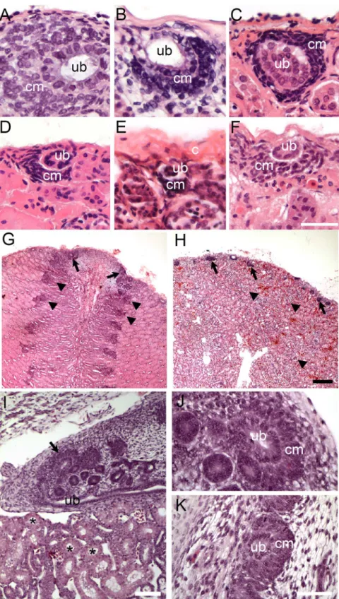
(U.aegyptia), Chinese water dragon (P.cocincinus), boa constrictor (B.constrictor) and western rat snake (P.obsoletus) (S1 Table). Nephrogenesis was detected as areas of condensed mesen-chyme surrounding a tip of terminal duct branch just under the renal capsule (Fig 1A–1H, also
S1 Fig). There was some variability in the size and the location of cap mesenchyme (especially in the case ofU.aegyptia,Fig 1E). At least some of the variability could be attributed to the freezing and drying artifact (specimens inFig 1C–1Hhave been stored frozen several weeks before processing). However, some of the variability may be reflective of the differences in the overall activity of these nephrogenic zones. It should be also noted that cap mesenchyme aggre-gates were often seen on the side of the ureteric bud or on the side opposite the capsule with respect to the ureteric bud (contrasting it to the mammalian embryonic development, where cap mesenchyme is most commonly positioned between the ureteric bud and the capsule). This can be only partially explained by random sectioning. Another partial reason is the differ-ence in overall geometry or the reptilian kidney, where ureter is often positioned in the subcap-sular location, extending parallel instead to perpendicular to the surface of the kidney.
However, there might be other explanations for the observed differences.
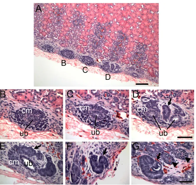
The overall histology of zones of nephrogenesis detected in juvenile and adult reptiles was similar to that observed during embryonic development (Fig 1I–1K). For example, in juvenile and adult alligators, condensed mesenchymal cells were detected surrounding tips of terminal duct branches (Fig 1A and 1G;Fig 2). This histology recapitulated embryonic nephrogenesis seen in mammals and reptiles, as demonstrated inFig 1I–1K. The progression of nephrogenesis in an adult alligator specimen was similar to embryonic nephron formation (Fig 2). Multiple zones of nephrogenesis were found under the renal capsule along the periphery of the kidney (Fig 2A). Nephrogenesis appeared to be asynchronous as we detected different stages of neph-ron formation in adjacent zones (Fig 2B–2D). Typical sequential forms of nephron develop-ment could be detected, including renal vesicle, S-shaped body and various stages of
glomerular maturation (Fig 2E–2G, alsoS2 Fig). Thus, we were able to detect histological evi-dence of postembryonic and adult nephrogenesis in a wide range of reptilian species, represent-ing all major reptilian groups (Crocodylia, Testudines and Squamata, summarized inFig 3).
For comparison, we sampled kidneys from two adult species of monotremes, the short-beaked echidna (Tachyglossusaculeatus) and the platypus (Ornithorhynchus anatinus) (Fig 3;
S3 Fig). Similar to other species of mammals, we did not find evidence of nephron formation in this basal mammalian group. This suggests that the ability to continually grow nephrons after birth was lost very early in the evolution of mammals, although more extensive sampling would be required to establish the timeline of the disappearance of nephrogenesis in mono-tremes as a function of age.
Postembryonic nephrogenesis is not universal among reptiles
Fig 1. Examples of reptilian post-embryonic nephrogenesis.(A-H) Kidney tissue sections stained with H&E showing zones of nephrogenesis. (A-F) High magnification of zones of nephrogenesis from juvenile American alligator (A.mississippiensis, A), adult turtles: red-eared slider (T.scripta, B) and painted turtle (C. picta, C), adult tegu (T.teguixin, D), adult Egyptian Mastigure (U.aegyptia, E), and adult boa constrictor (B. constrictor, F). Corresponding lower magnification images are shown inS1 Fig. Scale bar = 50μm. (G-H) Low magnification images showing tissue organization and variability in arrangement of zones of
nephrogenesis between American alligator (G) and red-eared slider (H). Arrows denote zones of
nephrogenesis, arrowheads point to mature glomeruli. Scale bar = 100μm. (I-K) Embryonic alligator kidney (stage 18–19). (I) Wide-field view of embryonic alligator mesonephros (bottom; asterisks label mesonephric
tubules) and metanephros (top). Zone of nephrogenesis is indicated by the arrow. Scale bar = 100μm. (J-K) High magnification of nephrogenesis in embryonic alligator metanephric kidney. Scale bar = 20μm. Ub = tip of the ureteric bud branch, cm = metanephric cap mesenchyme, c = capsule.
doi:10.1371/journal.pone.0153422.g001
Fig 2. Nephron formation in adult reptiles resembles embryonic nephrogenesis.(A-G) Adult alligator kidney tissue sections stained with H&E. (A) Wide field view of zones of nephrogenesis on the periphery of the kidney just under the capsule. Scale bar = 100μm. (B-D) Higher magnification of nephrogenic events as denoted in (A). Early stage nephron formation in (B) which progresses to developed condensed mesenchyme in (C) and a later stage
nephrogenesis event in (D) with condensing mesenchyme, ureteric bud-like branch tips (ub) and a newly formed immature nephron (arrow). (E-G) Nephrogenesis in adult alligator is reminiscent of embryonic nephron formation. (E) Metanephric cap mesenchyme undergoing MET to form early nephron structure (arrow). (F) S-shaped developing nephron (arrow) similar to S-shaped bodies detected during embryonic nephron formation. (G). Maturing glomeruli. Newly formed glomerulus (arrow) at the end of a newly formed tubule with progressively more mature glomeruli (arrowheads). Scale bar = 50μm. Ub = tip of the ureteric bud branch, cm = metanephric cap mesenchyme. The lower magnification images corresponding to (E-G) are shown inS2 Fig.
kidney size by nephrogenesis. Post-embryonic kidney growth in mammals is accomplished by nephron hypertrophy. To gain insight into whether the green anole uses a similar mechanism to increase renal size, we compared estimated glomerular size to body mass (Fig 4D). As expected for nephron hypertrophy, glomerular size increased in relation to body mass.
These data, along with the lack of new nephron endowment, suggest that the green anole, similar to mammals, uses nephron hypertrophy rather than nephrogenesis, to increase kidney size during adult growth. In contrast, when we estimated glomerular size in juvenile and adult American alligator (Fig 4E), which have robust nephrogenesis (Figs1and2), we did not detect an increase in glomerular size with increasing body length (up to 2.03 m,Fig 4E).
It is interesting to note that the green anole did not show evidence of adult nephrogenesis despite related species within the same suborder, which did display adult nephrogenesis (Fig 3). We detected nephrogenesis in adult Egyptian mastigure (Figs1and3), and nephrogenesis has been proposed to exist in the adult green iguana (I.iguana; [35] and Yarrow’s spiny tail liz-ard (Sceloporus jarrovii; [37]. Further examination is needed to determine if the green anole completely lacks adult nephrogenesis, similar to mammals, or if continual nephron endow-ment can be induced by nephron loss or kidney injury.
Frequency of nephrogenesis varies between reptilian groups
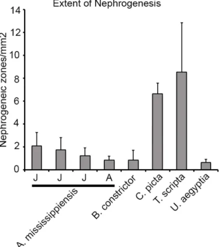
In our study, the majority of juvenile and adult reptilian species displayed evidence of nephro-genesis. However, the gross morphology and extent of new nephron formation varied. We counted zones of nephrogenesis in the specimens where adequate and reliable counts could be obtained, and compared them between species by normalizing to the effective surface area (Fig 5; seeMethods). The two adult turtle species,C.pictaandT.scripta, had the greatest average number of nephrogenic zones per unit surface area (6.7 ± 0.9 /mm2and 8.6 ± 4.3 /mm2
Fig 3. Species surveyed for post-embryonic nephrogenesis.Cladogram displaying species tested for presence of nephrogenesis in juvenile (J) or adult (A) kidney. Two species of monotremes,Tachyglossus aculeatus(short-beaked echidna) andOrnithorhynchus anatinus(platypus), were used for comparison as the most basal mammalian group. As with other mammals, no evidence of nephrogenesis was detected in the monotreme species. Post-embryonic nephrogenesis was detected in all major reptilian groups surveyed. Species names in light gray did not display evidence of nephrogenesis. NT = not tested.
doi:10.1371/journal.pone.0153422.g003
respectively,Fig 4). The juvenile alligator kidneys averaged between 1.3 and 2.1 /mm2, adult alligator (1.98 m), 0.86 ± 0.35 /mm2followed by the boa (B.constrictor), 0.9 ± 0.9 /mm2and the Egyptian mastigure (Uromastyx aegyptia), 0.7 ± 0.3 /mm2. Thus, there was significant vari-ation in the frequency of nephrogenesis between major reptilian groups. In addition, we found differences in the distribution of nephrogenesis events. Most species surveyed showed random distribution of nephrogenesis with respect to the surface of the kidney. In contrast, the kidney of the American alligator was highly organized [39] with rows of glomeruli leading to the apex
Fig 4. Kidney growth occurs by hypertrophy or nephrogenesis in reptiles.(A-C) Green anole (A.carolinesis) kidney measurements compared to body mass. (A) Scatter plot of kidney mass related to body mass with line of best fit. (B) Scatter plot of estimated glomerular number related to body mass. (C) Scatter plot of the ratio of glomerular number/kidney mass compared to body mass. Number of biological samples = 10. (D-E) Scatter plots of glomerular size compared to body mass in green anole (D) or body length in American alligator (A.mississippiensis) (E). (F) Histology of adult green anole kidney (left) and adult American alligator kidney (right). Arrow denotes zone of nephrogenesis in the American alligator. (g) = grams. (cm) = centimeters.
of each renal lobe (Fig 1H, arrowheads). We observed nephrogenesis events only at the points where rows of glomeruli met the capsule (Fig 1G, arrows), forming a distinct line of nephrogen-esis that could be detected grossly (S5 Fig). In comparison, in turtles, the kidney had glomeruli distributed throughout the cortex (Fig 1H, arrowheads), and the nephrogenesis events were distributed randomly under the capsule (Fig 1H, arrows).
Persistence of Six2-expressing kidney progenitor cell population
The metanephric mesenchyme (MM) in mammals expresses specific genes that mark and maintain the pool of kidney progenitor cells. One such gene is the transcription factor Six2, which labels the nephron progenitor population in the MM and is required for its maintenance [40,41]. If the areas of condensed mesenchyme identified in juvenile and adult reptiles are sites of continual nephron endowment, then those cells should express progenitor cell MM markers such as Six2.Fig 5. Extent of nephrogenesis in reptiles.The number of nephrogenic zones in each species was normalized to the effective surface area of each section analyzed (methods). The ratio of the total number of nephrogenic zones and the effective surface area was then used to compare the extent of post-embryonic nephrogenesis between different reptile species. Only the material with sufficient preservation to accurately estimate the number of nephrogenesis events was used for analysis. Species analyzed were:A.
mississippiensis(3 juvenile (J), 1 adult (A)),B.constrictor,C.picta,T.scripta,U.aegyptia. The
corresponding density of nephrogenesis: (A.mississippiensis: 2.1±1.1 (0.89m juvenile), 1.8±1.1 (1.04m juvenile), 1.3±0.7(1.63m juvenile), 0.86±0.35(1.98m adult).B.constrictor: 0.9±0.9.C.picta: 6.7±0.9.T. scripta: 8.6±4.3.U.aegyptia: 0.7±0.3).
doi:10.1371/journal.pone.0153422.g005
We stained sections from juvenile and adult alligator kidneys with a polyclonal antibody directed against human Six2 protein, which is over 90% identical in amino acid sequence to alligator Six2 (S6 Fig). On sections from juvenile and adult alligator kidneys, Six2 was specifi-cally localized to the condensed mesenchyme cells detected by histology (Fig 6A–6F). The Six2 positive mesenchymal cells were adjacent to a terminal ureteric bud branch at the location where rows of glomeruli met the capsule (Fig 6C and6F).Six2expression was also confirmed by RT-PCR from adult alligator kidney tissue (S6 Fig). The negative control sections stained without primary antibody (Six2) showed no significant fluorescence (Fig 6G–6I). The localiza-tion of the Six2 expressing mesenchyme in alligator seclocaliza-tions correlated with zones of nephro-genesis detected by histology, and was very reminiscent of embryonic nephronephro-genesis in mammals. For comparison, we stained embryonic day 15 (E15) mouse kidneys with Six2 anti-bodies (Fig 6J–6L). As expected, Six2 expression was localized to the cap mesenchyme cell pop-ulation at the periphery of the developing mouse kidney. In contrast, no Six2 was expressed in kidneys from adult mice (Fig 6M–6O). We also did not observe Six2 antibody staining in adult A.carolinensiskidneys (S7 Fig), although this result may need additional confirmation by mak-ing comparison to a stage in anolis development (juvenile or embryonic) durmak-ing which nephro-genesis can still be observed. Based on these findings, reptiles appear to maintain continual nephrogenesis throughout life, and this ability correlates with the presence of a progenitor cell population, as evidenced by the expression of Six2 transcription factor.
Discussion
Continual nephron formation in vertebrates
It has been thought that the metanephric kidney of amniotes forms a finite number of neph-rons just before or after birth. Here, we provide the first systematic study of continual nephro-genesis in the metanephric kidney of reptiles. Nephronephro-genesis was detected at the periphery of juvenile and adult kidneys in most of the reptile species surveyed (Figs1–3;S1andS2Figs). This is in sharp contrast to mammals, where nephrogenesis terminates at a specific point in development, for example, week 35 of gestation in humans [14], postnatal day 3 in mouse [15], and just prior to weaning in marsupials [16,17]. Similar to studied mammals, we did not detect evidence for continual nephron formation in two species of monotremes, even though the monotremes are likely the mammalian group most closely positioned to reptiles due to their intermediate phenotype, including the kidney [42].
Continual nephron formation has been detected in a number of vertebrates with a meso-nephric kidney, such as teleosts [8,9,25–27], amphibians [28], and elasmobrachii [31,33]. Pro-genitor cells competent for nephrogenesis are maintained in these species throughout
adulthood to continually add nephrons and increase organ size. The process of continual neph-ron formation in the mesonephros also becomes enhanced following an episode of injury. Acute kidney injury induced by gentamicin injection has been shown to increase nephrogen-esis in goldfish [32], medaka [27], and zebrafish [8,9]. Partial nephrectomy in the little skate also enhances new nephron formation (33). It appears that continual nephrogenesis is a com-mon theme in the mesonephric kidney as a mechanism to increase nephron number and recovery from injury.
Fig 6. Nephrogenic zones express kidney progenitor marker Six2.(A-I) Confocal immunofluorescence imaging of American alligator sections stained with Six2 antibodies. (A-C) Juvenile alligator kidney stained with Six2 (A) and wheat germ agglutinin (WGA) Alexa Fluor 555, used as a non-specific fluorescent counter-stain (B), merged image with DAPI in (C). (D-F) Adult alligator kidney stained with Six2 (D), WGA555 (E), and merged image with DAPI (F). (G-I) Juvenile alligator kidney stained with Alexa Fluor 488 secondary antibody alone (F), WGA555 (H), and merged image with DAPI in (I). (J-L) Kidney from mouse embryonic day 15 (E15) were similarly stained with Six2 antibodies (J) and WGA555 (K). Image merged with DAPI stain is shown in L. (M-O) Adult mouse kidney stained with Six2 antibodies (M) and WGA555 (N) using the same protocol. Image merged with DAPI stain is shown in O. The apparent cell clustering in (A,C, J and L) is due to freeze artifact. Ub = tip of the ureteric bud branch, cm = metanephric cap mesenchyme, c = capsule. Scale bar = 50μm.
doi:10.1371/journal.pone.0153422.g006
We have examined presence of juvenile and adult nephrogenesis in a number of reptilian spe-cies. Zones of nephrogenesis were detected in kidneys from species representing all major reptil-ian groups: Crocodylia, Testudines, and Squamata (Figs1–3;S1andS2Figs). Birds, which are archosaurian reptiles closely related to crocodylians, may also possess a metanephric kidney capa-ble of nephrogenesis post-embryonically. In the chicken (Gallus gallus), glomerular number con-tinues to increase for the first 12 weeks after birth [38] and the juvenile avian kidney also appears to possess nephrogenic zones similar to what is detected in other reptiles (our unpublished obser-vations). Continual nephron formation in vertebrates therefore appears to be the rule rather than the exception with examples of nephrogenesis found in fish, amphibians, and reptiles (including birds). For currently unknown reasons, mammals appear to have lost this ability very early in their evolution, as evidenced in the examined monotreme specimens where we did not detect evi-dence of continual nephrogenesis (although a more comprehensive study would be required to determine the time course of the disappearance of nephrogenesis in monotremes).
Evolutionary timing of the loss of nephrogenesis is difficult to constrain because of the lack of extant intermediates between the basal amniote and basal mammals. Synapsida (all verte-brates more closely related to mammals than to reptiles) diverged from Sauropsida more than 311 million years ago [43], but the monotreme-lineage did not diverge from other mammals for another 80 million years [44].
Nephrogenesis vs. hypertrophy
It has been suggested that continual nephrogenesis, especially in fish, is a mechanism for the kidney to increase filtration capacity in order to keep up with increasing body mass [24]. Mam-mals, on the other hand, respond to increasing body mass by increasing glomerular filtration pressure and increasing kidney mass by nephron hypertrophy [18]. Thus, continual nephro-genesis may be a mechanism to compensate for low blood pressure, such as in fish, while hypertrophy is an adaptation for species with high blood pressure such as in mammals [24]. We have identified both mechanisms in different species of reptile. The green anole does not demonstrate adult nephrogenesis (we were unable to detect histological evidence of nephro-genesis or increased glomerular number with increased body size) but instead utilizes hypertro-phy. Glomerular number remained relatively constant in relation to body mass in the green anole, while glomerular size increased (Fig 4). In the American alligator, however, nephrogen-esis was robust even in adult animals (Figs1and2) and glomerular size did not increase in rela-tion to body length (Fig 4E). Most reptile species have low blood pressure, although some species such as monitor lizards [45], alligators [46], and snakes [47] have relatively high blood pressure compared to other reptiles (some are in the range of human blood pressures). We have identified presence of adult nephrogenesis in boa constrictor and the American alligator, which have recorded blood pressures of 60-120/40-100 mmHg and 75/60 mmHg, respectively [46,47]. Based upon this data, blood pressure and glomerular filtration rate may not be the driving force determining nephrogenesis versus nephron hypertrophy as the mechanism underlying kidney growth.
Continual nephrogenesis and branching nephrogenesis
In developing mammalian kidney embryonic nephrogenesis is tightly linked to UB branching (thus the term branching nephrogenesis). It would be interesting to see if adult nephrogenesis in reptiles is similarly linked to collecting duct branching. We took advantage of the highly organized nature of the alligator kidney to try and address this question (Fig 7). It appears that branching of the collecting ducts occurs continually and sequentially with the newest branches located near the zones of nephrogenesis (Fig 7C, 7F, 7D and 7G). This is further illustrated in our 3D model of the alligator kidney anatomy (Fig 7H and 7I). Thus our results suggest that
Fig 7. Alligator kidney topology suggests tight correlation between nephrogenesis and branching of the collective system.(A-B) cross-section of near parallel collecting ducts that transverse the kidney in the sub-capsular location (asterisks), giving off near-perpendicular terminal branches that drain the nephrons (glomeruli can be seen in (A) inside the dotted area. (C-D) Longitudinal section through individual collecting ducts near their tips, where new nephrogenesis can be seen (further detailed in F-G). (E) Section taken parallel to and just under the capsule shows near-parallel row of collecting ducts that occasionally split along their course (white arrows). However, most branching events result in generation of terminal duct branches that are seen in cross-section. (F-G) Higher magnification longitudinal sections through individual collecting ducts near the tips. Black arrows point to zones of nephrogenesis. Arrowheads label glomeruli. White arrows show splitting of terminal collecting branches. (H-I) 3D model of Alligator kidney topology at a single lobe level (only the bottom half of the lobe is shown: it is mirrored on top). Each duct (red) gives off sequential terminal branches (grey), each draining a nephron (represented by yellow spheres). Examples of past branching events are marked with white arrows. At the tips of collecting ducts new nephrons are formed (red caps). Scale bars in (A-G) = 100μm.
doi:10.1371/journal.pone.0153422.g007
alligator kidney continues to undergo collecting duct branching as is adds new nephrons. We currently do not know whether this observation applies to other reptiles, however from consid-erations of symmetry, we would predict this is likely to be the case.
Persistence of metanephric progenitor cell niche
We surveyed presence of continual nephrogenesis in juvenile and adult reptiles by detecting presence of condensed mesenchyme on the periphery of kidneys just under the renal capsule (condensing cap mesenchyme), surrounding the tip of ureteric duct branch, which recapitu-lates nephron development during embryogenesis (Figs1and2). Using the alligator as a model, we were able to detect Six2 expression in the cap mesenchyme in both juvenile and adult animals (Fig 6). The localization of Six2 was very similar in adult alligators and embry-onic mouse kidneys. The Six2 transcription factor is critical for maintenance of the nephron progenitor cell population and in part controls appropriate differentiation as cells exit the pro-genitor cell pool [40,41,50]. The presence of Six2 expressing cells in the alligator suggests that reptiles are able to create nephrons de novo throughout life due to the persistence of the nephrogenic progenitor cells. Unlike other vertebrates, mammals lose the ability for continual nephrogenesis due to the loss of the Six2 expressing progenitor cells [15]. In order to maintain nephrogenesis, there must be a balance between self-renewal and differentiation of progenitor cells. The precise mechanism for maintaining Six2 positive mesenchymal cells in the kidney remains unknown, although it appears to involve signaling between compartments of the metanephric mesenchyme, adjacent ureteric bud epithelium, and renal capsule [51–55]. The presence of Six2 expressing mesenchyme in the nephrogenic zones in juvenile and adult alliga-tor kidneys suggests that the embryonic niche, or at least a modified niche, is stabilized in these animals. Alternatively, Six2-expressing cell population may arise de novo from yet another progenitor cell source before each nephrogenesis event.A.carolinensismay present a unique model to examine these processes. We were not able to detect evidence of adult nephrogenesis or increased nephron number in Gekkota and inA.carolinensis. Our sample of Gekkota was limited and may require further confirmation, but in adultA.carolinensis, we did not detect nephrogenesis despite exhaustive sampling and estimating nephron numbers. Interestingly, green anole is phylogenetically positioned among species demonstrating juvenile and adult nephrogenesis (Fig 3; [35,37]). In our samples ofA.carolinensiswe were not able to detect pres-ence of Six2 positive cells. This result correlates with the abspres-ence of nephrogenesis in adult green anole. However, this observation needs to be further confirmed by making comparison to a juvenile or embryonic stage that still shows evidence of nephrogenesis (we do not currently know at what point nephrogenesis ceases inA.carolinensis). We predict that disappearance of nephrogenesis will correlate with disappearance of Six2+ cell population.
On the other extreme, Testudines demonstrate unusually high rates of nephrogenesis com-pared to other examined reptiles. Mechanisms underlying these high rates of nephrogenesis are currently unknown. Another puzzling observation is the difference in distribution of nephrogenic zones. In particular, alligators show very orderly addition of new nephrons along
Thus, a comparative study of kidney development and continual nephrogenesis in reptiles may provide unique insights into tissue, cellular and molecular mechanisms required to gener-ate and maintain nephrogenic progenitor cell pools. Such study may provide crucial insights to understanding a developmental switch between nephrogenesis and nephron hypertrophy, and development of engineering tools for kidney regeneration.
Materials and Methods
Specimen collection and histology
The post-mortem frozen specimens ofC.picta(n = 1),G.gecko(n = 1),L.burtonis(n = 1),C. tropidosternum(n = 1),T.teguxin(n = 1),V.albigularis(n = 1),A.carolinensis(n = 1),U. aegyptia(n = 1),P.cocincinus(n = 1),B.constrictor(n = 1), were a generous gift by Jungle Bob's Reptile World, NY. All specimens obtained from Jungle Bob's Reptile World were pets that died of natural causes. Robert Smith, owner). As an alternative to incinerating them, Mr. Smith donated them to one of the authors (J.L. Conrad) for use in scientific studies. These pet trade specimens were used with permission.
A juvenile specimen ofP.obsoletus(n = 1) was found as a road-kill. Formalin fixed adult specimens ofT.scripta(n = 2) were purchased from (Ward’s Science, Rochester, NY). Addi-tional formalin fixed specimens ofA.carolinensis(n = 10) were purchased from (Nasco, WI). Adult, juvenile and embryonic specimens ofA.mississippiensis(n = 5) kidney were obtained as salvage specimens from alligators collected on Rockefeller Wildlife Refuge (Grand Chenier, LA) for other research projects in collaboration with staff biologists; embryos were preserved as part of an educational dissection exercise for high school students. The alligator kidneys were collected post-mortem by Dr. Ruth Elsey from three juvenile and two adult alligators as well as three archived embryos. The state agency is not university based, and does not have an IACUC. Scientific collecting permits were obtained from Louisiana Department of Wildlife and Fisheries (AV060315) and the collection was performed by employees of the Louisiana Department of Wildlife and Fisheries under supervision of Dr. Ruth Elsey in accordance with the agency’s Best Management Practices guidelines for use and care of alligators. Kidney sam-ples ofT.aculeatus(n = 2) andO.anatinus(n = 2) were obtained by sampling catalogued spec-imens at the American Museum of Natural History after obtaining a destructive sampling permit from the AMNH Department of Mammalogy. The specimens used for the study were: M-202818 and M-201412—Tachyglossus aculeatus; M-65819 and M-202081 -Ornithor-hynchus anatinus. We did not house or sacrifice any live animals for the purpose of this study.
Most kidneys were submitted entirely.B.constrictorandA.mississippiensiskidneys were sampled representatively, including proximal, mid and distal portions.T.aculeatusandO. ana-tinuskidneys were sampled representatively (about half of one kidney from each specimen).
Kidneys were dissected, fixed in formalin and processed for histology using standard tech-niques. Four-micron sections were stained with Hematoxylin and Eosin and examined using Olympus BX51 microscope.
Immunofluorescence and confocal microscopy
Tissue from freshly dissected kidneys was fixed in 4% formaldehyde overnight at 4°C, followed by washes in PBS. Tissue was then placed in a 30% sucrose/PBS solution at 4°C until the sucrose solution fully penetrated the tissue. The kidney tissue was then mounted in OCT for cryosectioning. 8μm sections were placed on slides for immunofluorescent staining. Antibody
staining and immunofluorescent detection was performed as described [56]. Rabbit polyclonal anti-human Six2 antibody was obtained from Proteintech (11562-1-AP) and was detected using Alexa488 anti-rabbit secondary antibody (Invitrogen). In addition, sections were also
stained with Alexa555 wheat germ agglutinin (Invitrogen) and DAPI (Roche). Images were captured using a Nikon C2 confocal microscope and processed in Adobe Photoshop.
Measurements and statistical analysis
Nephrogenesis counts were obtained manually in those specimens where accurate counts could be obtained (freeze artifact prevented obtaining nephrogenesis counts on all specimens), and normalized to the effective surface area of the kidney. To determine an effective surface area, we scanned each slide on which counts were obtained (using Epson Expression 1680 scanner), and measured surface perimeter using ImageJ. The resultant effective surface area was determined as S = (D+h)P, where D = average diameter of the zones of nephrogenesis,
h = section thickness, P = kidney section perimeter. The surface nephron density was deter-mined as a ratio of the number of nephrogenesis events per section divided by the effective sur-face area of the kidney section.
To estimate the total relative glomerular number, we performed total glomerular counts per section and divided by an effective section volume defined as V = (D+h)S, where D = average
glomerular diameter, h = section thickness, S = kidney section area (measured on scanned image using ImageJ). The resultant relative total glomerular number was estimated as
N = nW/dV, where n = glomerular number in the section, W = combined weight of the
kid-neys, V = effective volume of the section, d = kidney density. We could not assess value of‘d’
and assumed it to be 1g/ml. Thus, the total estimated glomerular numbers are likely slightly overestimated, but this should not impact the relative number estimates between kidneys of different size in the same species. Glomerular surface area was estimated as D^2, where D is a glomerular diameter. The actual surface area is virtually impossible to exactly measure, but it should remain proportionate to the square of the glomerular diameter, thus we can use D^2 as a surrogate measure of the glomerular surface area. All graphs were plotted in Excel, which was also used to calculate correlation coefficients and estimate slope line parameters.
Supporting Information
S1 Fig. Lower power images demonstrating zones of nephrogenesis (corresponding toFig 1).(A)A.mississippiensis(Fig 1A), (B)T.scripta(Fig 1B), (C)C.picta(Fig 1C), (D)T.teguxin (Fig 1D), (E)U.aegyptia(Fig 1E), (F)B.constrictor(Fig 1F). Arrows point to zones of nephro-genesis. Scale bar = 100μm.
(TIF)
S2 Fig. Lower power images demonstrating new nephron formation inA.mississippiensis. (A) corresponds to higher power images inFig 2E and 2G, (B) corresponds toFig 2F. Scale bars = 100μm.
(TIF)
S3 Fig. Monotreme adult kidney histology.(A-B) Adult kidney sections fromTachyglossus aculeatus(short-beaked echidna) obtained from two individual specimens stained with H&E. (C-D) Sections of adult kidney tissue isolated fromOrnithorhynchus anatinus(platypus). In all images, the outer cortex and renal capsule are shown. No evidence of nephrogenesis was detected in these species of monotremes, a trait shared with other mammals such as rodents and humans. Scale bar = 50μm.
(TIF)
examined specimens ofA.carolinensis, none showed evidence of nephrogenesis by histology (B, alsoFig 4F, left panel), or by estimating total glomerular number (Fig 4). Scale
bar = 100μm.
(TIF)
S5 Fig. Gross morphology of nephrogenic zones along renal lobes of the adult American alligator.Nephrogenic zones appear as opaque lines running along the periphery of each renal lobe (arrows). Inset: magnification of nephrogenic zones highlight by dotted lines.
(TIF)
S6 Fig. Six2 inAlligator mississippiensis. (A)Amino acid alignment of Six2 proteins from human and American alligator (XM_006272170). Red color indicates identical residues, dashes represent amino acid stretches present in one but not the other species, and blue indicates non-conserved residues. Six2 proteins from human and American alligator share over 90% amino acid identity. (B) RT-PCR ofSix2(XM_006272170) from adult American alligator kidney along withGAPDH(XM_006258364) loading control. To confirm correct amplicon for Six2, the fragment was gel purified and sequenced.
(TIF)
S7 Fig. Lack of Six2 antibody staining in adult kidneys ofAnolis carolinensis.
(XM_003225172).(A) Negative Six2 antibody staining (compare toFig 6Aand 6J). (B) Wheat germ agglutinin (WGA) staining (used here as a non-specific fluorescent counter-stain). (C) The result of merging (A) and (B). Scale bar = 50μm.
(TIF)
S1 Table. Species collected for analysis with corresponding body mass/length. (DOC)
Acknowledgments
We thank the American Museum of Natural History for allowing us to sample monotreme kid-neys, and Jungle Bob's Reptile World for donating a number of specimens of squamates used in this study. Special thanks to Eileen Westwig of the American Museum of Natural History for her great assistance.
Author Contributions
Conceived and designed the experiments: AV TC. Performed the experiments: TC AH AO AV PC SM. Analyzed the data: TC SR AV. Contributed reagents/materials/analysis tools: RE BB JC NS. Wrote the paper: TC AV.
References
1. Smith HW. The Evolution of the Kidney. Lectures of the Kidney Kansas City: University of Kansas Press; 1943. p. 3–23.
2. Drummond I. Making a zebrafish kidney: a tale of two tubes. Trends Cell Biol 2003 Jul; 13(7):357–365.
PMID:12837606
3. Bertram JF, Douglas-Denton RN, Diouf B, Hughson MD, Hoy WE. Human nephron number: implica-tions for health and disease. Pediatr Nephrol 2011 Sep; 26(9):1529–1533. doi:
10.1007/s00467-011-1843-8PMID:21604189
4. Wingert RA, Davidson AJ. The zebrafish pronephros: a model to study nephron segmentation. Kidney Int 2008 May; 73(10):1120–1127. doi:10.1038/ki.2008.37PMID:18322540
5. Costantini F, Kopan R. Patterning a complex organ: branching morphogenesis and nephron segmenta-tion in kidney development. Dev Cell 2010 May 18; 18(5):698–712. doi:10.1016/j.devcel.2010.04.008
PMID:20493806
6. Georgas KM, Chiu HS, Lesieur E, Rumballe BA, Little MH. Expression of metanephric nephron-pattern-ing genes in differentiatnephron-pattern-ing mesonephric tubules. Dev Dyn 2011 Jun; 240(6):1600–1612. doi:10.1002/
dvdy.22640PMID:21491542
7. Diep CQ, Peng Z, Ukah TK, Kelly PM, Daigle RV, Davidson AJ. Development of the zebrafish meso-nephros. Genesis 2015 Mar-Apr; 53(3–4):257–269. doi:10.1002/dvg.22846PMID:25677367 8. Zhou W, Boucher RC, Bollig F, Englert C, Hildebrandt F. Characterization of mesonephric development
and regeneration using transgenic zebrafish. Am J Physiol Renal Physiol 2010 Nov; 299(5):F1040–7.
doi:10.1152/ajprenal.00394.2010PMID:20810610
9. Diep CQ, Ma D, Deo RC, Holm TM, Naylor RW, Arora N, et al. Identification of adult nephron progeni-tors capable of kidney regeneration in zebrafish. Nature 2011 Feb 3; 470(7332):95–100. doi:10.1038/
nature09669PMID:21270795
10. Dressler GR. Advances in early kidney specification, development and patterning. Development 2009 Dec; 136(23):3863–3874. doi:10.1242/dev.034876PMID:19906853
11. Saxen L. Organogenesis of the Kidney. Cambridge, UK: Cambridge University Press; 1987.
12. Georgas K, Rumballe B, Valerius MT, Chiu HS, Thiagarajan RD, Lesieur E, et al. Analysis of early nephron patterning reveals a role for distal RV proliferation in fusion to the ureteric tip via a cap mesen-chyme-derived connecting segment. Dev Biol 2009 Aug 15; 332(2):273–286. doi:10.1016/j.ydbio.
2009.05.578PMID:19501082
13. Kao RM, Vasilyev A, Miyawaki A, Drummond IA, McMahon AP. Invasion of distal nephron precursors associates with tubular interconnection during nephrogenesis. J Am Soc Nephrol 2012 Oct; 23 (10):1682–1690. PMID:22904347
14. Gasser B, Mauss Y, Ghnassia JP, Favre R, Kohler M, Yu O, et al. A quantitative study of normal nephrogenesis in the human fetus: its implication in the natural history of kidney changes due to low obstructive uropathies. Fetal Diagn Ther 1993 Nov-Dec; 8(6):371–384. PMID:8286028
15. Hartman HA, Lai HL, Patterson LT. Cessation of renal morphogenesis in mice. Dev Biol 2007 Oct 15; 310(2):379–387. PMID:17826763
16. Nelson JE, Yuemin L, Gemmell RT. Development of the urinary system of the marsupial native cat Dasyurus hallucatus. Acta Anat (Basel) 1992; 144(4):336–342.
17. Buaboocha W, Gemmell RT. Development of lung, kidney and skin in the brushtail possum, Tricho-surus vulpecula. Acta Anat (Basel) 1997; 159(1):15–24.
18. Fine LG, Norman J. Cellular events in renal hypertrophy. Annu Rev Physiol 1989; 51:19–32. PMID:
2469382
19. Darmady EM, Offer J, Woodhouse MA. The parameters of the ageing kidney. J Pathol 1973 Mar; 109 (3):195–207. PMID:4719771
20. Nyengaard JR, Bendtsen TF. Glomerular number and size in relation to age, kidney weight, and body surface in normal man. Anat Rec 1992 Feb; 232(2):194–201. PMID:1546799
21. Yang L, Humphreys BD, Bonventre JV. Pathophysiology of acute kidney injury to chronic kidney dis-ease: maladaptive repair. Contrib Nephrol 2011; 174:149–155.
22. Saran R, Li Y, Robinson B, Ayanian J, Balkrishnan R, Bragg-Gresham J, et al. US Renal Data System 2014 Annual Data Report: Epidemiology of Kidney Disease in the United States. Am J Kidney Dis 2015 Jun; 65(6 Suppl 1):A7.
23. Takasato M, Little MH. The origin of the mammalian kidney: implications for recreating the kidney in vitro. Development 2015 Jun 1; 142(11):1937–1947. doi:10.1242/dev.104802PMID:26015537 24. Davidson AJ. Uncharted waters: nephrogenesis and renal regeneration in fish and mammals. Pediatr
Nephrol 2011 Sep; 26(9):1435–1443. doi:10.1007/s00467-011-1795-zPMID:21336813
25. Reimschuessel R, Bennett RO, May EB, Lipsky MM. Development of newly formed nephrons in the goldfish kidney following hexachlorobutadiene-induced nephrotoxicity. Toxicol Pathol 1990; 18(1 Pt 1):32–38. PMID:2362986
26. Reimschuessel R. A fish model of renal regeneration and development. ILAR J 2001; 42(4):285–291.
PMID:11581520
27. Watanabe N, Kato M, Suzuki N, Inoue C, Fedorova S, Hashimoto H, et al. Kidney regeneration through nephron neogenesis in medaka. Dev Growth Differ 2009 Feb; 51(2):135–143. doi:
10.1111/j.1440-169X.2009.01090.xPMID:19207184
29. Hentschel H, Walther P. Heterogenous distribution of glycoconjugates in the kidney of dogfish Scylior-hinus caniculus (L.) with reference to changes in the glycosylation pattern during ontogenetic develop-ment of the nephron. Anat Rec 1993 Jan; 235(1):21–32. PMID:8417626
30. Hentschel H, Storb U, Teckhaus L, Elger M. The central vessel of the renal countercurrent bundles of two marine elasmobranchs—dogfish (Scyliorhinus caniculus) and skate (Raja erinacea)—as revealed
by light and electron microscopy with computer-assisted reconstruction. Anat Embryol (Berl) 1998 Jul; 198(1):73–89.
31. Hentschel H. Renal blood vascular system in the elasmobranch, Raja erinacea Mitchill, in relation to kidney zones. Am J Anat 1988 Oct; 183(2):130–147. PMID:3202081
32. Salice CJ, Rokous JS, Kane AS, Reimschuessel R. New nephron development in goldfish (Carassius auratus) kidneys following repeated gentamicin-induced nephrotoxicosis. Comp Med 2001 Feb; 51 (1):56–59. PMID:11926303
33. Elger M, Hentschel H, Litteral J, Wellner M, Kirsch T, Luft FC, et al. Nephrogenesis is induced by partial nephrectomy in the elasmobranch Leucoraja erinacea. J Am Soc Nephrol 2003 Jun; 14(6):1506–1518.
PMID:12761251
34. Schulte K, Kunter U, Moeller MJ. The evolution of blood pressure and the rise of mankind. Nephrol Dial Transplant 2015 May; 30(5):713–723. doi:10.1093/ndt/gfu275PMID:25140012
35. Fox H. The Urinogenital system of reptiles. In: Gans C, Parsons T, editors. Biology of the reptilia Lon-don: Academic Press; 1977. p. 1–157.
36. Solomon SE. The morphology of the kidney of the green turtle (Chelonia mydas L.). J Anat 1985 May; 140 (Pt 3):355–369. PMID:4066475
37. Beuchat CA, Braun EJ. Allometry of the kidney: implications for the ontogeny of osmoregulation. Am J Physiol 1988 Nov; 255(5 Pt 2):R760–7. PMID:3189590
38. Wideman RF Jr. Maturation of glomerular size distribution profiles in domestic fowl (Gallus gallus). J Morphol 1989 Aug; 201(2):205–213. PMID:2474664
39. Moore BC, Hyndman KA, Cox A, Lawler A, Mathavan K, Guillette LJ Jr. Morphology and histochemistry of juvenile American alligator (Alligator mississippiensis) nephrons. Anat Rec (Hoboken) 2009 Oct; 292 (10):1670–1676.
40. Self M, Lagutin OV, Bowling B, Hendrix J, Cai Y, Dressler GR, et al. Six2 is required for suppression of nephrogenesis and progenitor renewal in the developing kidney. EMBO J 2006 Nov 1; 25(21):5214–
5228. PMID:17036046
41. Kobayashi A, Valerius MT, Mugford JW, Carroll TJ, Self M, Oliver G, et al. Six2 defines and regulates a multipotent self-renewing nephron progenitor population throughout mammalian kidney development. Cell Stem Cell 2008 Aug 7; 3(2):169–181. doi:10.1016/j.stem.2008.05.020PMID:18682239 42. Tsujii T, Inoue S, Takamiya H, Liszczynsky HR, Naora H, Seno S. Morphology of the kidney of the
platypus (Ornithorhynchus anatinus: Monotremata). Anat Rec 1992 Nov; 234(3):348–358. PMID:
1443663
43. van Tuinen M, Hadly EA. Error in estimation of rate and time inferred from the early amniote fossil record and avian molecular clocks. J Mol Evol 2004 Aug; 59(2):267–276. PMID:15486700
44. O'Leary MA, Bloch JI, Flynn JJ, Gaudin TJ, Giallombardo A, Giannini NP, et al. The placental mammal ancestor and the post-K-Pg radiation of placentals. Science 2013 Feb 8; 339(6120):662–667. doi:10.
1126/science.1229237PMID:23393258
45. Burggren W, Johansen K. Ventricular haemodynamics in the monitor lizard Varanus Exanthematicus: Pulmonary and systemic pressure separation. J Exp Biol 1982; 96:343–354.
46. Shelton G, Jones DR. The physiology of the alligator heart: the cardiac cycle. J Exp Biol 1991; 158:539–564.
47. Chinnadurai SK, Wrenn A, DeVoe RS. Evaluation of noninvasive oscillometric blood pressure monitor-ing in anesthetized boid snakes. J Am Vet Med Assoc 2009 Mar 1; 234(5):625–630. doi:10.2460/
javma.234.5.625PMID:19250041
48. Lui JC, Baron J. Mechanisms limiting body growth in mammals. Endocr Rev 2011 Jun; 32(3):422–440.
doi:10.1210/er.2011-0001PMID:21441345
49. Lowery LG. Prenatal growth of the pig. The American Journal of Anatomy 1911; 12(2):107–138. 50. Park JS, Ma W, O'Brien LL, Chung E, Guo JJ, Cheng JG, et al. Six2 and Wnt regulate self-renewal and
commitment of nephron progenitors through shared gene regulatory networks. Dev Cell 2012 Sep 11; 23(3):637–651. doi:10.1016/j.devcel.2012.07.008PMID:22902740
51. Karner CM, Das A, Ma Z, Self M, Chen C, Lum L, et al. Canonical Wnt9b signaling balances progenitor cell expansion and differentiation during kidney development. Development 2011 Apr; 138(7):1247–
1257. doi:10.1242/dev.057646PMID:21350016
52. Brown AC, Muthukrishnan SD, Guay JA, Adams DC, Schafer DA, Fetting JL, et al. Role for compart-mentalization in nephron progenitor differentiation. Proc Natl Acad Sci U S A 2013 Mar 19; 110 (12):4640–4645. doi:10.1073/pnas.1213971110PMID:23487745
53. Das A, Tanigawa S, Karner CM, Xin M, Lum L, Chen C, et al. Stromal-epithelial crosstalk regulates kid-ney progenitor cell differentiation. Nat Cell Biol 2013 Sep; 15(9):1035–1044. doi:10.1038/ncb2828
PMID:23974041
54. Barak H, Huh SH, Chen S, Jeanpierre C, Martinovic J, Parisot M, et al. FGF9 and FGF20 maintain the stemness of nephron progenitors in mice and man. Dev Cell 2012 Jun 12; 22(6):1191–1207. doi:10.
1016/j.devcel.2012.04.018PMID:22698282
55. Levinson RS, Batourina E, Choi C, Vorontchikhina M, Kitajewski J, Mendelsohn CL. Foxd1-dependent signals control cellularity in the renal capsule, a structure required for normal renal development. Devel-opment 2005 Feb; 132(3):529–539. PMID:15634693