Temperature: Novel Approaches
Cai-Rong Yang1,2, De-Qiang Miao1, Qing-Hua Zhang1, Lei Guo1, Jing-Shan Tong1, Yanchang Wei1, Xin Huang1, Yi Hou1, Heide Schatten3, ZhongHua Liu2*, Qing-Yuan Sun1*
1State Key Laboratory of Reproductive Biology, Institute of Zoology, Chinese Academy of Sciences, Beijing, People’s Republic of China,2College of Life Science, Northeast Agricultural University of China, Harbin, People’s Republic of China,3Department of Veterinary Pathobiology, University of Missouri, Columbia, Missouri, United States of America
Abstract
The objective of this study was to evaluate the feasibility of preserving porcine oocytes without freezing. To optimize preservation conditions, porcine cumulus-oocyte complexes (COCs) were preserved in TCM-199, porcine follicular fluid (pFF) and FCS at different temperatures (4uC, 20uC, 25uC, 27.5uC, 30uC and 38.5uC) for 1 day, 2 days or 3 days. After preservation, oocyte morphology, germinal vesicle (GV) rate, actin cytoskeleton organization, cortical granule distribution, mitochondrial translocation and intracellular glutathione level were evaluated. Oocyte maturation was indicated by first polar body emission and spindle morphology afterin vitroculture. Strikingly, when COCs were stored at 27.5uC for 3 days in pFF or FCS, more than 60% oocytes were still arrested at the GV stage and more than 50% oocytes matured into MII stages after culture. Almost 80% oocytes showed normal actin organization and cortical granule relocation to the cortex, and approximately 50% oocytes showed diffused mitochondria distribution patterns and normal spindle configurations. While stored in 199, all these criteria decreased significantly. Glutathione (GSH) level in the pFF or FCS group was higher than in the TCM-199 group, but lower than in the non-preserved control group. The preserved oocytes could be fertilized and developed to blastocysts (about 10%) with normal cell number, which is clear evidence for their retaining the developmental potentiality after 3d preservation. Thus, we have developed a simple method for preserving immature pig oocytes at an ambient temperature for several days without evident damage of cytoplasm and keeping oocyte developmental competence.
Citation:Yang C-R, Miao D-Q, Zhang Q-H, Guo L, Tong J-S, et al. (2010) Short-term Preservation of Porcine Oocytes in Ambient Temperature: Novel Approaches. PLoS ONE 5(12): e14242. doi:10.1371/journal.pone.0014242
Editor:Austin Cooney, Baylor College of Medicine, United States of America ReceivedJuly 24, 2010;AcceptedNovember 15, 2010;PublishedDecember 7, 2010
Copyright:ß2010 Yang et al. This is an open-access article distributed under the terms of the Creative Commons Attribution License, which permits unrestricted use, distribution, and reproduction in any medium, provided the original author and source are credited.
Funding:This study was supported by the National Natural Science Foundation of China (30930065, 30570944), and Major State Basic Research Program to QYS (2006CB944001). The funders had no role in study design, data collection and analysis, decision to publish, or preparation of the manuscript.
Competing Interests:The authors have declared that no competing interests exist. * E-mail: sunqy@ioz.ac.cn; liu086@yahoo.com
Introduction
Oocyte preservation and transport of oocytes are important aspects of research and experimentation which oftentimes needs to be performed at a convenient time or place that is different from the site of oocyte collection. As an excellent animal model, preservation of porcine oocytes also benefits studies on human oocytes because of the many similarities between porcine and human oocytes regarding physiology and immunology [1]. Maintaining meiotic arrest during preservation is critically important to improve the quality ofin vitromaturated oocytes.
At the present time, low temperature freezing and vitrification are the only practical methods for long-term preservation of oocytes and embryos. Conventional cryopreservation methods (slow-freezing) proved that porcine oocytes were highly sensitive to temperature below 15uC [2], and the formation of ice crystals from the lipid content of the cytoplasm was recognized as the main reason for the high sensitivity to low temperature [3]. Low temperature may also negatively affect subsequent nuclear and cytoplasmic reorganization of the GV stage oocytes [4] and even damage actin [5], mitochondria [6] and microtubules including those comprising the meiotic spindle [7]. In addition, cryoprotec-tant (CPA) toxicity and osmotic injury to the oocytes often occur
during the thawing/warming phase [1]. These problems ham-pered the application of oocyte cryopreservation. Therefore, it is essential to have a new method available for oocyte preservation based on physiological conditions without adding drugs or thawing/warming while not damaging developmental competence of porcine oocytes.
Compared to spermatozoa, oocytes are more fragile and difficult to store without freezing [8]. However, there are reports on short-term preservation of oocytes without freezing. An early report showed that porcine oocytes still displayed maturation capability and developmental capacity after 24 h preservation at 20uC in TCM-199 [9]. A previous study also demonstrated that at least a few percent of mouse oocytes stored at room temperature retained the potential for full-term development, but irreversible injuries not only damage the cytoplasm but also the spindle apparatus [8]. Likewise, it was also reported that mouse oocytes could be preserved at room temperature and parthenogenetic developmental competence was not affected by exposure of oocytes to room temperature for 1, 2 or 4 h in Dulbecco’s Phosphate Buffered Saline (DPBS) [10].
limited time [11]. Hence, an oocyte preservation method that maintains an extended meiotic arrest at a lower temperature without toxic or damaging effects has not yet been developed.
Porcine follicular fluid (pFF) is an important ingredient of ovary and oocytes are kept at the GV stages in vivo in the follicular fluid environment for a long time before gondotropin surge. A previous study reported that pFF contains a factor(s) which could inhibit porcine oocyte maturation [12]. Fetal calf serum (FCS) can be used to transport mitotic cells at an ambient temperature without evidently damaging the survival of cells. In our experience, somatic cells can be stored in the FCS and transported at an ambient temperature for 4–5 days and then used for cell culture. The cells grow well after such a long time transportation at an ambient temperature. Therefore, we hypothesized that pFF and FCS could be used to preserve oocyte for a couple of days at an ambient temperature.
The objective of this study was to determine how long could the pig oocytes be preserved in the pFF and FCS without freezing at different temperatures. To assess the quality of preserved oocytes, COC morphology, germinal vesicle (GV) arrest, actin organiza-tion, glutathione (GSH) content, redistribution of CGs and mitochondria after preservation, maturation capability and spindle configuration after culture, and early developmental competence after in vitro fertilization were evaluated. Our study is the first to preserve immature porcine oocytes in two kinds of fluids (pFF and FCS) for 3 d.
Results
Effects of oocyte preservation temperature on inhibition of meiotic resumption in different media
When porcine oocytes were preserved in pFF and FCS for 3 d, 65% oocytes in pFF and 64% oocytes in FCS were arrested at the GV stage at 27.5uC. However, only 4% was arrested at the GV stage in TCM-199. Although meiotic arrest was maintained successfully for 3 d in either pFF or FCS at 27.5uC, pFF had a wider effective temperature interval (from 20uC to 30uC) than FCS (from 27.5uC to 30uC). There were almost no oocytes arrested at the GV stage at 4uC and 38.5uC (Table 1). The effects of different media, time, and the interaction between them on GV maintenance were all significantly different (P,0.05).
Effects of preservation time on inhibition of meiotic resumption
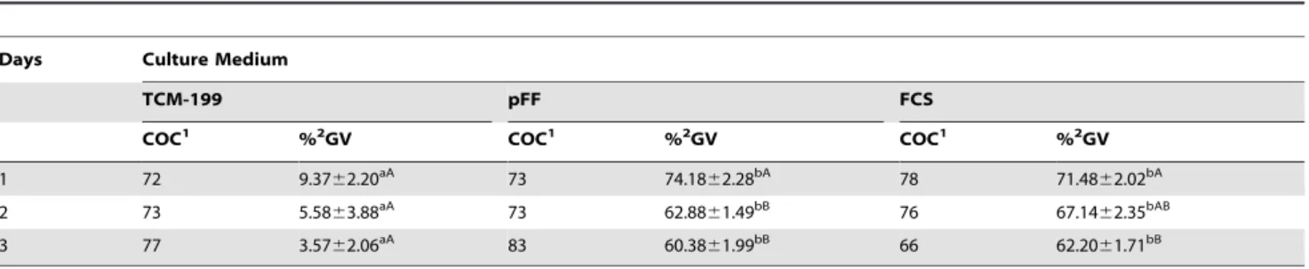
When porcine oocytes were preserved for different times (1 d, 2 d or 3 d) at 27.5uC, 74% oocytes in pFF and 71% in FCS were arrested at the GV stage for 1 d, while only 9% oocytes were arrested at the GV stage in TCM-199. The rate of oocytes at the GV stage dropped to 63% and 67% in pFF and FCS, respectively, after preservation for 2 d. There were no further significant changes in GV rates after preservation for 3 d (Table 2).
Maturation of porcine oocytes after preservation in different media
There was no difference between the maturation rate of the oocytes preserved in pFF and FCS (57% and 52%). However, in both groups the maturation rate was lower than in the control (69%). Only 7% of oocytes preserved in TCM-199 for 3 d matured to the MII stages (Table 3).
Effects of different preservation media on GSH level
When porcine oocytes were preserved in pFF and FCS at 27.5uC for 3 d, the content of intracellular GSH was 1.35 and 1.59 pmol/oocytes, respectively, which was significantly higher than those preserved in TCM-199 with a low level of 0.07 pmol/ oocytes, but still lower than in fresh oocytes probably due to the degeneration of parts of the oocytes preserved (Table 4).
Morphology of porcine oocytes after preservation in different media
As shown in Fig. 1, cumulus cells attached tightly to oocytes and remained unexpanded when preserved at 4uC and 27.5uC for 3 d, but cumulus cells detached from oocytes or expanded if preserved in TCM-199 and FCS at 38.5uC for 3 d. Darkening of cytoplasm was
observed in oocytes preserved in TCM-199 at 4uC, and COCs
degenerated in pFF at 38.5uC. There was no expansion or proliferation of the cumulus cells preserved in pFF and FCS at 27.5uC.
Effects of different preservation media on actin organizations
Two types of actin organizations were observed with laser confocal microscopy. Normal actin organization was prominently
Table 1.Effects of temperatures and media on GVBD inhibition of porcine oocytes after preservation for 3 d*.
Temperature(6C) Culture Medium
TCM-199 pFF FCS
COC1 %2GV COC1 %2GV COC1 %2GV
4 69 060aA 62 060aA 73 1.5961.59aA
20 73 060aA 60 17.26
68.55aB 66 1.52
61.52aA
25 64 3.6262.06aA 72 23.89
63.89bB 70 1.33
61.33aA
27.5 75 3.8062.22aA 86 65.3265.82bD 86 63.9064.70bC
30 105 9.0063.49aA 89 43.98
61.70bC 71 39.41
60.98bB
38.5 62 060aA 62 0
60aA 69 3.90
62.09aA
Data are presented as means6SEM from three replicated experiments.
*Table1 was analyzed with double factor variance analysis, because the effects of different media, time, and the interaction are all significant. So we did not consider the
main effects.
1Means oocytes treated in every group.
2Percentages are based on the total number of oocytes examined in every group. A, B, C, DValues with different superscripts are significantly different in each column (P,0.05). a, bValues with different superscripts are significantly different in each line (P
,0.05). doi:10.1371/journal.pone.0014242.t001
detected at the cell cortex of oocytes, while abnormal expression of actin showed discontinuous or incomplete distribution (Fig. 2A). Approximately 82% of oocytes showed normal actin distribution after preservation in pFF or FCS for 3 d, and only 7% of oocytes displayed intact distribution of actin in TCM-199. There was no significant difference between control oocytes and those preserved in pFF and FCS for 3 d (Fig. 2B).
Effects of different preservation media on distribution of cortical granules (CGs)
When porcine oocytes were preserved in TCM-199, pFF or FCS at 27.5uC for 3 d, CGs were redistributed to different degrees. CG distribution was classified into three categories: a) CGs were mainly distributed around the germinal vesicles with a portion of CGs having migrated into the centre of the cytoplasm (Fig. 3A a,b); b) CGs were distributed uniformly throughout the oocyte cytoplasm including the cortex (Fig. 3A c); c) CGs were distributed at the cortex with few or no sporadic CGs still located in the inner cytoplasm (Fig. 3A d).
Seventy-nine percent of fresh oocytes showed CG distribution in the central cytoplasm, similar to oocytes preserved in TCM-199 (71%); however, only 18% of oocytes preserved in pFF and 16% of oocytes preserved in FCS showed CG distribution at the central cytoplasm. Most of the oocytes (80%, 75%) displayed cortical area distribution patterns in the pFF and FCS groups (Fig. 3B).
Effects of different preservation media on mitochondria distribution
Three categories of mitochondrial distribution patterns were observed: a) oocytes showed strong staining of homogeneously
distributed peripheral mitochondria (Fig. 4A a); b) some mitochondria were translocated into half of the cytoplasimc region in many oocytes, but still some of them were distributed in the peripheral area (Fig. 4A b); c) mitochondria were distributed homogeneously in the central cytoplasm (Fig. 4A c).
Half (50%) of the fresh oocytes showed peripheral distribution of mitochondria, while only 25% of oocytes showed semi-peripheral distribution or diffuse distribution. When oocytes were preserved in TCM-199, pFF or FCS, there was no difference in semi-peripheral mitochondria distribution patterns. However, approximately 50% of oocytes displayed diffuse distribution in the pFF (53%) and FCS (48%) groups, while only 35% of oocytes in the TCM-199 group and 24% of oocytes in the control group showed such a pattern (Fig. 4B).
Effects of different preservation media on spindle configuration in porcine oocytes after in vitro maturation (IVM)
The normal spindle was barrel-shaped, with regular chromo-somal alignment, while abnormal spindle configurations showed disorganized microtubules along with scattered or fragmented chromosomes (Fig. 5A). Almost 60% oocytes displayed normal spindle configurations in the pFF and FCS groups, while only 25% oocytes displayed normal spindle configurations in the TCM-199 group. There was no significant difference between the control group and the pFF group or FCS group (Fig. 5B).
Table 2.The GVBD inhibition after preservation for different times in different media at 27.5uC.
Days Culture Medium
TCM-199 pFF FCS
COC1 %2GV COC1 %2GV COC1 %2GV
1 72 9.3762.20aA 73 74.18
62.28bA 78 71.48
62.02bA
2 73 5.5863.88aA 73 62.8861.49bB 76 67.1462.35bAB
3 77 3.5762.06aA 83 60.38
61.99bB 66 62.20
61.71bB
Data are presented as means6SEM from three replicated experiments.
1Means oocytes treated in every group.
2Percentages are based on the total number of oocytes examined in every group. A, BValues with different superscripts are significantly different in each column (P
,0.05).
a, bValues with different superscripts are significantly different in each line (P
,0.05). doi:10.1371/journal.pone.0014242.t002
Table 3.Maturation of oocytes after preservation at 27.5uC in different media.
Oocytes examined % matured oocytes
Control 94 68.7061.42a
TCM-199 67 7.4161.31b
pFF 73 56.6862.53c
FCS 69 52.2861.46c
Data are presented as means6SEM from three replicated experiments.
a, b, cValues with different superscripts are significantly different in each column
(P,0.05).
doi:10.1371/journal.pone.0014242.t003
Table 4.Effects of different preservation media on GSH level after preservation at 27.5uC for 3 d.
Oocytes examined GSH content (pmol/oocyte)
Control 90 2.9460.06a
TCM-199 88 0.0760.02b
pFF 90 1.3560.23c
FCS 89 1.5960.04c
Data are presented as means6SEM from three replicated experiments.
a, b, cValues with different superscripts are significantly different in each column
(P,0.05).
Effects of different preservation media on early embryo development
After preserving in different media, porcine oocytes were cultured in vitro. Following in vitro fertilization (IVF), there was
no difference between the cleavage rate of the oocytes preserved in pFF and FCS (56% and 60%, respectively) (P.0.05). However, significant differences were found in preserved groups and control group (72%) (P,0.05). Only 5% of oocytes preserved in TCM-199 for 3 d developed to 2-cell stage after IVM-IVF. However, there was no oocyte developed to the blastocyst stage in TCM-199 group. The incidence of embryos developing to the blastocyst stage in pFF (10%) or FCS (12%) group was lower than that of the control group (28%) (P,0.05). Although the mean cell numbers per blastocyst were 35 and 37, respectively in pFF and FCS groups, lower than the control, there was no significant difference between them (P.0.05) (Table 5).
Discussion
It is generally accepted that porcine oocyte preservation is highly significant for agriculture and biomedicine. It also has implications for human assisted reproduction technologies. However, many problems still exist concerning conventional porcine oocyte preservation methods, such as low temperature sensitivity, cryoinjuries, toxicity of preservation solution and low success rates. Therefore, it is important to determine new methods for short-term oocyte preservation and long distance transporta-tion, which not only avoids toxicity and cryoinjuries, but also allows synchronous maturation. Here, we report that 27.5uC is the optimal temperature for preservation of oocytes in pFF or FCS. After preservation for 3 d, oocytes are still viable, remain at the GV stage, and have undergone beneficial cytoplasmic changes.
Our study has addressed three questions. The first question involves in the temperature sensitivity and preservation medium. The second question relates to oocyte quality after preservation.
The third aspect deals with synchronization of nuclear and cytoplasmic maturation.
Temperature and medium are very important for oocyte preservation. It has been shown that offspring could be produced from mouse oocytes stored at room temperature in TYH, CZB-H and KSOM media with 0 to 0.5% Bovine serum albumin (BSA), not from oocytes stored at 5 or 37uC [8]. Compared with other species, porcine oocytes and embryos are more sensitive to low temperature [13]. Previous study demonstrated that the survival rate of porcine oocytes stored at 20uC was higher than at 37uC in TCM-199 with serum after 48 h [9]. Our results showed that 27.5uC was the optimal temperature for storing porcine oocytes in pFF or FCS. Porcine immature oocytes in the TCM-199 group showed the lowest rates of GVBD inhibition and further development to the MII stage after culture, while preservation of oocytes in pFF or FCS for 3 d at 27.5uC achieved significant GVBD inhibition and more than half of the oocytes completed nuclear maturation after culture. Follicular fluid and follicular shell fragments in the culture medium not only promote cytoplasmic maturation, but also contain a factor(s) which could inhibit porcine oocyte maturation [12]. Porcine oocytes were previously preserved by using pFF. After 4 h storage at room temperature, oocytes showed the lowest rates of cleavage and development to the morula/blastocyst stages after fertilization, because follicular fluid was sticky and turbid [10]. In our study pFF was centrifuged, and only the supernatant was used for preservation, so these problems were avoided, and oocytes can survive for 3 d. Our results indicate that FCS is also an appropriate medium for oocyte preservation at 27.5uC.
We next determined the oocyte quality after preservation on subcellular levels. Actin organization, CG migration, GSH content and mitochondria redistribution and spindle appearance were regarded as indicators of oocytes quality. It is well known that dynamic regulation of the actin cytoskeleton is important for maintenance of cellular structures and specific cellular functions
Figure 1. Morphologic changes of porcine cumulus-oocyte complexes (COCs) after preservation for 3 d.a1, a2, b1, b2, c1, c2, When preserved in TCM-199, pFF and FCS at 4uC or 27.5uC, cumulus cells attached tightly to oocytes and displayed no expansion; a3, c3, After preservation in TCM-199 and FCS at 38.5uC, almost all of the cumulus have shed from the oocytes; b3, After pFF preservation at 38.5uC, the whole COCs degenerated and showed black appearance; b2, c2, After preservation in pFF and FCS at 27.5uC, oocytes displayed dark ooplasm with compact cumulus cells. Scale bar = 200mm.
doi:10.1371/journal.pone.0014242.g001
[14,15]. When F-actin polymerization is inhibited, completion of oocyte meiotic maturation and embryo development are prevent-ed [16]. Traditional cryopreservation methods (vitrification) cause
a decrease in the percentage of oocytes with normal distribution of F-actin in both GV and metaphase II (MII) stage oocytes [5]. Our results proved that the percentage of oocytes with normal
Figure 2. Effect of different media on actin configuration in porcine oocytes after preservation.A. a, represents the normal actin pattern in porcine oocytes (white arrow pointing at the white circle denotes the germinal vesicle). b, c, and d represent abnormal actin patterns. In b, actin fragments formed an incomplete ring with large punctiform aggregation. In c, small punctiform actin aggregated as an incomplete ring. In d, only some actin aggregated near oocyte membrane. Green, microfilaments; Red, chromatin. Scale bar = 10mm. B. Ratios of oocytes with normal actin configuration in control group and different preservation groups. Porcine oocytes were preserved in TCM-199, pFF and FCS at 27.5uC for 3 d, then the actin configuration was stained with FITC-phalloidine. Normal and abnormal actin configuration were detected. The graph shows the mean6SEM of three independent experiments. The number ‘‘n’’ over the bars means the total treated oocyte in every group. The superscriptsa, bover the bars represent values of normal actin configuration that differ significantly (P,0.05).
distribution of F-actin was similar in GV stages among fresh, pFF and FCS groups, indicating that pFF and FCS do not disturb actin organization.
For many species, CG migration and mitochondrial distribution are regarded as clear indications of oocyte cytoplasmic maturation [17]. During oocyte growth and maturation, CGs migrate to the
Figure 3. Effect of different media on CG distribution in porcine oocytes after preservation.A. a, b, CGs perinuclear area distribution within the porcine oocytes; a, most of the CGs aggregate in the perinuclear area, with a few migrating to the central inner cytoplasm or cortical area; b is intermediate pattern between a and c, with most of the CGs still remaining in perinuclear area and some migrating to the central cytoplasm; c, CGs distributed uniformly in the inner cytoplasmic area, and a few migrated to the cortical area to form a discontinuous ring; d, CGs cortical area distribution; almost all of the CGs have migrated into the cortical area, and a few still remained in the central cytoplasm. Green, cortical granules. Scale bar = 10mm. (White arrows pointing at the white circle denotes the germinal vesicle). B. Ratios of different CG distribution patterns within the porcine oocytes after preservation in different media. Porcine oocytes were preserved in TCM-199, pFF and FCS at 27.5uC for 3 d, and then CG distribution was stained with FITC-labeled peanut agglutinin. Three categories of CG distribution were detected: perinuclear area; inner cyoplasmic area; cortical area. The graph shows the mean6SEM of three independent experiments. The number ‘‘n’’ in the bracket means the total treated oocyte in every group. The superscriptsa, bover the bars represent values that differ significantly in every categories of CG distribution (P,0.05). doi:10.1371/journal.pone.0014242.g003
cortex and form a continuous layer underneath the oolemma [18,19]. Any dysfunction or dislocation of cortical granules would decrease oocyte competence and has detrimental effects on embryo quality [20]. Our results indicate that most of the CGs migrated to the cortical area when oocytes were preserved in pFF or FCS for 3d. The migration of CGs toward the cortical region in oocytes preserved in the pFF or FCS group is a beneficial change for cytoplasmic maturation.
The activity and organization of mitochondria are necessary features among the diverse events involved in oocyte cytoplasmic maturation [21], and the homogeneous distribution of mitochon-dria in the cytoplasm is positively correlated with ATP content [22]. Dysfunction of mitochondria often appeared in vitrified oocytes. It was reported that the rate of oocytes with abnormal mitochondria distribution was increased after vitrification [6]. Indeed, in the pig, mitochondria were restricted to the oocyte periphery at GV stages, but migrated to the inner region of the cell during maturation both in vitro [23,24] and in vivo [25]. The
relocation of mitochondria occurs in oocytes with high develop-mental competence [26,27]. In contrast, little or no relocation was detected in low competence oocytes in which mitochondria persisted in a peripheral distribution pattern [28]. All these features suggest a linkage between low developmental ability and perturbed mitochondrial distribution. In our study, about half of the fresh oocytes showed peripheral distribution of mitochondria, while only one quarter showed semi-peripheral distribution or central cytoplasm distribution. On the contrary, approximately half of the oocytes displayed central cytoplasm distribution patterns in oocytes preserved in pFF or FCS for 3 d, indicating
progressive cytoplasmic maturation during the preservation period.
Cytoplasmic GSH content was also examined. GSH has been shown to be one of the markers for ooplasm quality. It regulates sperm nuclear decondensation and male pronuclear formation [29] and it protects cells against oxidative damage [30]. Low intracellular GSH may be responsible, in part, for the low developmental competence of porcine oocytes [31,32]. Our study shows that GSH content is decreased after preservation in pFF or FCS when compared to fresh oocytes, and that the TCM-199 group shows lowest GSH levels. The decrease in GSH content in preserved oocytes may be due to the degeneration of some preserved oocytes, but more than half of the oocytes maintained viability and developed into MII stages after 3 d preservation.
Oocytes need to undergo cytoplasmic maturation as well as nuclear maturation to become able to support successful development [33]. Synchronized cytoplasmic maturation and nuclear maturation are considered important for normal embryo development after fertilization [34]. A premature meiotic resumption without adequate cytoplasmic maturation was induced when oocytes were transferred into culture medium from follicles, which was one of the main reasons for poor quality. The peripheral migration of CGs and interior translocation of mitochondria during the preservation period may contribute to synchronization of cytoplasmic and nuclear maturation. Previous study found that the percentage of oocytes with normal spindle organization was decreased when vitrified GV oocytes were cultured to assess maturation [5]. Our results show that the percentage of normal spindles was not affected by preserving
Figure 4. Effects of different media on mitochondria distribution in porcine oocytes after preservation.A. a, peripheral mitochondrial distribution, with no distribution at the center of the oocytes; b, semi-peripheral mitochondrial distribution; c, mitochondrial relocation and uniform distribution in the inner region of oocyte cytoplasm. Red, Mito Tracker Red staining. Scale bar = 10mm. B. Ratios of different mitochondria distribution patterns within the porcine oocytes after preservation with different media. Porcine oocytes were preserved in TCM-199, pFF and FCS at 27.5uC for 3 d, and then mitochondria distribution was stained with MitoTracker Red CMXRos. Three categories of mitochondria distribution were detected: peripheral; semi-peripheral; diffuse. The graph shows the mean6SEM of three independent experiments. The number ‘‘n’’ in the bracket means the total treated oocyte in every group. The superscriptsa, b, cover the bars represent values that differ significantly in every categories of
Figure 5. Effects of different preservation media on spindle configuration in porcine oocytes after IVM.A. a, represents a normal spindle in porcine oocyte. b and c represent abnormal spindle. In b1, c1 arrows indicate abnormal spindle organization distribution. In b2, arrows indicate fragmented and displaced chromosomes. Green, tubulin; Red, chromatin. Scale bar = 10mm. B. Normal ratios of spindle configuration in porcine oocytes after preservation in different media following IVM. Porcine oocytes were preserved in TCM-199, pFF and FCS at 27.5uC for 3 d, then spindle was stained with FITC-a-tubulin after culture for 44 hin vitro. Normal and abnormal spindle configuration were detected. The graph shows the mean6SEM of three independent experiments. The number ‘‘n’’ over the bars means the total treated oocyte in every group. The superscriptsa, bover the bars represent
values of normal tubulin configuration that differ significantly between groups (P,0.05). doi:10.1371/journal.pone.0014242.g005
oocytes in pFF or FCS compared with the control group. Further studies have already clarified that oocytes preserved for 3 d in pFF or FCS have abilities to develop to term after IVM and IVF.
Taken together, our studies show that porcine oocytes could be
preserved in pFF or FCS at 27.5uC for 3 d without evident
cytoplasmic damage. More than half of the preserved oocytes retain the potential to mature in vitro, with normal spindle
formation, and about 10% oocytes developed to blastocysts with normal cell numbers after in vitro fertilization. Thus, we have developed a simple method for short-term (up to 3 d) preservation of porcine oocytesin vitro. Further research is necessary to clarify
whether these preserved oocytes can produce piglets after IVF.
Materials and Methods
Ethics statement
Porcine handling was conducted in accordance with policies promulgated by the Ethics Committee of the Institute of Zoology, Chinese Academy of Sciences. Porcine ovaries used in this study were obtained from AnDing slaughterhouse, a local slaughter-house of Beijing, P.R.China.
Chemicals and antibodies
All chemicals used in this study were purchased from Sigma Chemical Company (St. Louis, MO), unless otherwise noted. The GSH and GSSG Assay Kit was purchased from Beyotime Institute of Biotechnology (Beyotime, CN). Fetal calf serum was purchased from Gibco (Gibco, Grand Island, NY).
Preparation of pFF and FCS
pFF was prepared as follows: 1) The contents of follicles measuring 3–5 mm in diameter were aspirated with an 18-gaugle needle fixed to a 20-ml disposable syringe and pooled into a 50 ml conical tube (Falcon, Franklin Lakes, NJ). After sedimentation for about 15 min, the pellet was discarded; 2) The supernatant was centrifuged at 15,000 rpm for 15 min, then the supernatant was collected and frozen at 220uC. After thawing, the fluid was centrifuged followed by two cycles of freezing and thawing, each time discarding the pellet; 3) The supernatant was collected and filtrated for sterilization, and then stored at220uC until use.
FCS was purchased from Gibco (Grand Island, NY), and was subpackaged in 2-ml tube, then stored at220uC until use.
Collection and preservation of porcine oocytes
Porcine ovaries were obtained from prepubertal gilts at a local slaughterhouse and transported to the laboratory within 1 hour. The ovaries were maintained in 0.9% NaCl solution containing penicillin G (75 mg/mL) and streptomycin sulphate (50 mg/mL)
at 34uC,36uC for the entire time of the trip. Cumulus-oocyte
complexes (COCs) were aspirated from antral follicles (3–6 mm in diameter) of ovaries with an 18-gauge needle fixed to a 20 ml disposable syringe.
After three rinses in washing medium (TCM-199 medium
supplemented with 2.2% NaHCO3), COCs with uniform
cytoplasm and compact cumulus mass were selected for preser-vation. The COCs were washed three times in 50ml droplets of different fluids, then groups of 25 to 30 COCs were preserved in a 200ml drop of TCM-199, pFF and FCS at different preservative temperatures (4uC,20uC,25uC,27.5uC,30uC,38.5uC) in air condi-tion, covered with liquid paraffin oil in a biochemical incubator. The COCs were preserved for 1 d, 2 d or 3 d. Evaluation of morphological survival (homogenous cytoplasm, intact and compact cumulus mass) was carried out after preservation.
In vitro maturation (IVM)
The IVM of COCs was performed in accordance with the methods of our previous study [17,35,36,37]. The basic medium was TCM-199, supplemented with 75mg/ml potassium penicillin G, 50mg/ml streptomycin sulphate, 0.57 mM cysteine, 0.5mg/ml FSH, 0.5mg/ml LH, and 10 ng/ml EGF. Approximately 40 to 50 COCs were cultured in a 200ml drop of maturation medium in
4-well dishes, which was covered by liquid paraffin oil for up to 44 h
at 39uC in an atmosphere of 5% CO2 in air and saturated
humidity.
Evaluation of GSH
Intracellular content of GSH was measured as described by instruction supplied with the GSH and GSSG Assay Kit. Briefly, after preservation, cumulus cells were removed completely from oocytes by vortex and washed three times in phosphate buffered solution (PBS). Thirty oocytes composed one sample and three samples were assayed for each treatment. Oocytes were washed
two times with distilled water, then 5ml of 0.2 M sodium
phosphate containing 10 mM Na4-EDTA and 5ml of 1.25 M
phosphoric acid (Fluka, Buchs, Switzerland) were added to the 1.5 mL centrifuge tube containing 30 oocytes. Samples were vortexed thoroughly, and then frozen at270uC and thawed at 37uC in a water bath. This procedure was repeated at least three times for dissolving oocytes. Then the samples were placed in an ice bath for 5 minutes and centrifuged at 10,000 rpm for 10 minutes at 4uC. The tubes containing samples were stored at
270uC until assayed. All samples and solution preparations were carried out according to the directions of Total Glutathione Assay Kit (BiYunTian, The Institute of Biotech, S0052, Shanghai, China). Concentrations of GSH in the oocytes were determined by the 5, 59 dithio-bis (2-nitrobenzoic acid)-glutathione disulfide
Table 5.Effect of preserving medium on early development of IVM-IVF porcine oocytes after preservation at 27.5uC for 3 d.
Treatment Oocytes examined %1of oocytes cleaved (n) %1of blastocysts formation(n) No. cells/blastocyte
Control 227 71.9562.04a(164) 27.97
61.38a(64) 40.67 61.76a
TCM-199 28 5.2962.90b(3) 0
60b(0) 0
60b
pFF 183 56.4963.21c(103) 10.20
60.89c(19) 35.33 61.45a
FCS 209 59.6864.41c(125) 11.79
61.28c(25) 36.67 61.20a
Data are presented as means6SEM from three replicated experiments.
The values ‘‘n’’ in bracket represent the number of the oocytes cleaved at 48 hr and forming blastocysts at 7 d after IVF.
1Percentages are based on the total number of oocytes examined.
a, b, cValues with different superscripts are significantly different in each column (P
(DTNB-GSSG) reductase-recycling assay. Briefly, GSH content was measured according to methods described by Funahashi et al. [38] with minor modifications. First, 150ml of detection solution
was added to 96-well plates, and then 10ml of samples was
pipetted into each well. After the dishes were equilibrated at 25uC for 5 minutes, a total of 50ml 0.16 mg/mL NADPH was added to each well. The formation of 5-thio-2-nitrobenzonic acid, which has an absorption peak at 412 nm, was monitored continuously with a spectrophotometer for 25 minutes with reading recorded every 5 minutes. Standards (0.5, 1, 2, and 10mM) of glutathione
and a sample blank lacking glutathione were also assayed at the same time. The amount of glutathione in each sample was divided by the number of oocytes [39].
Assessment of nuclear status of porcine oocytes by orcein staining
After porcine COC preservation, oocytes were denuded of cumulus cells by vortexing, and then denuded oocytes were mounted on glass slides. We used vaseline and lanolin to keep the coverslip in contact with the oocytes without extensive pressure. For fixation, the slides were immersed in 25% (v/v) acetic acid in ethanol for at least 48 hours at room temperature (25uC). Oocytes were stained with 1% (w/v) orcein in 45% (v/v) acetic acid and examined under a light microscopy at a magnification of6100. Nuclear stages were clearly observed, oocytes containing filamen-tous or condensed chromatin enclosed in a nuclear envelope were classified as germinal vesicle (GV) stage. Oocyte maturation status after culture was also evaluated using the same protocol.
Evaluation of actin by confocal laser scanning microscopy
For microfilament detection, the zona pellucida were first removed from oocytes in acid Tyrode’s solution (pH 2.5), and then oocytes were fixed with 4% paraformaldehyde in PBS for at least 30 minutes at room temperature. They were permeabilized in PBS
containing 1% Triton X-100 overnight at 37uC, followed by
blocking in PBS containing 1% BSA (blocking solution) for 1 h at room temperature. After washing in PBS containing 0.1% Tween 20 and 0.01% Triton X-100 (washing solution) for 15 minutes, the oocytes were stained with FITC-phalloidin diluted 1:100 with washing solution for 2 hours at room temperature in a dark box. Then after washes in washing solution, nuclear status was determined after staining with PI (propidium iodide, 10mg/ml in PBS) for 10 minutes. Oocytes were washed extensively, mounted on glass slides, and observed under a laser-scanning confocal microscope (Zeiss LSM 510 META, Germany). At least 25 oocytes were examined for each time point, and each treatment was repeated at least three times.
The scorning system of actin was based on the method described in a previous study[40]. Two types of actin organizations were observed with laser confocal microscopy. Normal actin organization was prominently detected at the cell cortex of oocytes, while abnormal expression of actin showed discontinuous or incomplete distribution.
Evaluation of cortical granules by confocal laser scanning microscopy
The method for staining CGs was based on the procedures reported in our previous study [41]. After removing the zona pellucida in acidic Tyrode’s solution (pH 2.5), oocytes were fixed with 4% (w/v) paraformaldehyde in PBS for at least 30 minutes at room temperature, followed by washing in blocking solution (PBS containing 0.3% BSA and 100 mM glycine) three times for 5 min
each. After 5 minutes of permeabilization in PBS containing 0.1% Triton X-100, oocytes were washed two additional times in blocking solution (5 minutes each). They were then labeled with 100mg/ml FITC-labeled peanut agglutinin (Sigma) in PBS for 30 minutes in a dark box. Finally, the oocytes were washed three times in PBS containing 0.3% BSA and 0.01% Triton X-100. Nuclear status of the oocytes was determined after staining with 10mg/ml PI in PBS for 10 minutes, followed by washing three
times in PBS, and then mounted on glass slides. Oocytes were observed under a laser-scanning confocal microscope (Zeiss LSM 510 META, Germany). A total of 25 oocytes from each sample were scanned and recorded with confocal fluorescent microscopy. Each treatment was repeated at least three times.
The scorning system of cortical granule was based on a previous report [42]. Cortical granule migration was classified with the following categories: CGs were mainly distributed around the germinal vesicles with a small number of CGs having migrated into the centre of the cytoplasm; CGs were migrated throughout the oocyte cytoplasm uniformly including the cortex; CGs having migrated into the cortex with few or any sporadic CGs still distributed in the inner cytoplasm.
Evaluation of mitochondrial distribution by confocal laser scanning microscopy
Oocytes were stained for mitochondria according to the methods of our previous study [17]. A stock solution of MitoTracker Red CMXRos (Molecular Probes, Eugene, OR) fluorescence probe at a concentration of 1 mM was prepared in dimethyl sulfoxide and stored at220uC. Denuded oocytes at the GV stages were incubated in TCM-199 supplemented with
0.5mmol/L MitoTracker Red CMXRos in a dark environment
and humidified air with 5% CO2 for 30 minutes at 38.5uC,
followed by three washes with TCM-199 for 20 minutes, and then observed with a laser-scanning confocal microscope (Zeiss LSM 710 META, Germany).
The scorning system of mitochondrial was based on a previous report [28]. Three categories of mitochondrial distribution patterns were observed: a) oocytes showed strong staining of homogeneously distributed peripheral mitochondria; b) some mitochondria were translocated into half of the cytoplasimc region in many oocytes, but still some of them were distributed in the peripheral area; c) mitochondria were distributed homogeneously in the central cytoplasm.
Evaluation of spindle by confocal laser scanning microscopy
After maturation, only oocytes with a visible polar body were chosen for spindle detection. Oocytes were stained for spindle detection according to the methods of our previous studies [36,43]. Cumulus-free oocytes were fixed with 4% paraformaldehyde in PBS for at least 30 minutes at room temperature. They were permeabilized in PBS containing 1% Triton X-100 overnight at 37uC, followed by blocking in PBS containing 1% BSA (blocking solution) for 1 hour at room temperature. After washing in PBS containing 0.1% Tween 20 and 0.01% Triton X-100 (washing solution) for 15 minutes, oocyte microtubules were labeled by incubation in FITC-conjugated monoclonal anti-a-tubulin
anti-body (Sigma) diluted 1:100 with washing solution for 1 hour at room temperature in a dark box. After three washes in washing solution, nuclear status was determined after staining with PI (propidium iodide, 10mg/ml in PBS) for 10 minutes oocytes were washed extensively, and mounted on glass slides. Oocytes were observed under a laser-scanning confocal microscope (Zeiss LSM
510 META, Germany). Each treatment was repeated at least three times.
In vitro fertilization
Freshly ejaculated sperm-rich fraction was collected from fertile
boars, following short incubation at 39uC, the semen was
resuspended and washed 3 times in DPBS supplemented with 0.1% (w/v) BSA by centrifugation at 1500rpm for 4 min. The spermatozoa concentration was measured by a hemocytometer and the proportion of motile sperm was determined. The spermatozoa were diluted with modified Tris-buffered medium (mTBM) to optimal concentration. Cumulus-free matured oocytes were washed three times in mTBM and placed in prepared mTBM drops, covered with a mineral oil and incubated for 2 h at
38.5uC under 5% CO2 in air. Approximate 30 oocytes were
inseminated in 50ml drops at a final sperm concentration of
36105cells/ml for 5 hr co-incubation.
At 5 h after insemination, the oocytes were incubated in 200ml
of PZM-3 in a 4-well culture plate for 7 d at 38.5uC in an atmosphere of 5% CO2in air. Cleavage and blastocyst formation
of the oocytes were examined at 2 and 7 d after the start of culture, respectively. The blastocysts were stained with 5mg/ml
bisbenzi-mide (Hoechst 33342) to determine the number of nuclei by using a fluorescent microscope.
Statistical analysis
All data were obtained from at least three replicate trials for each experiment. All percentages from three repeated experiments
were expressed as mean 6 SEM. The percentage data were
subjected to arc-sine transformation and the transformed data were tested to verify ANOVA assumptions before being analyzed with ANOVA. All statistics were performed with analysis of variance and Fisher’s LSD analysis by SPSS version 10.0 (Installshield Software Corporation) except for table1. Table1 was analyzed with double factor variance analysis, because the effects of different media, temperatures and the interactions are all significant, so main effect was ignored. Different values were considered significant when the P value was less than 0.05.
Acknowledgments
We are grateful to Shi-Wen Li, Hua Qin for technical assistance, Drs. Zhen-Bo Wang, Mo Li, Sen Li, Zhao-Jia Ge, Xiang-Hong Ou, Shu-Tao Qi, Ya-Peng wang, Lei Chen, Jun-Yu Ma for insightful comments and suggestions on the experiment handling and manuscript preparation.
Author Contributions
Conceived and designed the experiments: CRY DQM QYS. Performed the experiments: CRY DQM QHZ LG YW. Analyzed the data: CRY DQM YW ZL QYS. Contributed reagents/materials/analysis tools: CRY JST YW XH YH. Wrote the paper: CRY HS QYS.
References
1. Zhou GB, Li N (2009) Cryopreservation of porcine oocytes: recent advances. Mol Hum Reprod 15: 279–285.
2. Didion BA, Pomp D, Martin MJ, Homanics GE, Markert CL (1990) Observations on the cooling and cryopreservation of pig oocytes at the germinal vesicle stage. J Anim Sci 68: 2803–2810.
3. Nagashima H, Kashiwazaki N, Ashman RJ, Grupen CG, Seamark RF, et al. (1994) Removal of cytoplasmic lipid enhances the tolerance of porcine embryos to chilling. Biol Reprod 51: 618–622.
4. Liu RH, Sun QY, Li YH, Jiao LH, Wang WH (2003) Maturation of porcine oocytes after cooling at the germinal vesicle stage. Zygote 11: 299–305. 5. Wu C, Rui R, Dai J, Zhang C, Ju S, et al. (2006) Effects of cryopreservation on
the developmental competence, ultrastructure and cytoskeletal structure of porcine oocytes. Mol Reprod Dev 73: 1454–1462.
6. Shi LY, Jin HF, Kim JG, Mohana Kumar B, Balasubramanian S, et al. (2007) Ultra-structural changes and developmental potential of porcine oocytes following vitrification. Anim Reprod Sci 100: 128–140.
7. Vajta G, Nagy ZP (2006) Are programmable freezers still needed in the embryo laboratory? Review on vitrification. Reprod Biomed Online 12: 779–796. 8. Wakayama S, Thuan NV, Kishigami S, Ohta H, Mizutani E, et al. (2004)
Production of offspring from one-day-old oocytes stored at room temperature. J Reprod Dev 50: 627–637.
9. Jiang Q (1992) The preservation of porcine oocytes at room temperature and their maturation in culture. Journal of Northeast Agricultural College 23: 3. 10. Kim HJ, Choi SH, Son DS, Cho SR, Choe CY, et al. (2006) Effect of exposure
duration of ovaries and oocytes at ambient temperature on parthenogenetic development of porcine follicular oocytes. J Reprod Dev 52: 633–638. 11. Han D, Zhao BT, Liu Y, Li JJ, Wu YG, et al. (2008) Interactive effects of low
temperature and roscovitine (ROS) on meiotic resumption and developmental potential of goat oocytes. Mol Reprod Dev 75: 838–846.
12. Tsafriri A, Channing CP (1975) An inhibitory influence of granulosa cells and follicular fluid upon porcine oocyte meiosis in vitro. Endocrinology 96: 922– 927.
13. Ledda S, Bogliolo L, Succu S, Ariu F, Bebbere D, et al. (2007) Oocyte cryopreservation: oocyte assessment and strategies for improving survival. Reprod Fertil Dev 19: 13–23.
14. Mitchison TJ, Cramer LP (1996) Actin-based cell motility and cell locomotion. Cell 84: 371–379.
15. Welch MD, Mallavarapu A, Rosenblatt J, Mitchison TJ (1997) Actin dynamics in vivo. Curr Opin Cell Biol 9: 54–61.
16. Wang WH, Abeydeera LR, Prather RS, Day BN (2000) Polymerization of nonfilamentous actin into microfilaments is an important process for porcine oocyte maturation and early embryo development. Biol Reprod 62: 1177– 1183.
17. Sha W, Xu BZ, Li M, Liu D, Feng HL, et al. (1111) Effect of gonadotropins on oocyte maturation in vitro: an animal model. Fertil Steril 93: 1650–1661. 18. Berg LK, Wessel GM (1997) Cortical granules of the sea urchin translocate early
in oocyte maturation. Development 124: 1845–1850.
19. Wessel GM, Conner SD, Berg L (2002) Cortical granule translocation is microfilament mediated and linked to meiotic maturation in the sea urchin oocyte. Development 129: 4315–4325.
20. Wang Q, Sun QY (2007) Evaluation of oocyte quality: morphological, cellular and molecular predictors. Reprod Fertil Dev 19: 1–12.
21. Van Blerkom J, Runner MN (1984) Mitochondrial reorganization during resumption of arrested meiosis in the mouse oocyte. Am J Anat 171: 335–355. 22. de Loos F, van Vliet C, van Maurik P, Kruip TA (1989) Morphology of
immature bovine oocytes. Gamete Res 24: 197–204.
23. Sun QY, Wu GM, Lai L, Park KW, Cabot R, et al. (2001) Translocation of active mitochondria during pig oocyte maturation, fertilization and early embryo development in vitro. Reproduction 122: 155–163.
24. Sun QY, Lai L, Wu GM, Park KW, Day BN, et al. (2001) Microtubule assembly after treatment of pig oocytes with taxol: correlation with chromosomes, gamma-tubulin, and MAP kinase. Mol Reprod Dev 60: 481–490.
25. Torner H, Brussow KP, Alm H, Ratky J, Pohland R, et al. (2004) Mitochondrial aggregation patterns and activity in porcine oocytes and apoptosis in surrounding cumulus cells depends on the stage of pre-ovulatory maturation. Theriogenology 61: 1675–1689.
26. Brevini TA, Vassena R, Paffoni A, Francisci C, Fascio U, et al. (2004) Exposure of pig oocytes to PCBs during in vitro maturation: effects on developmental competence, cytoplasmic remodelling and communications with cumulus cells. Eur J Histochem 48: 347–356.
27. Brevini TA, Vassena R, Francisci C, Gandolfi F (2005) Role of adenosine triphosphate, active mitochondria, and microtubules in the acquisition of developmental competence of parthenogenetically activated pig oocytes. Biol Reprod 72: 1218–1223.
28. Brevini TA, Cillo F, Antonini S, Gandolfi F (2007) Cytoplasmic remodelling and the acquisition of developmental competence in pig oocytes. Anim Reprod Sci 98: 23–38.
29. Yoshida M, Ishigaki K, Nagai T, Chikyu M, Pursel VG (1993) Glutathione concentration during maturation and after fertilization in pig oocytes: relevance to the ability of oocytes to form male pronucleus. Biol Reprod 49: 89–94. 30. Gasparrini B, Boccia L, Marchandise J, Di Palo R, George F, et al. (2006)
Enrichment of in vitro maturation medium for buffalo (Bubalus bubalis) oocytes with thiol compounds: effects of cystine on glutathione synthesis and embryo development. Theriogenology 65: 275–287.
31. Herrick JR, Brad AM, Krisher RL, Pope WF (2003) Intracellular adenosine triphosphate and glutathione concentrations in oocytes from first estrous, multi-estrous, and testosterone-treated gilts. Anim Reprod Sci 78: 123–131. 32. Brad AM, Bormann CL, Swain JE, Durkin RE, Johnson AE, et al. (2003)
Glutathione and adenosine triphosphate content of in vivo and in vitro matured porcine oocytes. Mol Reprod Dev 64: 492–498.
33. Gosden R, Krapez J, Briggs D (1997) Growth and development of the mammalian oocyte. Bioessays 19: 875–882.
35. Sun XS, Liu Y, Yue KZ, Ma SF, Tan JH (2004) Changes in germinal vesicle (GV) chromatin configurations during growth and maturation of porcine oocytes. Mol Reprod Dev 69: 228–234.
36. Xu BZ, Li M, Xiong B, Lin SL, Zhu JQ, et al. (2009) Involvement of calcium/ calmodulin-dependent protein kinase kinase in meiotic maturation of pig oocytes. Anim Reprod Sci 111: 17–30.
37. Miao YL, Sun QY, Zhang X, Zhao JG, Zhao MT, et al. (2009) Centrosome abnormalities during porcine oocyte aging. Environ Mol Mutagen 50: 666–671. 38. Funahashi H, Cantley TC, Stumpf TT, Terlouw SL, Day BN (1994) Use of low-salt culture medium for in vitro maturation of porcine oocytes is associated with elevated oocyte glutathione levels and enhanced male pronuclear formation after in vitro fertilization. Biol Reprod 51: 633–639.
39. Hao ZD, Liu S, Wu Y, Wan PC, Cui MS, et al. (2009) Abnormal changes in mitochondria, lipid droplets, ATP and glutathione content, and Ca(2+) release
after electro-activation contribute to poor developmental competence of porcine oocyte during in vitro ageing. Reprod Fertil Dev 21: 323–332.
40. Wang WH, Abeydeera LR, Han YM, Prather RS, Day BN (1999) Morphologic evaluation and actin filament distribution in porcine embryos produced in vitro and in vivo. Biol Reprod 60: 1020–1028.
41. Sun QY, Lai L, Bonk A, Prather RS, Schatten H (2001) Cytoplasmic changes in relation to nuclear maturation and early embryo developmental potential of porcine oocytes: effects of gonadotropins, cumulus cells, follicular size, and protein synthesis inhibition. Mol Reprod Dev 59: 192–198.
42. Liu XY, Mal SF, Miao DQ, Liu DJ, Bao S, et al. (2005) Cortical granules behave differently in mouse oocytes matured under different conditions. Hum Reprod 20: 3402–3413.
43. Li M, Li S, Yuan J, Wang ZB, Sun SC, et al. (2009) Bub3 is a spindle assembly checkpoint protein regulating chromosome segregation during mouse oocyte meiosis. PLoS One 4: e7701.