Antigen-Specific IP-10 Release Is a Sensitive
Biomarker of
Mycobacterium bovis
Infection
in Cattle
Sven D. C. Parsons1*, Kevina McGill2, Mairead B. Doyle2, Wynand J. Goosen1, Paul D. van Helden1, Eamonn Gormley2
1DST/NRF Centre of Excellence for Biomedical Tuberculosis Research/SAMRC Centre for Tuberculosis Research/Division of Molecular Biology and Human Genetics, Faculty of Medicine and Health Sciences, Stellenbosch University, Cape Town, South Africa,2School of Veterinary Medicine, University College Dublin (UCD), Dublin, Ireland
Abstract
The most widely used ante-mortem diagnostic tests for tuberculosis in cattle are the tuber-culin skin test and the interferon-gamma (IFN-γ) release assay, both of which measure cell-mediated immune responses toMycobacterium bovisinfection. However, limitations in the performance of these tests results in a failure to identify all infected animals. In attempting to increase the range of diagnostic tests for tuberculosis, measurement of the cytokine IP-10 in antigen-stimulated blood has previously been shown to improve the detection ofM.
tuberculosisandM.bovisinfection, in humans and African buffaloes (Syncerus caffer), respectively. In the present study, 60 cattle were identified by the single intradermal com-parative tuberculin test as tuberculosis reactors (n = 24) or non-reactors (n = 36) and the release of IFN-γand IP-10 in antigen-stimulated whole blood from these animals was mea-sured using bovine specific ELISAs. There was a strong correlation between IP-10 and IFN-γproduction in these samples. Moreover, measurement of the differential release of IP-10 in response to stimulation withM.bovispurified protein derivative (PPD) andM.aviumPPD distinguished between reactor and non-reactor cattle with a sensitivity of 100% (95% CI, 86%–100%) and a specificity of 97% (95% CI, 85%–100%). These results suggest that IP-10 might prove valuable as a diagnostic biomarker ofM.bovisinfection in cattle.
Introduction
Mycobacterium bovisis the principle causative agent of bovine tuberculosis (bTB), a chronic granulomatous disease that can result in reduced productivity and death in cattle. Moreover, becauseM.bovis-infected animals are a potential source of infection for humans the disease is subject to comprehensive control measures in order to limit both zoonotic transmission and economic losses. Such control is typically based on test-and-slaughter schemes, which require the accurate diagnosis of infected animals.
a11111
OPEN ACCESS
Citation:Parsons SDC, McGill K, Doyle MB, Goosen WJ, van Helden PD, Gormley E (2016) Antigen-Specific IP-10 Release Is a Sensitive Biomarker of
Mycobacterium bovisInfection in Cattle. PLoS ONE 11(5): e0155440. doi:10.1371/journal.pone.0155440
Editor:Thomas Jens Scriba, University of Cape Town, SOUTH AFRICA
Received:November 21, 2015
Accepted:March 29, 2016
Published:May 11, 2016
Copyright:© 2016 Parsons et al. This is an open access article distributed under the terms of the
Creative Commons Attribution License, which permits unrestricted use, distribution, and reproduction in any medium, provided the original author and source are credited.
Data Availability Statement:All relevant data are within the paper and its Supporting Information files.
The tuberculin skin test is the most widely used test to detectM.bovisinfection in cattle; however, the more sensitive interferon-gamma (IFN-γ) release assay (IGRA) is used as an ancillary ante-mortem test [1,2]. The latter assay detects the release of IFN-γfollowing anti-genic stimulation of whole blood withM.bovispurified protein derivative (PPDb) [1,2]. The PPDb comprises a complex mix of antigens that are not wholly specific toM.bovisand in order to take account of cross-reactive sensitization to other mycobacteria, the specificity of the assay is enhanced by comparing the PPDb-specific IFN-γresponse with that in response toM.
aviumPPD (PPDa). Alternatively, antigens that confer superior test specificity forM.bovis
andM.tuberculosis, i.e. early secretory antigenic target 6 kDa (ESAT-6) and culture filtrate protein 10 kDa (CFP-10), have been incorporated into IGRAs [3]. However, the sensitivity of these highly specific tests is lower than those using PPDb and PPDa [2,4,5].
One approach to improving the performance of such diagnostic assays is to identify addi-tional biomarkers of immune activation. In humans, a number of alternatives to IFN-γhave been evaluated for the diagnosis ofM.tuberculosisinfection [6,7]. Of these, interferon gamma-induced protein 10 (IP-10) has proven particularly noteworthy [7,8]. The production of IP-10 is strongly induced by IFN-γin human antigen presenting cells [9] and human and murine neutrophils [10,11]. Moreover, in cattle, transcription of the gene that encodes IP-10, i.e.
CXCL10, is induced by IFN-γin endothelial cells [12] and is highly correlated with the tran-scription of IFN-γmRNA in antigen-stimulated peripheral blood mononuclear cells [13]. In bovids, ESAT-6/CFP-10 stimulation of whole blood fromM.bovis-infected African buffaloes (Syncerus caffer) induced greater quantities of IP-10 than IFN-γ[14] and measurement of IP-10 significantly increased the sensitivity of diagnosis of this infection [15]. In contrast, in a study evaluating the diagnostic utility of this cytokine inM.bovis-infected cattle, IP-10 was not found to be a useful biomarker [13]. However, it was suggested that this might have been a con-sequence of limitations in the sensitivity of the enzyme-linked immunosorbent assay (ELISA) used to measure this molecule [13]. Notably, this study employed a human IP-10 ELISA [13] whereas the studies conducted in buffaloes used a commercially available bovine IP-10 assay [14,15]. Given that the measurement of antigen-induced IP-10 might increase the diagnostic sensitivity of tests ofM.bovisinfection, the aim of the present study was to resolve these con-flicting results by re-evaluating IP-10 as a biomarker forM.bovisinfection in cattle using a bovine-specific ELISA.
Materials and Methods
Animals
Cattle from herds naturally infected withM.bovisand cattle with no history ofM.bovis expo-sure were tested using the single intradermal comparative tuberculin test (SICTT) as part of the national bTB eradication programme. Animals were classified as either reactors (SICTT-positive) or non-reactors (SICTT-negative) and assigned to 3 experimental groups. Group 1 consisted of twelve reactors, which were relocated and maintained at an approved research farm facility. Water and food of the highest quality were provided at all times and animals were cared for and monitored daily, with veterinary oversight according to approved standard oper-ating procedures. There was no deviation from normal housing practices and animals were never kept in isolation. When required, cattle were humanely slaughtered at an approved slaughterhouse. Following slaughter, these animals presented with gross macroscopic lesions consistent with advanced bTB and were confirmed as bTB-positive. Hereafter, an additional 12 reactor and 16 non-reactor cattle were randomly selected from anM.bovis-infected herd in the national herd (Group 2) and were included in the study together with 20 non-reactor cattle with no knownM.bovisexposure (Group 3). Heparinised blood samples for cytokine analysis
were collected from all animals by a Senior Veterinary Inspector of the Irish Department of Agriculture, Food & the Marine under a research license issued by the Department of Health & Children. The study was approved by the UCD Animal Research Ethics Committee (AREC-P-11-49-Gormley) and all diagnostic testing was conducted in accordance with regulations of the EU trade Directive 64/432/EEC and the Irish bTB eradication programme.
Interferon gamma release assay
For each animal, 1.5 ml aliquots of heparinized blood in 24-well Costar tissue culture plates (Corning Inc., Corning, NY, USA) were incubated for 24 h with, respectively, a 100μl solution of PPDb (final conc 20μg/ml blood) (Thermo Fisher Scientific Prionics AG, Schlieren, Switzer-land), a 100μl solution of PPDa (10μg/ml blood) (Thermo Fisher Scientific Prionics AG), and 100μl phosphate buffered saline (PBS) as a non-stimulating control. For all bTB-positive ani-mals, duplicate samples were incubated as described above for 48 h and additionally, 250μl of blood was cultured in 96-well plates (Corning Inc.) with 25μl of PC-EC peptide cocktail at a final concentration of 5μg/ml blood (Thermo Fisher Scientific Prionics AG) for both 24 and 48 h. All blood cultures were incubated at 37°C in a humidified atmosphere with 5% CO2
before harvesting of plasma supernatants following centrifugation. The production of IFN-γin each sample was quantified in duplicate by sandwich ELISA [16] using a Bovigam1
ELISA kit (Thermo Fisher Scientific Prionics AG) and the relative IFN-γconcentrations were recorded as the optical density (OD) measured at 450 nm. Bovigam test results were calculated as the OD derived from the PPDb-stimulated sample minus that derived from the PPDa-stimulated sam-ple. A Bovigam result which was greater or equal to 0.1 was regarded as positive as per the manufacturer’s recommendations.
IP-10 ELISA
The concentration of IP-10 in plasma samples generated as described above was measured as follows. Anti-bovine CXCL10 antibody (Kingfisher Biotech Inc., St Paul, MN, USA) in PBS (1μg/ml; 100μl/well) was incubated overnight at 4°C in 96-well Nunc Maxisorb microtitre plates (Thermo Fisher Scientific, Waltham, MA, USA). Between subsequent steps, wells were washed with Bovigam1Wash Buffer (Thermo Fisher Scientific Prionics AG) and all further reactions were done at room temperature. Wells were blocked for 1 h with 200μl Assay Buffer consisting of 0.1% bovine serum albumin (Roche, Basel, Switzerland) and 0.05% Tween (Sigma-Aldrich, St. Louis, MO, USA) in PBS. After blocking, plasma samples (diluted 1:5 in Assay Buffer) were incubated in duplicate wells for 2 h (100μl/well). Additionally, samples from selected cattle were incubated in parallel with a dilution series of recombinant bovine CXCL10 (Kingfisher Biotech Inc.). Hereafter, plates were incubated with biotinylated anti-bovine CXCL10 antibody (Kingfisher Biotech Inc.), diluted in Assay Buffer (0.2μg/ml) for 1 h (100μl/well) followed by incubation with Streptavidin-horse radish peroxidase (R & D sys-tems, Minneapolis, MN, USA), diluted 1:200 in Assay Buffer, for 30 min (100μl/well). After a final wash step, Bovigam1Chromogen substrate was added to each well for 30 min (100μl/ well) before colour reactions were stopped with 50μl Bovigam1Stop Solution (both Thermo Fisher Scientific Prionics AG). The optical density (OD) of each reaction was measured at 450 and 620 nm. For all animals, plasma IP-10 concentrations in each sample were recorded as the OD value measured at 450 nm minus that measured at 620 nm.
Statistical analysis
M.bovis-specific IP-10 release was calculated as the concentration of plasma IP-10 from whole blood stimulated with PC-EC peptides minus that in unstimulated blood (ΔEC) and the con-centration of plasma IP-10 in blood incubated with PPDb corrected for the levels obtained fol-lowing incubation with PPDa (ΔPPD). For selected animals, the OD values of a standard dilution series of recombinant IP-10 were used to quantify IP-10 concentrations in PPDb- and PPDa-stimulated blood samples by linear regression analysis. For these samples, the correla-tion between IP-10 and IFN-γrelease was calculated as Spearman's rank correlation coefficient. Within animal groups, plasma IP-10 release under each experimental condition was compared using the Wilcoxon signed rank test. To investigate the diagnostic potential of IP-10, the accu-racy of a test utilizing theΔPPD results for reactor and non-reactor cattle was determined using receiver operating characteristic (ROC) curve analysis. The optimal cut off value for ΔPPD was calculated as Youden’s Index [17]. All analyses were carried out using GraphPad Prism Version 5.00 (GraphPad Software, Inc., La Jolla, CA, USA).
Results
Effect of antigen stimulation and incubation time on IP-10 release
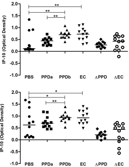
In an initial study we measured IP-10 production in whole blood from 12 bTB-positive cattle (Group 1) which was stimulated with antigen for 24 h and 48 h. Following incubation for 24 h, IP-10 release in blood stimulated with PPDb and PC-EC peptides was significantly greater when compared with that released in blood incubated with PBS (Fig 1a). The median IP-10 response in PPDb-stimulated samples was also significantly greater than in blood stimulated with PPDa. In contrast, there was no significant difference in IP-10 responses in blood samples incubated with PPDa and PBS (Fig 1a). At this time-point, all 12 bTB-positive animals showed greater IP-10 release in response to PPDb stimulation compared to PPDa stimulation while 11/ 12 animals showed greater IP-10 release in PC-EC peptide-stimulated blood compared with unstimulated blood (Fig 1a).
Following 48 h of incubation, median levels of IP-10 release remained significantly greater in PPDb- and PC-EC-stimulated blood compared to blood co-incubated with PBS and in blood stimulated with PPDb compared to that stimulated with PPDa (Fig 1b). At this time point, IP-10 release was greater in response to PPDb than to PPDa in 11/12 animals and greater in PC-EC peptide-stimulated blood than unstimulated blood in 9/12 animals (Fig 1b). For most animals, the release of IP-10 in blood cultured with PBS increased between 24 h and 48 h and median levels were significantly greater after 48 h of incubation when compared with the earlier time point (Fig 2).
Antigen-specific IP-10 release in whole blood from reactor and
non-reactor cattle
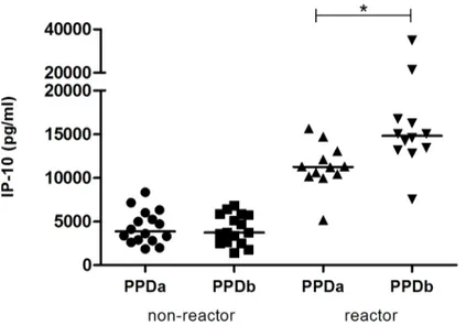
In order to further characterize the antigen-specific release of IP-10, quantitative IP-10 levels were compared in PPDb- and PPDa-stimulated blood from an additional 12 reactor and 16 non-reactor cattle from a herd with knownM.bovisinfection (Group 2). In the reactor group, the release of IP-10 was significantly greater in response to stimulation with PPDb (7500–
34800 pg/ml) than in response to PPDa (5200–15600 pg/ml) (Fig 3). In contrast, in the non-reactor cattle, IP-10 responses to PPDb and PPDa ranged from 1400–8300 pg/ml and there was no significant difference in IP-10 responses to these antigens (Fig 3).
of the ELISA). Within these limits, concentrations of IFN-γand IP-10 were strongly correlated (r = 0.65, n = 41, p<0.0001;Fig 4).
Diagnostic potential of measuring
M
.
bovis
-specific IP-10 release
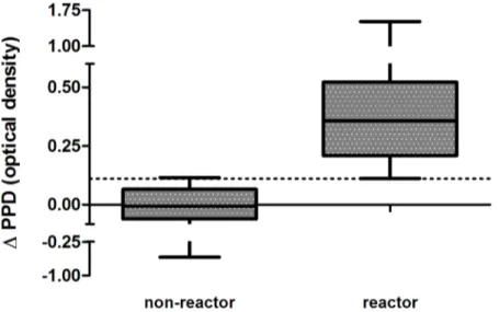
The potential diagnostic value of IP-10 was evaluated usingΔPPD results for all animals includ-ing an additional 20M.bovis-unexposed cattle. All SICTT-positive cattle were Bovigam-positive and all SICTT-negative animals were Bovigam-negative. Using ROC analysis, theΔPPD results for the 26 reactor and 36 non-reactor animals were compared and showed extremely good agreement with the Bovigam assay and SICTT status of all animals (AUC = 1.00, p<0.0001,S1
Fig). An optimal IP-10ΔPPD cut-off value of 0.11 distinguished between reactors and non-reac-tors with a sensitivity of 100% (95% CI, 86%–100%) and a specificity of 97% (95% CI, 85%–
100%) (Fig 5). This cut-off value assigned all but one of the Bovigam/SICTT-negative animals as
Fig 1. IP-10 release in response to antigenic stimulation after (A) 24 h and (B) 48 h.Whole blood from bTB-positive cattle (n = 12) was incubated at 37°C with saline (PBS),M.aviumPPD (PPDa),M.bovisPPD (PPDb) and ESAT-6/CFP-10 peptides (EC). IP-10 release in whole blood in response to PPDb and EC was significantly greater than that in blood co-incubated with PBS and significantly greater in response to PPDb than in response to PPDa. Median values and the differential IP-10 responses to PPDa and PPDb (ΔPPD) and PBS and EC (ΔEC) are shown (*, p<0.01;**, p<0.05).
ΔPPD-negative (35/36). Changing theΔPPD cut-off to 0.13 was the lowest value that had a specificity of 100% (95% CI, 90%–100%) and this identified reactor cattle with a sensitivity of 96% (95% CI, 79%–100%).
Discussion
The most sensitive immunodiagnostic assays ofM.bovisinfection in animals are those that detect the activation of memory T-lymphocytes in response to stimulation with tuberculin antigens [2]. In this study we have shown that the measurement of IP-10 release in whole
Fig 2. Spontaneous IP-10 release in whole blood from reactor cattle.Whole blood from cattle (n = 12) was incubated with phosphate buffered saline for 24 and 48 h. IP-10 release showed a significant increase over time (*<0.005).
doi:10.1371/journal.pone.0155440.g002
Fig 3. IP-10 release in response to antigenic stimulation in reactor and non-reactor cattle.Whole blood from non-reactor (n = 16) and reactor (n = 12) cattle was incubated withM.aviumPPD (PPDa) andM.bovis PPD (PPDb) for 24 h at 37°C. For reactor cattle, IP-10 release in response to PPDb was significantly greater than that in response to PPDa. Median values are shown (*, p<0.0005).
blood of cattle is a sensitive marker of antigen recognition and immune activation and a prom-ising diagnostic biomarker ofM.bovisinfection.
The release of IP-10 was highly correlated with that of IFN-γand after 24 h of blood culture, all reactor cattle showed consistently greater PPDb-induced IP-10 responses when compared with stimulation by PPDa. Moreover, as has been reported for humans [18] and African buffa-loes [14], antigen-induced IP-10 was released at far greater concentrations than have previ-ously been reported for IFN-γ[19,20]. Together, these findings are evidence that IP-10 is a useful measure of antigen-specific cell mediated immunity in cattle. Because IP-10 is produced
Fig 4. Correlation between IFN-γand IP-10 release.Whole blood from non-reactor and reactor cattle was
stimulated with eitherM.bovisPPD orM.aviumPPD for 24 h at 37°C. The release of IFN-γand IP-10 was highly correlated in these samples (r = 0.65; p<0.0001).
doi:10.1371/journal.pone.0155440.g004
Fig 5. Antigen-specific IP-10 release distinguishes between non-reactor and reactor cattle.Whole blood from non-reactor (n = 36) and reactor cattle (n = 24) was incubated withM.bovisPPD andM.avium PPD for 24 h at 37°C. The difference in IP-10 release in these samples (ΔPPD) was used to calculate, by ROC analysis, an optimal cut off value of 0.11 (dotted line). This distinguished between these groups with a sensitivity of 100% and a specificity of 97%.
in response to IFN-γ, we considered that the optimal incubation time of an assay measuring this cytokine might be different than that for an IGRA. However, in the bTB-positive cohort, the differential in the IP-10 response to PPDb and PPDa detected after 24 h of blood stimula-tion diminished after 48 h of incubastimula-tion. It is therefore possible that a shorter incubastimula-tion time might provide for even greater resolution between these responses, though this remains to be determined. Nonetheless, measurement ofΔPPD after 24 h allowed for the highly accurate dis-crimination between Bovigam/SICTT-positive and Bovigam/SICTT-negative animals and indicates the potential of IP-10 as a highly specific diagnostic biomarker ofM.bovisinfection in cattle. Notably, selection of aΔPPD threshold with a specificity of 100%, i.e. 0.13, distin-guished between these groups with a sensitivity of 96% suggesting that this marker does not significantly compromise diagnostic specificity in cattle. This finding is in agreement with human studies that have shown IP-10 assays to have a specificity of close to 100% [8]. A more sensitive cut-off value of 0.11 detected all Bovigam/SICTT-positive cattle and one additional ΔPPD-positive individual. While the true infection status of this animal was not determined, this result may indicate the potential for the IP-10 assay to increase the accuracy of diagnosis of
M.bovisinfection in cattle.
The release of IP-10 also shows promise as a marker of immune sensitization to the highly specificM.bovisproteins ESAT-6 and CFP-10. Importantly, measurement of IP-10, in combi-nation with IFN-γ, has been shown to improve the detection of ESAT-6/CFP-10 sensitization in humans and African buffaloes [6,15]. In cattle, use of these antigens would be particularly useful for specifically detectingM.bovis-infected animals that have been vaccinated withM.
bovisBCG [21] and for testing herds with high rates of false positive reactions to standard PPD assays [2]. However, in our study, the measurement of ESAT-6/CFP-10-specific IP-10, with reference to levels of this cytokine in unstimulated blood, was in some cases compromised by its release in control samples. The spontaneous production of IP-10 in blood from cattle has previously been reported and may reflect thein vivoinduction of this cytokine [13]. This is consistent with findings from human studies that have identified a peripheral whole blood gene transcription signature in tuberculosis patients that is dominated by a neutrophil-driven interferon-inducible gene profile [22].
In other instances in the present study, IP-10 release in unstimulated blood exceeded that in response to ESAT-6/CFP-10 and PPD. While the mechanism of this phenomenon is unclear, the activation of antigen-specific memory lymphocytes may have resulted in reduced IP-10 production via the release of inhibitory cytokines, e.g. interleukin-10 [23]. Also, numerous pro-teases are secreted from activated leukocytes and proteolysis of plasma IP-10 might obscure detection of its release [24].
An earlier study with cattle experimentally infected withM.bovisfound that IP-10 responses to PPDb did not exceed the respective responses to medium alone at any time point over the course of the infection [13]. This finding, in part, may have been a consequence of the high lev-els of IP-10 measured in unstimulated control samples in that study. However, limitations in the availability of suitable samples, and the use of a human ELISA, may have influenced these earlier findings [13]. In the present study we used a bovine-specific ELISA to measure IP-10 and showed that significant release of this protein occurs following antigenic stimulation of whole blood of cattle.
IP-10, which is produced in abundance in response to IFN-γ, might serve to amplify anM.bovis -specific signal in an infected animal.
In summary, IP-10 release in stimulated whole blood is strongly correlated with that of IFN-γand is a robust biomarker of antigen-specific immunological memory in cattle. How-ever, the spontaneous release of the cytokine in unstimulated blood requires further investiga-tion in order to understand its origin and regulainvestiga-tion as it might limit the performance of the assay when utilizingM.bovis-specific antigens. Nonetheless, the release of IP-10 in response to PPDb and PPDa antigens is a valuable biomarker ofM.bovisinfection in cattle and measure-ment of this cytokine might improve the diagnostic performance of whole blood stimulation assays.
Supporting Information
S1 Fig. Measurement of antigen-specific IP-10 release shows extremely good agreement with the Bovigam assay and single intradermal comparative tuberculin test (SICTT). Whole blood from Bovigam/SICTT-negative (n = 36) and Bovigam/SICTT-positive cattle (n = 24) was incubated withM.bovisPPD andM.aviumPPD for 24 h at 37°C and the differ-ence in IP-10 release in these samples (ΔPPD) was determined by ELISA. TheΔPPD results for cattle from each group were compared by ROC curve analysis and IP-10 test outcomes showed extremely good agreement with the reference tests.
(TIF)
Acknowledgments
We are grateful to Anthony Duignan SVI (DAFM) for collecting blood samples from reactor cattle and to staff from DAFM Longtown farm for their assistance in handling animals. We would also like to thank Prof Steve Gordon and Louise Britton at UCD for providing surplus plasma samples from non-infected control animals.
Author Contributions
Conceived and designed the experiments: SDCP PvH EG. Performed the experiments: SDCP KM MBD WG. Analyzed the data: SDCP EG. Contributed reagents/materials/analysis tools: PvH EG. Wrote the paper: SDCP PvH EG.
References
1. Gormley E, Doyle MB, Fitzsimons T, McGill K, Collins JD. Diagnosis ofMycobacterium bovisinfection in cattle by use of the gamma-interferon (Bovigam1
) assay. Vet Microbiol. 2006; 112: 171–179. doi:10.
1016/j.vetmic.2005.11.029PMID:16321478
2. Schiller I, Oesch B, Vordermeier HM, Palmer MV, Harris BN, Orloski KA, et al. Bovine tuberculosis: a review of current and emerging diagnostic techniques in view of their relevance for disease control and eradication. Transbound Emerg Dis. 2010; 57: 205–220. doi:10.1111/j.1865-1682.2010.01148.x
PMID:20561288
3. Bass KE, Nonnecke BJ, Palmer MV, Thacker TC, Hardegger R, Schroeder B, et al. Clinical and diag-nostic developments of a gamma interferon release assay for use in bovine tuberculosis control pro-grams. Clin Vaccine Immunol. 2013; 20: 1827–1835. doi:10.1128/CVI.00519-13PMID:24132602
4. Pollock JM, Girvin RM, Lightbody KA, Neill SD, Clements RA, Buddle BM, et al. Assessment of defined antigens for the diagnosis of bovine tuberculosis in skin test-reactor cattle. Vet Rec. 2000; 146: 659–
665. doi:10.1136/vr.146.23.659PMID:10883854
6. Chegou NN, Heyckendorf J, Walzl G, Lange C, Ruhwald M. Beyond the IFN-γhorizon: biomarkers for immunodiagnosis of infection withMycobacterium tuberculosis. Eur Respir J. 2014; 43: 1472–1486.
doi:10.1183/09031936.00151413PMID:24311770
7. Ruhwald M, Bjerregaard-Andersen M, Rabna P, Eugen-Olsen J, Ravn P. IP-10, 1, 2, MCP-3, and IL-1RA hold promise as biomarkers for infection withM.tuberculosisin a whole blood based T-cell assay. BMC Res Notes. 2009; 2: 19. doi:10.1186/1756-0500-2-19PMID:19193208
8. Ruhwald M, Dominguez J, Latorre I, Losi M, Richeldi L, Pasticci MB, et al. A multicentre evaluation of the accuracy and performance of IP-10 for the diagnosis of infection withM.tuberculosis. Tuberculosis. 2011; 91: 260–267. doi:10.1016/j.tube.2011.01.001PMID:21459676
9. Ruhwald M, Aabye MG, Ravn P. IP-10 release assays in the diagnosis of tuberculosis infection: current status and future directions. Expert Rev Mol Diagn. 2012; 12: 175–187. doi:10.1586/erm.11.97PMID:
22369377
10. Cassatella MA, Gasperini S, Calzetti F, Bertagnin A, Luster AD, McDonald PP. Regulated production of the interferon-γ-inducible protein—10 (IP-10) chemokine by human neutrophils. Eur J Immunol. 1997;
27: 111–115. doi:10.1002/eji.1830270117PMID:9022006
11. Molesworth-Kenyon SJ, Oakes JE, Lausch RN. A novel role for neutrophils as a source of T cell-recruit-ing chemokines IP-10 and Mig durcell-recruit-ing the DTH response to HSV-1 antigen. J Leukoc Biol. 2005; 77: 552–559. doi:10.1189/jlb.0904485PMID:15629884
12. Taubert A, Hermosilla C. Bovine recombinant IFNγinduces endothelial cell gene transcription of immu-noregulatory molecules and upregulates PMN and PBMC adhesion on bovine endothelial cells. Vet Res Commun. 2008; 32: 35–47. doi:10.1007/s11259-007-9001-2PMID:17516142
13. Waters WR, Thacker TC, Nonnecke BJ, Palmer MV, Schiller I, Oesch B, et al. Evaluation of gamma interferon (IFN-γ)-induced protein 10 responses for detection of cattle infected withMycobacterium bovis: comparisons to IFN-γresponses. Clin Vaccine Immunol CVI. 2012; 19: 346–351. doi:10.1128/
CVI.05657-11PMID:22237891
14. Goosen WJ, Cooper D, Warren RM, Miller MA, van Helden PD, Parsons SDC. The evaluation of candi-date biomarkers of cell-mediated immunity for the diagnosis ofMycobacterium bovisinfection in African buffaloes (Syncerus caffer). Vet Immunol Immunopathol. 2014; 162: 198–202. doi:10.1016/j.vetimm.
2014.10.008PMID:25464825
15. Goosen WJ, Cooper D, Miller MA, van Helden PD, Parsons SDC. IP-10 is a sensitive biomarker of anti-gen recognition in whole-blood stimulation assays used for the diagnosis ofMycobacterium bovis infec-tion in African buffaloes (Syncerus caffer). Clin Vaccine Immunol. 2015; 22: 974–978. doi:10.1128/CVI.
00324-15PMID:26108287
16. Rothel JS, Jones SL, Corner LA, Cox JC, Wood PR. A sandwich enzyme immunoassay for bovine interferon-gamma and its use for the detection of tuberculosis in cattle. Aust Vet J. 1990; 67: 134–137.
PMID:2115767
17. Youden WJ. Index for rating diagnostic tests. Cancer. 1950; 3: 32–35. PMID:15405679
18. Ruhwald M, Bjerregaard-Andersen M, Rabna P, Kofoed K, Eugen-Olsen J, Ravn P. CXCL10/IP-10 release is induced by incubation of whole blood from tuberculosis patients with ESAT-6, CFP10 and TB7.7. Microbes Infect. 2007; 9: 806–812. doi:10.1016/j.micinf.2007.02.021PMID:17533146
19. Thom M, Morgan JH, Hope JC, Villarreal-Ramos B, Martin M, Howard CJ. The effect of repeated tuber-culin skin testing of cattle on immune responses and disease following experimental infection with Mycobacterium bovis. Vet Immunol Immunopathol. 2004; 102: 399–412. doi:10.1016/j.vetimm.2004.
08.005PMID:15541793
20. Hope JC, Thom ML, Villarreal-Ramos B, Vordermeier HM, Hewinson RG, Howard CJ. Exposure to Mycobacterium aviuminduces low-level protection fromMycobacterium bovisinfection but compro-mises diagnosis of disease in cattle. Clin Exp Immunol. 2005; 141: 432–439. doi:10.1111/j.1365-2249.
2005.02882.xPMID:16045732
21. Vordermeier M, Gordon SV, Hewinson RG.Mycobacterium bovisantigens for the differential diagnosis of vaccinated and infected cattle. Vet Microbiol. 2011; 151: 8–13. doi:10.1016/j.vetmic.2011.02.020
PMID:21411245
22. Berry MPR, Graham CM, McNab FW, Xu Z, Bloch SAA, Oni T, et al. An interferon-inducible neutrophil-driven blood transcriptional signature in human tuberculosis. Nature. 2010; 466: 973–977. doi:10.
1038/nature09247PMID:20725040
23. Re F, Strominger JL. IL-10 released by concomitant TLR2 stimulation blocks the induction of a subset of Th1 cytokines that are specifically induced by TLR4 or TLR3 in human dendritic cells. J Immunol. 2004; 173: 7548–7555. doi:10.4049/jimmunol.173.12.7548PMID:15585882
24. Mortier A, Gouwy M, Van Damme J, Proost P. Effect of posttranslational processing on the in vitro and in vivo activity of chemokines. Exp Cell Res. 2011; 317: 642–654. doi:10.1016/j.yexcr.2010.11.016