Distinctive Patterns of Evolution of the
Globin
Gene (HBD) in Primates
Ana Moleirinho1,2,3
*, Alexandra M. Lopes1,2, Susana Seixas1,2, Ramiro Morales-Hojas4, Maria J. Prata1,2,3, António Amorim1,2,3
1Instituto de Investigação e Inovação em Saúde, Universidade do Porto, Porto, Portugal,2IPATIMUP– Institute of Molecular Pathology and Immunology, University of Porto, Porto, Portugal,3Department of Biology, Faculty of Sciences, University of Porto, Porto, Portugal,4Genetics and Genomics Group, The Pirbright Institute, Compton Laboratory, Compton, Berkshire, United Kingdom
Abstract
In most vertebrates, hemoglobin (Hb) is a heterotetramer composed of two dissimilar globin chains, which change during development according to the patterns of expression ofα- and
β-globin family members. In placental mammals, theβ-globin cluster includes three early-expressed genes,ε(HBE)-γ(HBG)-ψβ(HBBP1), and the late expressed genes,δ(HBD) and
β(HBB). WhileHBBencodes the major adultβ-globin chain,HBDis weakly expressed or totally silent. Paradoxically, in human populationsHBDshows high levels of conservation typical of genes under strong evolutionary constraints, possibly due to a regulatory role in the fetal-to-adult switch unique of Anthropoid primates. In this study, we have performed a comprehensive phylogenetic and comparative analysis of the two adultβ-like globingenes in a set of diverse mammalian taxa, focusing on the evolution and functional divergence of
HBDin primates. Our analysis revealed that anthropoids are an exception to a general pat-tern of concerted evolution in placental mammals, showing a high level of sequence conser-vation atHBD, less frequent and shorter gene conversion events. Moreover, this lineage is unique in the retention of a functional GATA-1 motif, known to be involved in the control of the developmental expression of theβ-like globingenes. We further show that not only the mode but also the rate of evolution of theδ-globingene in higher primates are strictly asso-ciated with the fetal/adultβ-cluster developmental switch. To gain further insight into the possible functional constraints that have been shaping the evolutionary history ofHBDin primates, we calculated dN/dS (ω) ratios under alternative models of gene evolution. Al-though our results indicate thatHBDmight have experienced different selective pressures throughout primate evolution, as shown by differentωvalues between apes and Old World Monkeys + New World Monkeys (0.06 versus 0.43, respectively), these estimates corrobo-rated a constrained evolution forHBDin Anthropoid lineages, which is unlikely to be related to protein function. Collectively, these findings suggest that sequence change at theδ -globingene has been under strong selective constraints over 65 Myr of primate evolution, likely due to a regulatory role in ontogenic switches of gene expression.
OPEN ACCESS
Citation:Moleirinho A, Lopes AM, Seixas S, Morales-Hojas R, Prata MJ, Amorim A (2015) Distinctive Patterns of Evolution of theδ-GlobinGene (HBD) in Primates. PLoS ONE 10(4): e0123365. doi:10.1371/journal.pone.0123365
Academic Editor:Francesc Calafell, Universitat Pompeu Fabra, SPAIN
Received:June 6, 2014
Accepted:March 2, 2015
Published:April 8, 2015
Copyright:© 2015 Moleirinho et al. This is an open access article distributed under the terms of the
Creative Commons Attribution License, which permits unrestricted use, distribution, and reproduction in any medium, provided the original author and source are credited.
Data Availability Statement:All relevant data are within the paper and its Supporting Information files.
Funding:This work was supported by the Portuguese Foundation for Science and Technology (FCT) (PTDC/SAU-MET/110323/2009). AM and AML are supported by fellowships from FCT (SFRH / BD / 73508 / 2010 and SFRH / BPD / 73366 / 2010, respectively). SS is supported by POPH-QREN–
Introduction
Hemoglobin (Hb), found in the circulating red blood cells of all vertebrates, is the major oxy-gen-transporting molecule, playing a key role in the cellular aerobic metabolism [27]. In mam-mals, Hb is a heterotetramer composed of twoα-like and twoβ-like globin chains that are differentially expressed during development, such that functionally distinct Hb isoforms are synthesized in embryonic and adult erythroid cells [27–29].
These globin chains are encoded by members of theα-andβ-globingene families, which arose via tandem duplication of an ancestral, single-copy globin gene approximately 450–500 Mya, in the common ancestor of jawed vertebrates [14,23,25,35,78]. The two paralogous gene families exhibit a number of significant differences in gene content among jawed vertebrate taxa. These differences are especially pronounced in the case of theβ-globincluster, in which distinct repertoires of mammalianβ-like globingenes originated by independent lineage-specif-ic dupllineage-specif-ications followed by functional divergence [33–35,52–54,57,78,79]. In both monotremes and marsupials, theβ-globin genecluster contains a single pair of genes, the early expressedε -globin and the late expressedβ-globin[53]. In contrast, within the eutherian stem, further tan-dem duplications gave rise to a cluster of fiveβ-like globingenes, containing early-expressed genes, located at the 5’end of the clusterε-(HBE)-γ(HBG)-ψβ(HBBP1), and late expressed genes,δ(HBD) andβ(HBB), at the 3’end, consistent with the orientation in contemporary species [26,31,53]. The fine tuning of the level and timing of expression of each of these genes relies on interactions with the locus control region (LCR), located from approximately 6 to 18 kb upstream ofHBE[4,11,84].
Over the course of eutherian evolution the structure of theβ-globin genecluster has been dy-namic and the late-expressedHBDandHBBparalogs have experienced different evolutionary fates. In the majority of mammals, the adult form of Hb (α2β2) contains similarβ-chain
sub-units which are encoded by one or more copies of theHBBgene [52]. Contrastingly, the δ-globingene, although present in almost all eutherian species examined to date, is frequently pseudogenized [19,24,26,30,54]. In a few species, a transcriptionally active but weakly express-ed copy of theδ-globingene was maintained, encoding theδ-globin chain of the minor fraction of the adult Hb (α2δ2), known as HbA2,which is thus assumed to be physiologically irrelevant
[45,46,76]. In fact, theδ-globin chain is absent in Old World Monkeys (OWM) [45,46] and ranges from 1% concentration in hominoids [10] to 40% in the galago [80], reaching 6% in New World Monkeys (NWM) [74] and 18% in tarsiers [40]. Surprisingly, in some eutherians
HBDshows a level of sequence conservation typical of genes under strong evolutionary con-straints [91]. In humans for example,HBDwas found to have lower nucleotide diversity than
HBB, suggesting that purifying selection has shaped the evolutionary history ofHBD[49,87] through an unrecognized role not associated with oxygen transport [46,74,86].
The involvement ofHBDin the fetal/adult Hb switch was proposed decades ago [5,55] and since then some studies have provided evidence supporting this hypothesis [6,49,67]. In fact, the fetal to adult Hb switch of anthropoid primates is unique. Furthermore, while both Anthro-poids and Prosimians possess aγ-globin gene, its switch after birth only takes place in the major anthropoid branch, the catarrhines, occurring earlier in NWM, whereas in Prosimians it is only expressed at the embryonic stage [38]. Therefore, phylogenetic and comparative geno-mic analysis across placental mammals with distinct repertoires ofβ-like genes and corre-sponding expression programs should provide clues to the evolution and putative functional divergence of theδ-globingene.
However, the evolutionary history of the eutherianHBDis quite complex due to unusually frequent sequence exchanges through extensive gene conversion and unequal recombination with its neighbor,β-globin[19,31,40,54,80], resulting in extensive sequence homogenization
funders had no role in study design, data collection and analysis, decision to publish, or preparation of the manuscript.
and hampering the assignment of orthologous relationships amongHBDandHBBgenes. In-deed,HBDwas initially thought to be the result of a recent duplication in primate evolution, approximately 40 MY [18], but recently an older origin has been proposed [30,53]. Under such scenario of controversy, a revisit of the evolutionary history of the adultδ-globingene can help elucidate the origin of this gene family, which in spite of many efforts is not yet fully under-stood [31,37,40,45,46,53,60,74,80].
Here, we perform a comprehensive phylogenetic and comparative analysis of the two adult
β-like globingenes in a wide range of mammalian taxa, with a special focus on primates. Our results further document reticulation in the topology of the evolutionary history ofδ-globin
gene, demonstrating that it has behaved as an evolutionary palimpsest, with repeated and par-tially overlappingβ/δsequence transfers obscuring orthology. Additionally, we show that the
δ-globingene is highly conserved in Anthropoids, with a particularly strong signal of purifying selection in Great Apes. Sequence conservation at this locus is unlikely related to protein func-tion and may reflect mutafunc-tional constraints on regulatory regions involved in the fetal-to-adult developmental expression switch of theβ-globincluster.
Materials and Methods
DNA Sequence data and gene identification
To obtain DNA sequences spanning the entireHBDandHBBgenes, we used Blat queries to in-terrogate the genome assemblies of several mammalian species available in the UCSC Genome Browser website (http://genome-euro.ucsc.edu/index.html). Whenever theHBBsequence was available, we used it to identify its paralogous sequences (the first hit); alternatively we used the humanHBBandHBDsequences to identify their corresponding orthologs. Due to a history of concerted evolution, in some cases high sequence identity betweenHBDandHBBproduced ambiguous results. In these cases the genome coordinates were used to distinguish between the two genes, since the order of theβ-like globingenes has been maintained throughout mammali-an evolution. Additional genomic data was obtained either from the High Throughput Geno-mic Sequences database (HTSG), Trace Archives or by direct sequencing to complete sequence gaps and to include further mammalian species. Detailed information on sampling is listed in
S1 Table. Following all these steps of manual curation, our sample included 29 sequences from the three major subclasses of mammals: 1 Prototheria (Ornithorhynchus anatinus), 1 Metha-teria (Monodelphis domestica) and 27 Eutheria, including representatives of the following superordinal groups (1 Xenarthra, 7 Laurasiatheria and 19 Euarchontoglires). We also included one avian species (Gallus gallus) as outgroup.
Evolutionary analysis
ML analyses were performed with RAxML [75] run in the CIPRES Science Gateway [48]. Phylogenetic analysis of the complete gene was run with the following partitions: 1) 5’ UTR + Exon 1, 2 and 3 + Intron 1; and 2) Intron 2 + 3’UTR. The GTR+G+I model was im-plemented for each of these. ML analysis of the CDS was performed with three partitions cor-responding to the three codon positions. The GTR+G model was implemented for each partition. The resulting trees were evaluated with 1000 bootstrap replicates.
BI analyses were run in MrBayes 3.2 [64] in the CIPRES Science Gateway. The complete gene analysis was performed with three partitions: 1) 5’UTR + Exon 1, 2 and 3 + Intron 1; 2) Intron 2; and 3) 3’UTR. The implemented models of evolution were the K80+G, GTR+G+I and HKY+G+I for the first, second and third partitions, respectively. BI with the CDS sequence was performed with the data matrix partitioned according to codon position; the models of evo-lution implemented were the K80+G for the first codon position and the GTR+G for the second and third positions. Two independent runs of 10 million (complete gene analysis) and 5 million (CDS analysis) generations with 8 chains each (7 heated and one cold) were set up. Trees were sampled every 200 (complete gene) or 100 (CDS) generation and the first 12500 trees (25% of the sample) were discarded as burn-in. Convergence and burn-in were assessed using Tracer 1.6 [61], MCMC Trace Analysis Packagehttp://tree.bio.ed.ac.uk/software/tracer/).
Additionally, a third phylogenetic analysis was performed using models of codon substitu-tion with the coding sequence only as input. This method allows us to take into considerasubstitu-tion any potential divergence in codon usage across the mammalian lineage and differences in selective pressure among gene copies, which included pseudogenised copies. Analyses were run using CodonPhyML [21] with the GY CF3x4 [22] model of codon substitution and specifying the M3 model of selective pressure [90]. An initial tree was obtained using
BioNJ + GYECMK07 and the topology was searched using the subtree pruning and regrafting (SPR) heuristic search. Branch support was obtained using the approximate Likelihood Ra-tion Test (aLRT) as implemented in the software. To identify potential recombinaRa-tion events in our primate data set we used the recombination detection package (RDP3) [43], applying a set of seven statistical methods, which includes RDP [42], GENECONV [56], Bootscan [44], Maxchi [72], Chimaera [59], SiSscan [20] and 3Seq [8]. Briefly, two of these are phylogenetic methods, which infer recombination when different parts of the genome result in discordant topologies (RDP and Bootscan), while the other five are nucleotide substitution methods, which examine the sequences either for a significant clustering of substitutions or for a fit to an expected statistical distribution: MaxChi, Chimaera, Geneconv, 3Seq and SiScan. The lat-ter primarily uses genetic similarity estimates but also takes some phylogenetic information into account. To uncover both species specific and ancient events of gene conversion, we con-ducted the analysis with the two paralogous genes for all species simultaneously. Settings for all methods executed in RDP3 were as follows: Sequences were considered to be linear, the p-value cutoff was set to 0.05, and the standard Bonferroni correction was used. In addition, phylogenetic relationships were recovered for each fragment showing signs of gene conver-sion, in Anthropoids and Catarrhines, following the same procedure described above. ML analyses were performed using the GTR+G model without sequence partitioning. The result-ing trees were evaluated with 1000 bootstrap replicates.
Search for open reading frames and promoter analysis
alignment for eutherianHBBproteins andHBDopen reading frame were performed using ClustalW [83] implemented in Geneious version 5.5 created by Biomatters (available from
http://www.geneious.com/). To identify conserved motifs previously shown to be essential for
HBBandHBDexpression, promoter sequences located 5’to the twoβ-like globingenes (~200bp) were aligned using the same approach as above. To confirm transcription factor binding site matches identified within the alignments, we used MatInspector implemented in the Genomatix Software Suite (http://www.genomatix.de/index.html).
Evolutionary rate estimates and selection tests
Pairwise sequence divergence was deduced from Jukes and Cantor distance calculated with DnaSP v.5.10 [65]. In our estimates we scored each insertion or deletion, regardless of length, as one difference as in [13]. Divergence times between species were obtained with TimeTree [32]. Maximum-likelihood estimates of dN/dS (ω; dS—synonymous substitution rate and dN —non-synonymous substitution rate) were carried out using the codeml program from the software package Phylogenetic Analysis by Maximum Likelihood—PAML version 4.8 [89]. To investigate the selective pressures that have shaped the evolution ofHBBandHBDgenes we first calculated dN/dS ratios (M0 model) for each gene separately in the entire mammalian phylogeny. Next, to test the hypothesis of variable selective pressures amongHBDin primates, we performed nested branch models using either the one-ratio model calculated for the whole anthropoid phylogeny, the two-ratio estimated for Great Apes and other primates and three-ratio inferred for Great Apes, OWM and NWM [7,88]. Althoughωvalues below 1 (ω<1) are
generally considered as an evidence of purifying selection, to reject the hypothesis of neutral evolution all models were compared with a null model whereωwas fixed to 1 (ω= 1). The sig-nificance was obtained with likelihood ratio tests (LRT) which were calculated as twice the vari-ation of the likelihoods (-2Δl) with aχ2distribution. For the calculation ofHBDωvalues, pseudogene sequences were only included after the removal of positions affected by premature stop codons and frameshift mutations. In the specific case of lemur species, theHBDsequences were excluded from the analysis given their hybridψβ/δnature [37].
Results
Evolutionary history of the two adult
β-like genes in mammals
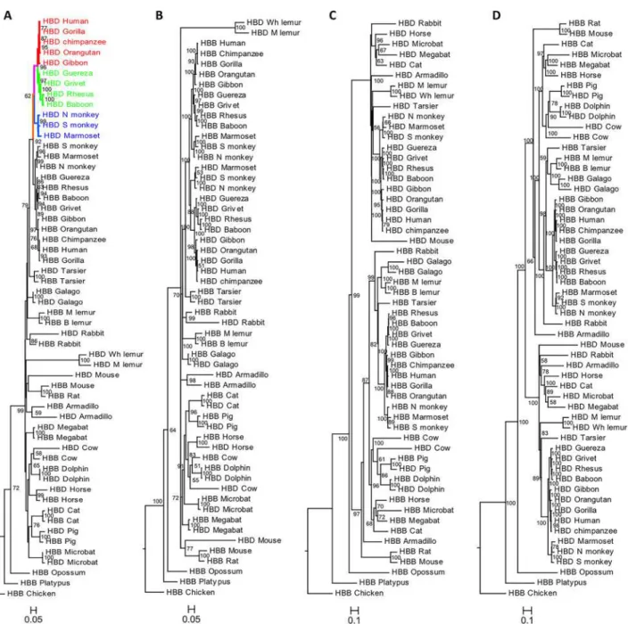
We conducted a phylogenetic analysis of the adultβ-like globingenes,HBDandHBB, in a di-verse dataset, including monotremes, marsupials and placental mammals, and one avian spe-cies, which was used as outgroup. The phylogenies obtained with ML, BI and the codon model approach were similar when either the complete gene sequence or the CDS were used (Fig 1A–
1DandS4 Fig).
However, trees obtained using the complete gene were different from those estimated with the CDS only (Fig 1andS4 Fig). In the phylogenies based on the CDS the twoβ-like paralo-gous genes from the same species cluster together, consistent with a process of interparalogue gene conversion, referred to as“concerted evolution”and previously described in several taxa [19,31,40,52,80]. The exceptions to this general pattern occur in Anthropoid primates (mon-keys and apes) whereHBDandHBBare grouped into two reciprocally monophyletic groups, and in the lemur species. This could lead to the erroneous interpretation thatHBDarose re-cently through a duplication in primate evolution, as initially thought [18], but more likely reflects a gene conversion event in the common ancestor of those primate lineages. In the lemur species, the exception is easily explained by the fact that in the ancestry of lemurs a hy-bridψβ/δpseudogene was created by unequal crossing-over between misalignedHBDand
no evidence for interparalog gene conversion betweenHBDandHBBorthologs (HBD-T1
andHBB-T1), in agreement with previous results [33]. Phylogenetic analyses based on the entire gene sequence lead to better-supported trees, which more reliably replicate the species tree, recovering the true evolutionary history of gene duplication. This phylogeny is consis-tent with previous results supporting a duplication event ofHBBandHBDgenes after the marsupial/eutherian split [53]. Both ML and BI trees contain two major sister clades, one comprising theHBDgene copies of most species and a second clade mainly with theHBB
genes. Within theHBBclade, theHBDgenes from galago, cow, pig and dolphins are clustered Fig 1. Phylograms depicting relationships among adultβ-likegenes in mammals.The phylogeny reconstructions were performed using two methods: A), C) Maximum Likelihood and B), D) Bayesian Inference; trees were estimated using the A), B) coding sequence and C), D) complete gene sequence. Branch support values are given on the internodes. Red branches represent the Great Apes, green the OWM and blue the NWM; the pink and orange branches represent the common branch of Catarrhines and Anthropoids, respectively.
with each of theirHBBparalogs. In galago, it is well documented that the ancestralHBDgene was almost completely converted by theHBBgene, explaining the monophyletic pattern ob-served [80]. According to a recent analysis the phylogenetic incongruence seen in the latter three species is likely due to independent gene conversion events that followed an extensive unequal cross-over spanningHBDintron 2 in the stem lineage of cetartiodactyls [19]. Our re-sults also confirm that the interparalog conversion is largely restricted to the coding regions of globin genes [19,26,30,31,40,45,53], because the incorporation of the phylogenetic infor-mation of non-coding sequence, including intronic and 5’and 3’flanking sequence, proved to be especially useful in assigning orthologous relationships.
Analysis of gene structure
In all species examined theHBBgene retains an intact ORF with conserved donor/acceptor splice sites encoding a polypeptide of 144–146 amino acids (S1 Fig). By contrast, theHBDgene has accumulated several inactivating mutations throughout the mammalian phylogeny. These include the introduction of premature stop codons, small insertions and deletions and muta-tions in the consensus donor (GT) or acceptor (AG) splice sites (S2 Fig). Moreover,HBBand
HBDalso display different across species conservation patterns (S3 Fig). TheHBBpromoter is highly conserved and contains conserved consensus TATA, CAAT and EKLF (Erythroid Krüppel-like factor) binding motifs in most species examined. Only cat and cow show substitu-tions in the EKLF consensus sequence (S3A Fig) but present an upstream EKLF binding ele-ment, identified by MatInspector, which is likely to replace the possible disrupted EKLF motif. A lower conservation inHBDpromoter region is readily apparent from the difficulty in obtain-ing a good multiple alignment for all species. We detected three species, galago, cat and micro-bat, in whichHBDhas aβ-like promoter, acquired through independent gene conversion events (S3A Fig), as previously shown [19,80]. In anthropoids, high sequence homology was observed at theHBDpromoter region (S3B Fig), with a conserved consensus TATA binding motif, a functional GATA-1 motif [47] close to the mutated CAAC box, and lack of the EKLF binding element in all these species. These features of theHBDpromoter region have been shown to be responsible for the low expression of the adultδ-globingene [63,81,82]. The re-maining species (S3C Fig) all share the major defect in the proximalδ-promoter, the absence of a consensus EKLF-binding motif, but do not have the GATA-1 motif common to all otherδ -like promoters. The lack of various conserved motifs in theHBDpromoters that are crucial for
β-like globin expression suggests that they are transcriptionally inefficient in tarsier, mouse, rabbit, dolphin, cow, pig, horse, megabat and armadillo.
Recombination events in primates
In the phylogenetic analyses we did not detect evidence of recombination events betweenHBD
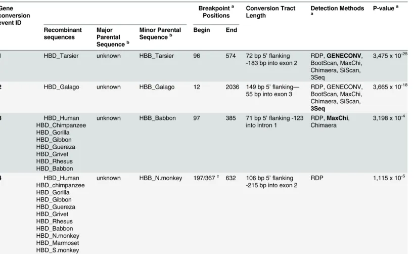
andHBBin anthropoid primates. This result is in agreement with other studies that proposed aδ-βgene conversion in the anthropoid stem [40]. However, it has been suggested that further gene conversions occurred independently in catarrhine and platyrrhine lineages [37,45,60]. Al-though we did not find phylogenetic evidence for gene conversions within these primate line-ages, we cannot exclude the possibility of short-tract gene conversion events not detectable by phylogenetic analysis. Therefore, we sought to re-examine the possibility of lineage-specific gene conversion taking advantage of a more comprehensive sample of primate species and the use of multiple methods implemented in the software package RDP3 [43] for detecting recom-bination signals and putative recombinant sequences. We found robust signals for four inde-pendent interparalog gene conversion events, in which portions ofHBBwere copied ontoHBD
Two of these gene conversion events, 1 and 2, were detected by all seven methods, and cor-roborate independent gene conversions previously identified in tarsier and galago [40,80]. A third event was detected, corresponding to the conversion of the firstHBDexon and intron by
HBBsequences, in catarrhines (OWM and Great Apes). Since this event was detected in ortho-logousHBDcopies of all catarrhines represented, it most likely took place before the divergence of OWM and Great Apes, along the pink branch inFig 1A. The event number 4 corresponds to a more extensive conversion tract present in all anthropoids (platyrrhines and catarrhines), suggesting that those sequences have all descended from an anthropoid ancestor sequence in which the recombination event occurred (orange branch inFig 1A). Thisδ-βconversion Table 1. Summary of gene conversion analysis for primateHBDandHBBparalogues.
Gene conversion event ID Breakpointa Positions Conversion Tract Length Detection Methods a P-value a Recombinant sequences Major Parental Sequenceb Minor Parental
Sequenceb Begin End
1 HBD_Tarsier unknown HBB_Tarsier 96 574 72 bp 5’flanking
-183 bp into exon 2
RDP,GENECONV, BootScan, MaxChi, Chimaera, SiScan, 3Seq
3,475 x 10-25
2 HBD_Galago unknown HBB_Galago 12 2036 149 bp 5’flanking—
55 bp into exon 3
RDP, GENECONV, BootScan, MaxChi, Chimaera, SiScan, 3Seq
3,665 x 10-18
3 HBD_Human HBD_Chimpanzee HBD_Gorilla HBD_Gibbon HBD_Guereza HBD_Grivet HBD_Rhesus HBD_Babbon
unknown HBB_Babbon 97 385 71 bp 5’flanking -123 into intron 1
RDP,MaxChi, Chimaera
3,198 x 10-4
4 HBD_Human HBD_chimpanzee HBD_Gorilla HBD_Gibbon HBD_Guereza HBD_Grivet HBD_Rhesus HBD_Babbon HBD_N.monkey HBD_Marmoset HBD_S.monkey
unknown HBB_N.monkey 197/367c 632 106 bp 5 ’flanking -215 bp into exon 2
RDP 1,115 x 10-5
aIn cases where multiple methods detected the same or a similar conversion event, we reported the breakpoint positions and the method yielding the lowest average Bonferroni corrected p-value, which is shown in bold; breakpoint positions refer to the nucleotide positions in the full alignment of the PrimateHBBandHBDsequences.
bThe major parental and minor parental sequences correspond to the parent contributing to the larger fraction and to the minor fraction of the recombinant sequence, respectively. In all 4 events the major parental sequence is unknown given that the presence of a parent and a recombinant in the alignment is sufficient for a recombination event to be detected by these methods.
cThe breakpoint for the recombination event number 4 varies depending on the species in which it was detected: 197 in Platyrrhines (N.monkey, Marmoset and S.monkey) and 367 in Catarrhines (Human, Chimpanzee, Gorilla, Gibbon, Guereza, Grivet, Rhesus, Babbon), leading to different gene conversion tract length predictions for these groups.
appears to extend from the 5’promoter region till the end of the second exon, however in catar-rhines the signal for this older gene conversion is restricted to the second exon, because a sub-sequent event (identified as number 3) overprinted part of the older one.
In order to assess whether the conversion events 3 and 4 have been correctly identified, we constructed and compared two phylogenetic trees, one with the region containing evidence for an older gene conversion in both platyrrhines and catarrhines (nucleotide 367–632), and a sec-ond one with the portion of the alignment between the inferred breakpoints in event 3 (nucleo-tide 97–385) where a second, more recent conversion event occurred in the catarrhine stem (S5 Fig). The topology of the first tree (S5A Fig) is consistent with a gene conversion in the com-mon ancestor of Anthropoids, in agreement with results obtained with other methods
[31,40,45,73]. In contrast, in the second tree (S5B Fig) theHBDandHBBgenes group together within the catarrhine and platyrrhine lineages, as expected under the hypothesis of a conver-sion event restricted to catarrhines. This tree topology could also indicate that parallel gene conversion events have occurred in the stem of catarrhines and platyrrhines; however, no gene conversion event in platyrrhines has been detected with any of the methods used in our analy-sis. Nevertheless, this alternative hypothesis remains disputable given that an older conversion event occurring between still very closely related sequences would be very difficult to detect. Therefore, although our results do not fully support previous evidence thatHBDhas been in-volved in a conversion in platyrrhines [60], it cannot be ruled out. It is noteworthy that albeit two independent gene conversion events occurred in different Anthropoid lineages, the GATA-1 motif in theHBDpromoter region remained intact.
Evolutionary rates and functional constraints in primates
From our previous analysis, it is apparent that whileHBDhas diverged at markedly different rates in different primate lineages, as shown by the variable branch lengths in the phylogenetic trees (Fig 1), anthropoidHBDs share a high sequence identity not only in their coding region but also at the promoter. As a first measure of the rate ofHBDevolution, we compared the ge-netic distance between humans and 13 other primate species, for both adultβ-like globingenes.
HBBP1was also included in the analysis due to the unusually slow substitution rates previously reported for this pseudogene estimated by comparison of human, gorilla and chimpanzee se-quences [13]. Genetic distances were then plotted against the corresponding divergence times for each pairwise comparison, and the linear regression trend line was estimated for each group, as shown inFig 2.
From the slope of the trend lines and the r2values we are able to compare the rate of intron and exon evolution and its constancy over time, respectively. The results presented inFig 2and
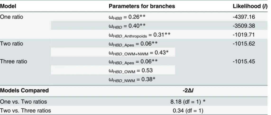
S2 Tableshow that, overall, exons evolved at a lower rate than introns, except forHBBP1, in which nucleotide differences are more homogeneously distributed between exons and introns, as expected for a pseudogene. In Prosimians, there is a trend towards increasing evolutionary rates, more pronounced in non-coding regions (introns andHBPP1exons). Remarkably, the rate of evolution ofHBDexons has remained relatively constant across primate evolution (r2= 0.95) and is comparable to that ofHBBexons, even though in higher primatesHBDis either silent or contributes to only a very small fraction of adult Hb. To gain further insight into the possible functional constraints that have shaped the evolutionary history ofHBDin primates, we calculat-ed dN/dS (ω) ratios under alternative models of gene evolution (table 2).
First, we estimated singleωvalues for the entire mammalian phylogeny (M0 model), for
ei-therHBBorHBDgenes. The observedωvalues were significantly lower than 1 (ωHBB= 0.26
the case ofHBDorthologs, the detected signal of purifying selection may be an outcome of gene conversion by theHBBgene in multiple lineages, mostly in the coding region. As illegiti-mate recombination betweenHBDandHBBhas been noticeably reduced since the last com-mon ancestor of Anthropoids 65.5 Mya, we examined the extent of the selective pressures exerted in this specific clade. Overall, the significance of the low value obtained under the one-ratio model (ωHBD_Anthropoids= 0.31) rejects the hypothesis of a neutral evolution ofHBDin
an-thropoids. Then, to examine whetherHBDhas been subject to variable selective constraints Fig 2. Genetic distance vs divergence times between human and different primate species (Anthropoids and Prosimians) forβ-likegenes.Circles and dotted lines correspond to introns while diamonds and solid lines correspond to exons. Divergence times between humans and other species were obtained with TimeTree [32] and are as follows: Ptr: 6,3 Myr; Ggo 8,8 Myr; Ppy: 15.7 Myr; Nle 20,4 Myr; OWM (Mcc, Panu, Cgue and Caa): 29 Myr; NWM (Sbol, Cjac and Anan): 42,6 Myr; Tsyr: 65,5 Myr; Ogar: 74 Myr. Linear regression trend lines were set to intercept the origin.
doi:10.1371/journal.pone.0123365.g002
Table 2. Parameter Estimates and Likelihood Scores under Different Branch Models.
Model Parameters for branches Likelihood (l)
One ratio ωHBB= 0.26** -4397.16
ωHBD= 0.40** -3509.38
ωHBD_Anthropoids= 0.31** -1019.71
Two ratio ωHBD_Apes= 0.06** -1015.62
ωHBD_OWM+NWM= 0.43*
Three ratio ωHBD_Apes= 0.06** -1015.45
ωHBD_OWM= 0.53 ωHBD_NWM= 0.38*
Models Compared -2Δl
One vs. Two ratios 8.18 (df = 1)*
Two vs. Three ratios 0.34 (df = 1)
NOTE—ωHBBandωHBD,ωfor allHBBandHBDlineages, respectively;ωHBD_Anthropoids,ωfor all AnthropoidHBDlineages;ωHBD_Apes,ωfor Great apeHBDlineages;ωHBD_OWM+NWM,ωfor all OWM and NWMHBDlineages;ωHBD_OWMandωHBD_NWM,ωfor OWM and NWMHBDlineages, respectively; df— degrees of freedom.
*Significant P<0.01 **Significant P<0.001
among different anthropoid clades (Fig 1A), we applied the two-ratio model separating the phylogenetic group of Great Apes from all remaining primates (OWM and NWM). In addi-tion, we also applied the three-ratio model, in whichωwas allowed to vary across Great Apes,
OWM and NWM clades. The comparison of the one-ratio and the two-ratio models showed that the two-ratio presents an improved fit to anthropoidHBDevolution and that it did not differ significantly from the three-ratio model. Nevertheless, in both two-ratio and three-ratio models the Great Apes clade showed an extremely lowωvalue (ωHBD_Apes= 0.06), while the
OWM+NWM branches present a higherω(ωHBD_OWM+NWM= 0.43), but still suggesting a
constrained evolution. Although our results indicate thatHBDmight have experienced differ-ent selective pressures throughout primate evolution, these estimates corroborated a high con-servation ofHBDin Anthropoid lineages that is unlikely related to protein function, since in most primate species this gene is either weakly expressed or not transcribed at all.
Discussion
In humans and in chimpanzees, unusually high levels ofHBDsequence conservation, when compared to functional paralogs, have been described [49]. Such pattern of conservation has been difficult to reconcile with the negligible expression of HbA2. Moreover, the evolutionary
history ofHBDis complex and orthologous relationships amongHBDand its paralog gene (HBB) have been obscured by a history of recurrent gene conversion and unequal crossing overs, throughout eutherian evolution [19,52–54]. Here we gained insight into the evolutionary history ofHBDand its likely regulatory role in the fetal-to-adult switch unique of Anthropoids, by performing a comprehensive phylogenetic and comparative analysis of the two adultβ-like globingenes in a wide range of mammalian taxa. The results from our phylogenetic reconstruc-tion are in agreement with previous findings which demonstrated thatHBDduplication oc-curred before the radiation of Eutheria [53]. The obtained tree topology is also consistent with a history of concerted evolution betweenHBDandHBBthat has created chimericβ/δfusion genes in multiple, independent lineages [19,31,40,54,80]. However, our results show that pri-mates represent an exception to this common trend, given that phylogenetic relationships are maintained throughout this lineage, suggesting that illegitimate recombination betweenHBD
evolution do not seem a plausible explanation for the signal of purifying selection detected, since to date,HBA2has no recognized physiological function [62,76,77].
Interestingly, a role forHBDandHBBP1in the regulatory mechanisms coordinating the fetal-to adult switch has been proposed in early independent studies [5,13,24,26,55]. Taking into account that the mechanism of Hb switch is common to all simian primates [38], we might expect to find similar patterns of conservation and diversity in orthologHBDsequences for a 65.5 Myr time frame. Accordingly, the patterns of conservation we have now uncovered perfectly overlap withHBGduplication and the acquisition of a fetally expressed hemoglobin in anthropoid primates. Noteworthy, we detected in all anthropoid primates a conserved func-tional GATA-1 motif in the promoter ofHBD, which has remained intact despite recurrent gene conversion events overlapping the promoter region among these lineages. Considering thatHBDhas very low expression levels in anthropoids, the conservation of a functional GATA-1 binding motif suggests other functional constraints rather than positive regulation of
δ-globingene expression. Indeed Gaudry, et al. [19] demonstrated that only late expressedβ -like globingenes retaining anHBB-like promoter are efficiently transcribed. Developmental regulation of gene expression at theβ-globincluster involves the formation of chromatin loops mediated by several transcription factors and cofactors [66,68]. It has been shown that GATA-1, along with other cofactors, is required for efficient long-range chromatin interactions be-tween LCR andβ-like globingenes, namely at the time ofγ- toβ- globin switch [9,39,85]. Im-portantly, is has also been demonstrated that theHBDupstream region harbors a binding site for BCL11A, which is a biochemically validated and fundamental switching factor necessary for fetal hemoglobin silencing [67,69]. Remarkably, strong interactions between the LCR and the region encompassing bothHBDandHBBP1were uncovered by chromosome conforma-tion (3C and 5C) analyses at theβ-globinlocus [6,17,70]. Collectively, these findings suggest thatHBDandHBBP1might be involved in chromatin looping in the human-globin cluster, a crucial mechanism for temporal coordination of gene expression [16,36]. Interestingly, the ob-served differences in the rate of evolution between the branches leading to Great Apes and the common branch of OWM and NWM suggest different selective pressures, which may reflect alternative mechanisms of controlling expression in theβ-globincluster among Anthropoids. Selective constraints on the protein function cannot be completely ruled out, although evidence of functional relevance of HbA2is lacking. HbA2has features that are nearly identical to those
Supporting Information
S1 Fig. Sequence alignment for eutherian HBB proteins.
(PDF)
S2 Fig. Sequence alignment of eutherianHBDopen reading frame.Blue and purple filled boxes mark the exons and donnor/acceptor splice sites, respectively. Dots represent nucleotide identities to the human sequence that was set as reference. Coloured nucleotides indicate changes to the human sequence and aminoacid alterations are marked by filled coloured boxes. The lemur species were excluded from the analysis given that their hybridψβ/δpseudogene
[37] generates multiple misalignments. (PDF)
S3 Fig. Sequence alignment of eutherianHBBandHBDpromoters.A)HBBandHBB-like HBDpromoters; B) AnthropoidHBDpromoters and C)HBDpromoters lacking the TF bind-ing motifs which are conserved inHBB-like andHBD-like promoters. Conserved binding mo-tifs are indicated in grey boxes. Again, the lemur species were excluded from the analysis. (PDF)
S4 Fig. Phylograms depicting relationships among adultβ-likegenes in mammals.The phy-logenic tree, based on the coding sequence, was constructed using the Goldman–Yang codon model. Branch support values, obtained using the approximate Likelihood Ration Test (aLRT), are given on the internodes.
(PDF)
S5 Fig. Maximum Likelihood phylograms depicting relationships amongβ-likegenes of Anthropoids.The phylogeny reconstructions were based on A) the portion of the alignment that contain evidence of the anthropoid gene conversion (nucleotide 367–632) and B) the por-tion of the alignment between the inferred breakpoint in event 3 (nucleotide 97–385). Boot-strap branch support (1000 replicates) are given on the internodes.
(PDF)
S1 Table. Listing ofHBBandHBDsequences used for phylogeny reconstructions. (PDF)
S2 Table. Evolutionary rates based on Jukes Cantor distance.
(PDF)
Author Contributions
Conceived and designed the experiments: AM AA. Performed the experiments: AM. Analyzed the data: AM AML SS MJP AA RMH. Contributed reagents/materials/analysis tools: AM AML SS RMH. Wrote the paper: AM AML SS MJP AA RMH.
References
1. Aguileta G, Bielawski JP, Yang Z. Gene conversion and functional divergence in the beta-globin gene family. J Mol Evol. 2004; 59: 177–189. PMID:15486692
2. Armougom F, Moretti S, Poirot O, Audic S, Dumas P, Schaeli B, et al. Expresso: automatic incorpo-ration of structural information in multiple sequence alignments using 3D-Coffee. Nucleic Acids Res. 2006; 34: W604–608. PMID:16845081
3. Artimo P, Jonnalagedda M, Arnold K, Baratin D, Csardi G, de Castro E, et al. ExPASy: SIB bioinformat-ics resource portal. Nucleic Acids Research. 2012; 40: W597–W603. doi:10.1093/nar/gks400PMID:
4. Bank A. Regulation of human fetal hemoglobin: new players, new complexities. Blood. 2006; 107: 435– 443. PMID:16109777
5. Bank A, Mears JG, Ramirez F. Disorders of human hemoglobin. Science. 1980; 207: 486–493. PMID:
7352255
6. Beauchemin H, Trudel M. Evidence for a bigenic chromatin subdomain in regulation of the fetal-to-adult hemoglobin switch. Mol Cell Biol. 2009; 29: 1635–1648. doi:10.1128/MCB.01735-08PMID:19114559
7. Bielawski JP, Yang Z. Maximum likelihood methods for detecting adaptive evolution after gene duplica-tion. J Struct Funct Genomics. 2003; 3: 201–212. PMID:12836699
8. Boni MF, Posada D, Feldman MW. An Exact Nonparametric Method for Inferring Mosaic Structure in Sequence Triplets. Genetics. 2007; 176: 1035–1047. PMID:17409078
9. Bottardi S, Ross J, Bourgoin V, Fotouhi-Ardakani N, Affar el B, Trudel M, et al. Ikaros and GATA-1 com-binatorial effect is required for silencing of human gamma-globin genes. Mol Cell Biol. 2009; 29: 1526– 1537. doi:10.1128/MCB.01523-08PMID:19114560
10. Boyer S, Crosby E, Noyes A, Fuller G, Leslie S, Donaldson L, et al. Primate hemoglobins: Some se-quences and some proposals concerning the character of evolution and mutation. Biochemical Genet-ics. 1971; 5: 405–448. PMID:4999925
11. Bulger M, Groudine M. Looping versus linking: toward a model for long-distance gene activation. Genes Dev. 1999; 13: 2465–2477. PMID:10521391
12. Burge C, Karlin S. Prediction of complete gene structures in human genomic DNA. J Mol Biol. 1997; 268: 78–94. PMID:9149143
13. Chang LY, Slightom JL. Isolation and nucleotide sequence analysis of the beta-type globin pseudo-gene from human, gorilla and chimpanzee. J Mol Biol. 1984; 180: 767–784. PMID:6098690
14. Czelusniak J, Goodman M, Hewett-Emmett D, Weiss ML, Venta PJ, Tashian RE. Phylogenetic origins and adaptive evolution of avian and mammalian haemoglobin genes. Nature. 1982; 298: 297–300. PMID:6178039
15. de Bruin SH, Janssen LHM. Comparison of the oxygen and proton binding behavior of human hemoglo-bin A and A2. Biochimica et Biophysica Acta (BBA)—Protein Structure. 1973; 295: 490–494.
16. Dean A. On a chromosome far, far away: LCRs and gene expression. Trends Genet. 2006; 22: 38–45. PMID:16309780
17. Dostie J, Richmond TA, Arnaout RA, Selzer RR, Lee WL, Honan TA, et al. Chromosome Conformation Capture Carbon Copy (5C): a massively parallel solution for mapping interactions between genomic el-ements. Genome Res. 2006; 16: 1299–1309. PMID:16954542
18. Efstratiadis A, Posakony JW, Maniatis T, Lawn RM, O'Connell C, Spritz RA, et al. The structure and evolution of the human beta-globin gene family. Cell. 1980; 21: 653–668. PMID:6985477
19. Gaudry MJ, Storz JF, Butts GT, Campbell KL, Hoffmann FG. Repeated Evolution of Chimeric Fusion Genes in theβ-Globin Gene Family of Laurasiatherian Mammals. Genome Biology and Evolution. 2014. doi:10.1093/gbe/evu097
20. Gibbs MJ, Armstrong JS, Gibbs AJ. Sister-scanning: a Monte Carlo procedure for assessing signals in recombinant sequences. Bioinformatics. 2000; 16: 573–582. PMID:11038328
21. Gil M, Zanetti MS, Zoller S, Anisimova M. CodonPhyML: Fast Maximum Likelihood Phylogeny Estima-tion under Codon SubstituEstima-tion Models. Molecular Biology and EvoluEstima-tion. 2013. doi:10.1093/molbev/ mst034
22. Goldman N, Yang Z. A codon-based model of nucleotide substitution for protein-coding DNA se-quences. Mol Biol Evol. 1994; 11: 725–736. PMID:7968486
23. Goodman M, Czelusniak J, Koop BF, Tagle DA, Slightom JL. Globins: a case study in molecular phy-logeny. Cold Spring Harb Symp Quant Biol. 1987; 52: 875–890. PMID:3454296
24. Goodman M, Koop BF, Czelusniak J, Weiss ML. The eta-globin gene. Its long evolutionary history in the beta-globin gene family of mammals. J Mol Biol. 1984; 180: 803–823. PMID:6527390
25. Goodman M, Moore GW, Matsuda G. Darwinian evolution in the genealogy of haemoglobin. Nature. 1975; 253: 603–608. PMID:1089897
26. Hardies SC, Edgell MH, Hutchison CA 3rd. Evolution of the mammalian beta-globin gene cluster. J Biol Chem. 1984; 259: 3748–3756. PMID:6706976
27. Hardison R. Evolution of hemoglobin and its genes. Cold Spring Harb Perspect Med. 2012; 2: a011627. doi:10.1101/cshperspect.a011627PMID:23209182
29. Hardison R. Organization, evolution and regulation of the globin genes. In: Steinberg MH, Forget BG, Higgs DR, Nagel RL, editors. Disorders of Hemoglobin: Genetics, Pathophysiology, and Clinical Man-agement. Cambridge: Cambridge University Press; 2001. PMID:12779271
30. Hardison RC. Comparison of the beta-like globin gene families of rabbits and humans indicates that the gene cluster 5'-epsilon-gamma-delta-beta-3' predates the mammalian radiation. Mol Biol Evol. 1984; 1: 390–410. PMID:6599973
31. Hardison RC, Margot JB. Rabbit globin pseudogene psi beta 2 is a hybrid of delta- and beta-globin gene sequences. Mol Biol Evol. 1984; 1: 302–316. PMID:6599969
32. Hedges SB, Dudley J, Kumar S. TimeTree: a public knowledge-base of divergence times among organ-isms. Bioinformatics. 2006; 22: 2971–2972. PMID:17021158
33. Hoffmann FG, Opazo JC, Storz JF. New genes originated via multiple recombinational pathways in the beta-globin gene family of rodents. Mol Biol Evol. 2008; 25: 2589–2600. doi:10.1093/molbev/msn200
PMID:18780876
34. Hoffmann FG, Storz JF. The alphaD-globin gene originated via duplication of an embryonic alpha-like globin gene in the ancestor of tetrapod vertebrates. Mol Biol Evol. 2007; 24: 1982–1990. PMID:
17586601
35. Hoffmann FG, Storz JF, Gorr TA, Opazo JC. Lineage-specific patterns of functional diversification in the alpha- and beta-globin gene families of tetrapod vertebrates. Mol Biol Evol. 2010; 27: 1126–1138. doi:10.1093/molbev/msp325PMID:20047955
36. Holwerda S, de Laat W. Chromatin loops, gene positioning, and gene expression. Front Genet. 2012; 3: 217. doi:10.3389/fgene.2012.00217PMID:23087710
37. Jeffreys AJ, Barrie PA, Harris S, Fawcett DH, Nugent ZJ, Boyd AC. Isolation and sequence analysis of a hybrid delta-globin pseudogene from the brown lemur. J Mol Biol. 1982; 156: 487–503. PMID:
6214636
38. Johnson RM, Gumucio D, Goodman M. Globin gene switching in primates. Comp Biochem Physiol A Mol Integr Physiol. 2002; 133: 877–883. PMID:12443943
39. Keys JR, Tallack MR, Zhan Y, Papathanasiou P, Goodnow CC, Gaensler KM, et al. A mechanism for Ikaros regulation of human globin gene switching. Br J Haematol. 2008; 141: 398–406. doi:10.1111/j. 1365-2141.2008.07065.xPMID:18318763
40. Koop BF, Siemieniak D, Slightom JL, Goodman M, Dunbar J, Wright PC, et al. Tarsius delta- and beta-globin genes: conversions, evolution, and systematic implications. J Biol Chem. 1989; 264: 68–79. PMID:2491855
41. Lanfear R, Calcott B, Ho SY, Guindon S. Partitionfinder: combined selection of partitioning schemes and substitution models for phylogenetic analyses. Mol Biol Evol. 2012; 29: 1695–1701. doi:10.1093/ molbev/mss020PMID:22319168
42. Martin D, Rybicki E. RDP: detection of recombination amongst aligned sequences. Bioinformatics. 2000; 16: 562–563. PMID:10980155
43. Martin DP, Lemey P, Lott M, Moulton V, Posada D, Lefeuvre P. RDP3: a flexible and fast computer pro-gram for analyzing recombination. Bioinformatics. 2010; 26: 2462–2463. doi:10.1093/bioinformatics/ btq467PMID:20798170
44. Martin DP, Posada D, Crandall KA, Williamson C. A modified bootscan algorithm for automated identifi-cation of recombinant sequences and recombination breakpoints. AIDS Res Hum Retroviruses. 2005; 21: 98–102. PMID:15665649
45. Martin SL, Vincent KA, Wilson AC. Rise and fall of the delta globin gene. J Mol Biol. 1983; 164: 513– 528. PMID:6188843
46. Martin SL, Zimmer EA, Kan YW, Wilson AC. Silent delta-globin gene in Old World monkeys. Proc Natl Acad Sci U S A. 1980; 77: 3563–3566. PMID:6251467
47. Matsuda M, Sakamoto N, Fukumaki Y. Delta-thalassemia caused by disruption of the site for an ery-throid-specific transcription factor, GATA-1, in the delta-globin gene promoter. Blood. 1992; 80: 1347– 1351. PMID:1515647
48. Miller MA, Pfeiffer W, Schwartz T. Creating the CIPRES Science Gateway for inference of large phylo-genetic trees; 2010. pp. 1–8.
49. Moleirinho A, Seixas S, Lopes AM, Bento C, Prata MJ, Amorim A. Evolutionary constraints in the beta-globin cluster: the signature of purifying selection at the delta-beta-globin (HBD) locus and its role in develop-mental gene regulation. Genome Biol Evol. 2013; 5: 559–571. doi:10.1093/gbe/evt029PMID:
23431002
51. Notredame C, Higgins DG, Heringa J. T-Coffee: A novel method for fast and accurate multiple se-quence alignment. J Mol Biol. 2000; 302: 205–217. PMID:10964570
52. Opazo JC, Hoffmann FG, Storz JF. Differential loss of embryonic globin genes during the radiation of placental mammals. Proceedings of the National Academy of Sciences. 2008; 105: 12950–12955. doi:
10.1073/pnas.0804392105PMID:18755893
53. Opazo JC, Hoffmann FG, Storz JF. Genomic evidence for independent origins ofβ-like globin genes in monotremes and therian mammals. Proceedings of the National Academy of Sciences. 2008; 105: 1590–1595. doi:10.1073/pnas.0710531105PMID:18216242
54. Opazo JC, Sloan AM, Campbell KL, Storz JF. Origin and Ascendancy of a Chimeric Fusion Gene: The β/δ-Globin Gene of Paenungulate Mammals. Molecular Biology and Evolution. 2009; 26: 1469–1478. doi:10.1093/molbev/msp064PMID:19332641
55. Ottolenghi S, Giglioni B, Comi P, Gianni AM, Polli E, Acquaye CT, et al. Globin gene deletion in HPFH, delta (o) beta (o) thalassaemia and Hb Lepore disease. Nature. 1979; 278: 654–657. PMID:450068
56. Padidam M, Sawyer S, Fauquet CM. Possible emergence of new geminiviruses by frequent recombina-tion. Virology. 1999; 265: 218–225. PMID:10600594
57. Patel VS, Cooper SJ, Deakin JE, Fulton B, Graves T, Warren WC, et al. Platypus globin genes and flanking loci suggest a new insertional model for beta-globin evolution in birds and mammals. BMC Biol. 2008; 6: 34. doi:10.1186/1741-7007-6-34PMID:18657265
58. Petronella N, Drouin G. Purifying selection against gene conversions in the folate receptor genes of pri-mates. Genomics. 2014; 103: 40–47. doi:10.1016/j.ygeno.2013.10.004PMID:24184359
59. Posada D, Crandall KA. Evaluation of methods for detecting recombination from DNA sequences: com-puter simulations. Proc Natl Acad Sci U S A. 2001; 98: 13757–13762. PMID:11717435
60. Prychitko T, Johnson RM, Wildman DE, Gumucio D, Goodman M. The phylogenetic history of New World monkey beta globin reveals a platyrrhine beta to delta gene conversion in the atelid ancestry. Mol Phylogenet Evol. 2005; 35: 225–234. PMID:15737593
61. Rambaut A, Suchard MA, Xie D, Drummond AJ. Tracer v1.6; 2014. Available:http://beast.bio.ed.ac.uk/ Tracer.
62. Ranney HM, Lam R, Rosenberg G. Some properties of hemoglobin A2. American Journal of Hematolo-gy. 1993; 42: 107–111. PMID:8416283
63. Ristaldi MS, Casula S, Porcu S, Marongiu MF, Pirastu M, Cao A. Activation of the delta-globin gene by the beta-globin gene CACCC motif. Blood Cells Mol Dis. 1999; 25: 193–209. PMID:10575545
64. Ronquist F, Teslenko M, van der Mark P, Ayres DL, Darling A, Hohna S, et al. MrBayes 3.2: efficient Bayesian phylogenetic inference and model choice across a large model space. Syst Biol. 2012; 61: 539–542. doi:10.1093/sysbio/sys029PMID:22357727
65. Rozas J. DNA sequence polymorphism analysis using DnaSP. Methods Mol Biol. 2009; 537: 337–350. doi:10.1007/978-1-59745-251-9_17PMID:19378153
66. Sankaran VG, Orkin SH. The switch from fetal to adult hemoglobin. Cold Spring Harb Perspect Med. 2013; 3: a011643. doi:10.1101/cshperspect.a011643PMID:23209159
67. Sankaran VG, Xu J, Byron R, Greisman HA, Fisher C, Weatherall DJ, et al. A Functional Element Nec-essary for Fetal Hemoglobin Silencing. New England Journal of Medicine. 2011; 365: 807–814. doi:10. 1056/NEJMoa1103070PMID:21879898
68. Sankaran VG, Xu J, Orkin SH. Advances in the understanding of haemoglobin switching. Br J Haema-tol. 2010; 149: 181–194. doi:10.1111/j.1365-2141.2010.08105.xPMID:20201948
69. Sankaran VG, Xu J, Ragoczy T, Ippolito GC, Walkley CR, Maika SD, et al. Developmental and species-divergent globin switching are driven by BCL11A. Nature. 2009; 460: 1093–1097. doi:10.1038/ nature08243PMID:19657335
70. Sanyal A, Lajoie BR, Jain G, Dekker J. The long-range interaction landscape of gene promoters. Na-ture. 2012; 489: 109–113. doi:10.1038/nature11279PMID:22955621
71. Schechter AN. Hemoglobin research and the origins of molecular medicine. 3927–3938 p; 2008 72. Smith JM. Analyzing the mosaic structure of genes. J Mol Evol. 1992; 34: 126–129. PMID:1556748
73. Song G, Hsu CH, Riemer C, Zhang Y, Kim HL, Hoffmann F, et al. Conversion events in gene clusters. BMC Evol Biol. 2011; 11: 226. doi:10.1186/1471-2148-11-226PMID:21798034
74. Spritz RA, Giebel LB. The structure and evolution of the spider monkey delta-globin gene. Mol Biol Evol. 1988; 5: 21–29. PMID:2833675
76. Steinberg MH, Adams JG 3rd. Hemoglobin A2: origin, evolution, and aftermath. Blood. 1991; 78: 2165– 2177. PMID:1932737
77. Steinberg MH, et al. Disorders of Hemoglobin. Cambridge University Press; 2009.
78. Storz JF, Opazo JC, Hoffmann FG. Gene duplication, genome duplication, and the functional diversifi-cation of vertebrate globins. Mol Phylogenet Evol. 2013; 66: 469–478. doi:10.1016/j.ympev.2012.07. 013PMID:22846683
79. Storz JF, Opazo JC, Hoffmann FG. Phylogenetic diversification of the globin gene superfamily in chor-dates. IUBMB Life. 2011; 63: 313–322. doi:10.1002/iub.482PMID:21557448
80. Tagle DA, Slightom JL, Jones RT, Goodman M. Concerted evolution led to high expression of a prosim-ian primate delta globin gene locus. J Biol Chem. 1991; 266: 7469–7480. PMID:2019578
81. Tang DC, Ebb D, Hardison RC, Rodgers GP. Restoration of the CCAAT box or insertion of the CACCC motif activates [corrected] delta-globin gene expression. Blood. 1997; 90: 421–427. PMID:9207479
82. Tang DC, Rodgers GP. Activation of the human delta-globin gene promoter in primary adult erythroid cells. Br J Haematol. 1998; 103: 835–838. PMID:9858241
83. Thompson JD, Higgins DG, Gibson TJ. CLUSTAL W: improving the sensitivity of progressive multiple sequence alignment through sequence weighting, position-specific gap penalties and weight matrix choice. Nucleic Acids Res. 1994; 22: 4673–4680. PMID:7984417
84. Tolhuis B, Palstra RJ, Splinter E, Grosveld F, de Laat W. Looping and interaction between hypersensi-tive sites in the achypersensi-tive beta-globin locus. Mol Cell. 2002; 10: 1453–1465. PMID:12504019
85. Vakoc CR, Letting DL, Gheldof N, Sawado T, Bender MA, Groudine M, et al. Proximity among distant regulatory elements at the beta-globin locus requires GATA-1 and FOG-1. Mol Cell. 2005; 17: 453– 462. PMID:15694345
86. Vincent KA, Wilson AC. Evolution and transcription of old world monkey globin genes. J Mol Biol. 1989; 207: 465–480. PMID:2760921
87. Webster MT, Clegg JB, Harding RM. Common 5' beta-globin RFLP haplotypes harbour a surprising level of ancestral sequence mosaicism. Hum Genet. 2003; 113: 123–139. PMID:12736816
88. Yang Z. Likelihood ratio tests for detecting positive selection and application to primate lysozyme evolu-tion. Mol Biol Evol. 1998; 15: 568–573. PMID:9580986
89. Yang Z. PAML 4: Phylogenetic Analysis by Maximum Likelihood. Molecular Biology and Evolution. 2007; 24: 1586–1591. PMID:17483113
90. Yang Z, Nielsen R, Goldman N, Pedersen AM. Codon-substitution models for heterogeneous selection pressure at amino acid sites. Genetics. 2000; 155: 431–449. PMID:10790415
91. Zhang J. Evolution by gene duplication: an update. Trends in Ecology & Evolution. 2003; 18: 292–298. 92. Zhao Z, Hewett-Emmett D, Li WH. Frequent gene conversion between human red and green opsin