386
Srp Arh Celok Lek. 2011 May-Jun;139(5-6):386-389 DOI: 10.2298/SARH1106386R
ПРИКАЗ БОЛЕСНИКА / CASE REPORT UDC: 616.366-003.7:616.36-008.6
Correspondence to: Nedeljko RADLOVIĆ University Children’s Hospital Tiršova 10, 11000 Belgrade Serbia
n.radlovic@beotel.net
Association of Hereditary Elliptocytosis and
Gilbert’s Syndrome as the Cause of Biliary Calculosis:
Case Report
Nedeljko Radlović1,2, Dragana Ristić1, Radivoj Brdar1,2, Nenad Janić1,2, Zoran Leković1,
Dragana Janić1,2, Željko Smoljanić1, Lidija Dokmanović1,2, Miodrag Jovanović3 1University Children’s Hospital, Belgrade, Serbia;
2Faculty of Medicine, University of Belgrade, Belgrade, Serbia;
3Clinic for Abdominal and Endocrine Surgery, Military Medical Academy, Belgrade, Serbia
INTRODUCTION
Cholelithiasis is a rare disease in children (0.13-0.31%) [1, 2]. Although it can be seen already during the first postpartum months, and even intrauterally, the highest incidence has been recorded in the period during and after puberty [1, 2, 3]. Except for chronic haemolytic condi-tions, cystic fibrosis, resection of the terminal ileum, long-term total parenteral nutrition, anorexia nervosa, cholecystitis, anomalies of the ductus choledocus, chronic hepatitis and hyper-cholesterolemia, the essential factors for the development of cholelithiasis are familial predis-position, obesity, a rapid weight-loss, Gilbert’s syndrome, as well as being of female gender at the onset of puberty [2-9]. Pigmented gallstones, particularly in younger children, are consider-ably more frequent than cholesterol or mixed ones, as well as multiple compared to solitary gallstones [3, 10, 11]. If they are impregnated with calcium salts, which is not rare in children, this can be verified, not only by ultrasound, but also by native x-ray imaging [2]. Although it can be asymptomatic and even reversible, both in children and adults cholelithiasis, except for biliary colic, often atypical and difficult to detect in the youngest age, can lead to serious compli-cations, such as choledocholithiasis, cholangio-hepatitis, pancreatitis, rupture of ductus
choled-ocus, cholecystitis, cholecystic perforation and other [3, 6]. Therapy of choice of symptomatic cholelithiasis is cholecystectomy which is now most often performed by laparoscopy [3].
If considered pathogenetically, biliary calcu-losis presents a complex and mostly insuffi-ciently clarified problem [3, 6]. Except for a too high concentration of cholesterol and/or biliru-bin, desolubilization of biliary content is induced by the deficit or inadequate content of bile acid and phospholipids, a high bilirubin content in the form of bilirubin-monoglucuronide, hypo-tonia and gallbladder dyskinesia, obstruction of ductus cysticus and choledocus, the pres-ence of inflammatory detritus or mucus and other factors [3, 12, 13]. In conditions where these factors occur associated the risk of bili-ary calculosis is significantly increased [14-20]. This can be particularly seen in disorders with a moderate lithogenic potential, such as a compen-sated clinical form of hereditary spherocytosis and Gilbert’s syndrome, which is the case of the patient we are presenting.
CASE REPORT
A 15-year-old male referred due to cholelihti-asis and etiologically unexplained non-conju-gated hyperbilirubinemia. The enclosed data SUMMARY
Introduction Biliary calculosis is rare in children. It occurs associated with different haemolytic and non-haemolytic disorders, which are sometimes also combined.
Case Outline A 15-year-old male was hospitalized due to biliary calculosis and non-conjugated hyper-bilirubinemia. A mild non-conjugated hyperbilirubinemia, without anaemia and other symptoms of liver dysfunction, was registered at age 8 years, and 7 years later cholelithiasis with transitory choledocho-lithiasis. The finding of ellyptocytes in blood smear, which was also verified in mother, normal haemo-globin count and the absence of diseases followed by secondary dysmorphic erythrocytes of this type, indicated a clinically milder (compensated) hereditary ellyptocytosis, while more than a double increase of non-conjugated serum bilirubin fracture after a three-day hypocaloric diet (400 kcal per day) showed the concurrent presence of Gilbert’s syndrome. In the laparascopically removed gallbladder a larger number of small pigmented calculi were disclosed.
Conclusion Gilbert’s syndrome is an essential precipitating factor of biliary calculosis in patients with chronic haemolytic condition. Thus, in all cases of biliary calculosis and non-conjugated hyperbilirubi-nemia with absent clinical and laboratory parameters of liver disorders and anaemia, except in compen-sated haemolytic disease and Gilbert’s syndrome as isolated disorders, a possibility of their association should be taken into consideration.
387
www.srp-arh.rs
Srp Arh Celok Lek. 2011;139(5-6):386-389
showed that the first episode of non-conjugated hyperbil-irubinemia (60 µmol/L), without either anaemia or clinical and laboratory findings of liver disease, was registered at age 8 years. During further period the patient was without any problems until 3 months before arrival to our hospital when he had an attack of intensive epigastric pain asso-ciated with nausea, vomiting, jaundice, acholic stools and darkened urine. Physical examination, except for icterus, mild dehydration, moderate epigastric sensitivness and spleen palpable 1 cm below the costal margin, revealed no other pathological changes. Laboratory blood analy-sis showed a high serum bilirubin level (total 423, conju-gated 164 µmol/L), gamma-glutamyl transpeptidase (115 U/L), alkakaline phosphatase (1248 U/L) and transaminase (AST 229, ALT 435 U/L). Bile colour of urine was positive (urobilinogen ++, bilirubin +), while haemoglobin count was normal (150 g/L). In blood smear, except for reticu-locytosis (3.51%) and a mild erythrocyte anisocytosis, no other abnormalities were found. Other laboratory find-ings, including the serological markers for hepatitis A, B and C, microscopic agglutination-lysis-test for leptospi-rosis, serum cholesterol level, ferritin, lactate dehydroge-nase (LDH), ceruloplasmin, C-reactive protein (CRP), as well as Coombs test, haemoglobin electrophoresis, urine and blood amylase levels, and erythrocyte sedimenta-tion, were all normal. Beside moderate splenomegaly and
multiple cholelithiasis, abdominal ultrasound also showed a suspected calculus in the ductus choledocus, so that the child was sent to our hospital for further investigation.
According to the data obtained by parents, he was the child of the second normal term pregnancy. During the first 3 months he had breast-feeding jaundice, but with-out anaemia neither at that period nor later. His growth and development were normal. Except for these findings, he had no other health problems.
On admission the child was without subjective prob-lems, normally developed and nourished, with signs of full sexual maturity. Complete physical findings were normal, except for a mild icterus of the sclera, hard palate, fron-tal chest skin, as well as of the left cosfron-tal arch of the palpa-ble spleen. Abdominal ultrasound confirmed the pres-ence of multiple cholelithiasis and moderate splenomeg-aly, but without the elements of choledolithiasis (Figure 1). Haemoglobin level was normal (147 g/L), while in the peripheral blood smear, except for high reticulocytosis (4.2%), there was a significant elliptocyte count (Figure 2). Erythocyte osmotic resistance was normal, and qual-itative test to glucose-6-phosphate dehydrogenase defi-ciency was negative. Except for non-conjugated hyperbil-irubinemia (114 µmol/L), other laboratory blood findings were within referent values, including conjugated bilirubin fraction, gamma-glutamyl transpeptidase, LDH, iron, ferri-tin, amylase and CRP. Also, erythrocyte count with mean corpuscular volume (MCV), white blood cell (WBC), WBC formula and platelet counts were normal.
Asymptomatic elliptocytosis was also confirmed in mother, while father’s peripheral blood smear was normal. According to the data obtained by parents, the patient’s older brother was healthy. Also, biliary calculosis was not registered in any of the second degree relatives.
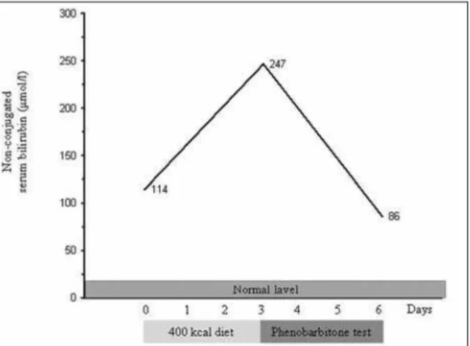
The concurrent presence of Gilbert’s syndrome was determined by a hypocaloric diet test (Graph 1). After a three-day hyporcaloric diet (400-kcal per day) non-conju-gated bilirubinemia was increased 2.1-fold. Next, a three-day phenobarbiton test (2 mg/kg/three-day) was performed after which the non-conjugated serum bilirubin fracture decreased 2.76-fold.
Figure 1. Abdominal ultrasound, multiple cholelithiasis
Figure 2. Peripheral blood smear of our patient. Visible normal erythro-cyte and considerably increased elliptoerythro-cyte counts.
388
doi: 10.2298/SARH1106386R
Radlović N. et al. Association of Hereditary Elliptocytosis and Gilbert’s Syndrome as the Cause of Biliary Calculosis: Case Report
Having in mind symptomatic cholelithiasis with tran-sitory choledolithiasis, the child underwent laparoscopic cholecystectomy. The obtained stones were of pigmented character, multiple and of tiny size. The intervention and postoperative course were normal. Two years and 3 months after surgery the patient was without subjective problems. Except for a moderate splenomegaly, abdominal ultra-sound was normal. Except for non-conjugated hyperbil-irubinemia (143 µmol/L), other serum findings (conju-gated bilirubin fraction, gamma-glutamyl transpeptidase, alkali phosphatase, cholesterol, AST, ALT, LDH, ferritin, amylase, LDH and CRP) were within referent values. In blood smear, beside elliptocytosis, there were 0.9% of retic-ulocytes, while haemoglobin, MCV and other parameters were normal.
DISCUSSION
The paper presents an adolescent with biliary calculosis caused by association of a compensated clinical form of hereditary elliptocytosis and Gilbert’s syndrome. A mild asymptomatic non-conjugated hyperbilirubinemia, with normal haemoglobin level and other liver function find-ings were registered at age 8 years, while at age 15 years this association resulted in cholelithiasis with an episode of choledolithiasis.
Hereditary elliptocytosis represents a rare autosomal dominant membranopathy followed by elliptoid appear-ance and increased fragility of erythrocytes [21]. It occurs in 0.3-0.5 per 1000 newborns, and in about 90% of cases it passes asymptomatically [22, 23]. In about 95% of patients it develops due to gene mutation responsible for α- and β-spectrin expression, i.e. polypeptides which in tetra-meric form compose the basis of cell cytoskeleton [23, 24]. Mutations bound to the protein 4.1 and glycoforin C are rare [21, 24]. If the mutation occurs on one allele only, the disease passes asymptomatically, while in cases when it is bilateral it features moderate or more severe haemo-lytic anaemia [21-24]. Although without genetic confir-mation, based on the permanent finding of elliptocytes in blood smear, which was also verified in mother, as well as the fact that the child did not have either overmarked or prolonged neonatal jaundice, both at early and later age, it can be concluded that our patient had a heterozygotic, i.e. a milder clinical form of hereditary elliptocytosis [21, 23, 24]. In addition, the hereditary nature of the disor-der is also supported by the absence of elements indicat-ing other conditions that are followed by the presence of elliptocytes, such as the deficiency of iron, folic acid and vitamin B12 [21, 22, 23].
Contrary to hereditary elliptocytosis, Gilbert’s syndrome is a frequent disorder. It occurs in 3-10% of general
popula-tion featuring a benign and mild non-conjugated hyperbil-irubinemia potentiated by hunger, fever and physical strain [25]. A low non-conjugated bilirubin clearance is primar-ily caused by autosomal recessive defect in the promoter region of the UGT1A1 gene (2q37) responsible for the expression of bilirubin uridine-diphosphate glucurono-syl transferase (UGPGT), a hepatic microsomal enzyme of key significance for bilirubin conjugation [26]. This results in decreased synthesis of bilirubin UDPGT which reduces to about 30% compared to the normal level that in turn leads to a lower capacity of bilirubin conjugation with glucuronic acid [27]. The additional pathogenetic signif-icance are also a shorter life span of erythrocytes that is seen in about 50% of cases, as well as the defect in uptake and transport of non-conjugated bilirubin at the hepato-cyte level [28]. In the expression of Gilbert’s syndrome sex hormones, particularly androgens, play the major role, which explains its occurrence at the onset of puberty, as well as a 2-fold higher incidence in sexually mature males as compared to females [29, 30]. In addition, adult males are characterized by higher erythrocyte and muscular mass [29]. The presence of Gilbert’s syndrome in our patient was confirmed by the occurrence of more than double increase of non-conjugated serum bilirubin fraction after a three-day hypocaloric diet test [28].
As well known, haemolytic conditions and Gilbert’s syndrome present risk factors for the development of bili-ary calculosis, and also that it is probably more frequent in cases of their association [14-19]. The risk factor for the development of biliary calculosis in the first case is bili-rubin hyperproduction and in the latter its elimination in the form of low water-soluble bilirubin monoglucuronide [14-19]. Although hereditary elliptocytosis and Gilbert’s syndrome are congenital disorders, our patient did not develop biliary calculosis before the end of puberty, i.e. in the condition of marked androgenous suppression of bilirubin UDPGT, as well as additionally higher biliru-bin production caused by the increased erythrocyte and muscular mass [29, 30]. As expected, in our patient the stones were composed of bilirubin.
389
www.srp-arh.rs
Srp Arh Celok Lek. 2011;139(5-6):386-389
REFERENCES
1. Ganesh R, Muralinath S, Sankaranarayanan VS, Sathiyasekaran M. Prevalence of cholelithiasis in children – a hospital-based observation. Indian J Gastroenterol. 2005; 24(2):85.
2. Guralnick S. Cholelithiasis and cholecystitis. Pediatr Rev. 2009; 30(9):368-9.
3. Beoderick A. Gallbladder disease. In: Kleinman RE, Sanderson IR, Goulet O, Sherman PM, Mieli-Vergani G, Shneider BL, editors. Walker’s Pediatric Gastroenterointestinal Disease. Hamilton: BC Decker Inc; 2008. p.1173-83.
4. Wesdorp I, Bosman D, de Graaff A, Aronson D, van der Blij F, Taminiau J. Clinical presentations and predisposing factors of cholelithiasis and sludge in children. J Pediatr Gastroenterol Nutr. 2000; 31:411-7.
5. Kaechele V, Wabitsch M, Thiere D, Kessler AL, Haenle MM, Mayer H, et al. Prevalence of gallbladder stone disease in obese children and adolescents: influence of the degree of obesity, sex, and pubertal development. J Pediatr Gastroenterol Nutr. 2006; 42(1):66-70. 6. Herzog D, Bouchard G. High rate of complicated idiopathic
gallstone disease in pediatric patients of a North American tertiary care center. World J Gastroenterol. 2008; 14(10):1544-8.
7. Simić D, Djurišić N. Nutritional support in short bowel syndrome. Srp Arh Celok Lek. 2003; 131(1-2):77-81.
8. Yanagisawa S, Oue T, Odashima T, Kuda M, Tanabe Y, Yokomori K. Cholelithiasis and choledocholithiasis associated with anomalous junction of the cystic duct in a child. J Pediatr Surg. 2007; 42(10):E17-9.
9. Barness LA, Opitz JM, Gilbert-Barness E. Obesity: genetic, molecular, and environmental aspects. Am J Med Genet A. 2007;
143A(24):3016-34.
10. Stringer MD, Soloway RD, Taylor DR, Riyad K, Toogood G. Calcium carbonate gallstones in children. J Pediatr Surg. 2007; 42(10):1677-82.
11. Gowda DJ, Agarwal P, Bagdi R, Subramanian B, Kumar M, Ramasun-daram M, et al. Laparoscopic cholecystectomy for cholelithiasis in children. J Indian Assoc Pediatr Surg. 2009; 14(4):204-6.
12. Čolović R, Milosavljević T, Zogović S. The Mirizzi syndrome – from the first description until today. Acta Chir Iugosl. 2001; 48(1):65-9. 13. Lambou-Gianoukos S, Heller SJ. Lithogenesis and bile metabolism.
Surg Clin North Am. 2008; 88(6):1175-94.
14. del Giudice EM, Perrotta S, Nobili B, Specchia C, d’Urzo G, Iolascon A. Coinheritance of Gilbert syndrome increases the risk for developing gallstones in patients with hereditary spherocytosis. Blood. 1999; 94(7):2259-62.
15. Galanello R, Piras S, Barella S, Leoni GB, Cipollina MD, Perseu L, et al. Cholelithiasis and Gilbert’s syndrome in homozygous beta-thal-assaemia. Br J Haematol. 2001; 115(4):926-8.
16. Borgna-Pignatti C, Rigon F, Merlo L, Chakrok R, Micciolo R, Perseu L, et al. Thalassemia minor, the Gilbert mutation, and the risk of gallstones. Haematologica. 2003; 88(10):1106-9.
17. Haverfield EV, McKenzie CA, Forrester T, Bouzekri N, Harding R, Serjeant G, et al. UGT1A1 variation and gallstone formation in sickle cell disease. Blood. 2005; 105(3):968-72.
18. Chaar V, Kéclard L, Diara JP, Leturdu C, Elion J, Krishnamoorthy R, et al. Association of UGT1A1 polymorphism with prevalence and age at onset of cholelithiasis in sickle cell anemia. Haematologica. 2005; 90(2):188-99.
19. Wasmuth HE, Keppeler H, Herrmann U, Schirin-Sokhan R, Barker M, Lammert F. Coinheritance of Gilbert syndrome-associated UGT1A1 mutation increases gallstone risk in cystic fibrosis. Hepatology. 2006; 43(4):738-41.
20. Hooman N, Otoukesh H, Talachian E, Hallaji F, Mehrazma M. Common bile duct stone associated with hemolytic uremic syndrome. Arch Iran Med. 2007; 10(3):401-3.
21. Segel GB. Hereditary elliptocytosis. In: Kliegman RM, Behrman RE, Jenson HB, Stanton BF, editors. Nelson Textbook of Pediatrics. Philadelphia: Saunders; 2007. p.2023-4.
22. Hoffman R, Benz E, Shattil S, Furie B, Cohen H. Hoffman Hematol-ogy: Basic Principles and Practice. 4th ed. Philadelphia: Churchill Livingstone; 2005.
23. An X, Mohandas N. Disorders of red cell membrane. Br J Haematol. 2008; 141(3):367-75.
24. Gallagher PG. Hereditary elliptocytosis: spectrin and protein 4.1R. Semin Hematol. 2004; 41(2):142-64.
25. Bosma PJ. Inherited disorders of bilirubin metabolism. J Hepatol. 2003; 38:107-17.
26. Bosma PJ, Chowdhury JR, Bakker C, Gantla S, de Boer A, Oostra BA, et al. The genetic basis of the reduced expression of bilirubin UDP-glucuronosiltransferase 1 in Gilbert’s syndrome. N Engl J Med. 1995; 333:1171-5.
27. Black M, Billing BH. Hepatic bilirubin UDP-glucuronyl transferase activity in liver disease and Gilbert’s syndrome. N Engl J Med. 1969; 280:1266-71.
28. Tanner S. Jaundice. In: Tanner S, editor. Pediatric Hepatology. Edinburgh: Churchill Livinstone; 1989. p.20-49.
29. Radu P, Atsmon J. Gilbert’s syndrome – clinical and pharmacologi-cal implications. Isr Med Assoc J. 2001; 3(8):593-8.
30. Radlović N, Leković Z, Mladenović M, Ristić D, Radlović V, Lekić V, et al. Gilbert’s syndrome in children – our experience. Srp Arh Celok Lek. 2007; 135(5-6):317-20.
КРА ТАК СА ДР ЖАЈ
Увод Би ли јар на кал ку ло за је рет ка код де це. Јавља се у скло-пу раз ли чи тих хе мо ли зних и не хе мо ли зних по ре ме ћа ја, не-ка да и ком би но ва них.
При каз бо ле сни ка Пет на е сто го ди шњи де чак је при мљен на бол нич ко ле че ње због би ли јар не кал ку ло зе и не ко нју го-ва не хи пер би ли ру би не ми је. Бла га асимп то мат ска не ко нју-го ва на хи пер би ли ру би не ми ја, без ане ми је и дру гих по ка за-те ља дис функ ци је је тре, ди јаг но сти ко ва на му је у осмој го-ди ни, а се дам го го-ди на ка сни је хо ле ли ти ја за с про ла зном хо-ле до хо ли ти ја зом. На лаз елип то ци та у раз ма зу кр ви, ко ји је по твр ђен и код мај ке, нор ма лан ни во хе мо гло би на и из о ста-нак обо ље ња пра ће них се кун дар ном ди смор фи јом ери тро-ци та овог ти па ука зи ва ли су на кли нич ки бла жу (ком пен зо-ва ну) хе ре ди тар ну елип то ци то зу, а ви ше на дво стру ко
по-ве ћа ње не ко нју го ва не фрак ци је се рум ског би ли ру би на на-кон тро днев ног хи по ка ло ријског те ста (400 kcal днев но), на исто вре ме но по сто ја ње Жил бе ро вог син дро ма. У жуч ној ке-си ци, од стра ње ној ла па ро скоп ски, на ђен је ве ћи број ма лих пиг мент них кал ку лу са.
За кљу чак Жил бе ров син дром је ва жан пра те ћи чи ни лац би-ли јар не кал ку ло зе код бо ле сни ка с хро нич ним хе мо би-ли зним ста њем. Оту да у свим слу ча је ви ма би ли јар не кал ку ло зе и не ко нју го ва не хи пер би ли ру би не ми је, при из о стан ку кли-нич ко-ла бо ра то риј ских по ка за те ља оште ће ња је тре и ане-ми је, сем на ком пен зо ва но хе мо ли зно обо ље ње и Жил бе-ров син дром као изо ло ва не по ре ме ћа је, тре ба у об зир узе-ти и мо гућ ност њи хо ве удру же не по ја ве.
Кључ не ре чи: би ли јар на кал ку ло за; хе ре ди тар на елип то-ци то за; Жил бе ров син дром
Уд у е о т
е ед т
е
ел пто то е
л е о о
д о
к о
у ок
л ј
е
к лкуло е
:
п к
оле
к
Недељко Радловић1,2, Драгана Ристић1, Радивој Брдар1,2, Ненад Јанић1,2, Зоран Лековић1, Драгана Јанић1,2, Жељко Смољанић1, Лидија Докмановић1,2, Миодраг Јовановић3
1Универзитетска дечја клиника, Београд, Србија; 2Медицински факултет, Универзитет у Београду, Београд, Србија; 3Клиника за абдоминалну и ендокрину хирургију, Војномедицинска академија, Београд, Србија