FÁBIO RIBEIRO BRAGA
CONTROLE BIOLÓGICO DE NEMATODIOSES INTESTINAIS DE
EQUINOS POR FUNGOS NEMATÓFAGOS.
Tese apresentada à Universidade Federal de Viçosa, como parte das exigências do Programa de Pós-Graduação em Medicina Veterinária, para obtenção do título de
Doctor Scientiae.
VIÇOSA
FÁBIO RIBEIRO BRAGA
CONTROLE BIOLÓGICO DE NEMATODIOSES INTESTINAIS DE
EQUINOS POR FUNGOS NEMATÓFAGOS.
Tese apresentada à Universidade Federal de Viçosa, como parte das exigências do Programa de Pós-Graduação em Medicina Veterinária, para obtenção do título de Doctor
Scientiae.
APROVADA: 28 de fevereiro de 2011.
____________________________ ___________________________ Prof. José Humberto de Queiroz Prof. Ricardo Toshio Fujiwara (Co-orientador)
____________________________ ___________________________ Prof. Walter dos Santos Lima Dr. John Furlong
_____________________________________ Prof. Jackson Victor de Araújo
“A persistência é o caminho para o êxito”
Charles Chaplin
AGRADECIMENTOS
A Deus, pelas bênçãos, pela minha vida e por sempre estar ao meu lado em todos os meus caminhos e horas.
À Universidade Federal de Viçosa, pela oportunidade única de crescimento profissional e pessoal.
A Coordenação de Aperfeiçoamento de Pessoal de Nível Superior - CAPES pela concessão da bolsa de estudo que viabilizou meus estudos e pesquisa.
Ao eterno e paterno Professor Jackson Victor de Araújo, pela amizade, confiança depositada, ensinamentos, respeito e por acreditar que seria capaz de realizar este projeto. E acima de tudo, pelo grande exemplo a ser seguido e pela singular orientação. À Gracilene Maria Almeida Muniz Braga, minha linda e maravilhosa esposa, pois sem ela não poderia chegar ao final dessa jornada. Agradeço pelo seu amor, carinho e compreensão que sempre me proporcionam dias melhores e acima de tudo pelo meu maior presente, meu filho, tão emocionadamente esperado.
A todos os professores, servidores e amigos do Departamento de Veterinária da Universidade Federal de Viçosa. Em especial a secretária Rosinéia Aparecida da Cunha Andrade por todo respeito, carinho de mãe, confiança e grande ajuda no decorrer dessa jornada.
Aos meus grandes amigos José Geraldo de Oliveira (Tuim) e Ademir Alves, pela ajuda, respeito, e por serem como verdadeiros pais durante a realização da minha pesquisa. Aos meus pais Ricardo Neves Braga e Ângela Ribeiro Braga, meu muito obrigado por sempre acreditar em mim, pelos ensinamentos, pelo amor incondicional e respeito. E acima de tudo, por estarem presentes em todos os momentos da minha vida. Gostaria de expressar toda a minha gratidão.
Aos eternos amigos da Pós-Graduação, por todos os momentos bons compartilhados e por terem proporcionado uma convivência maravilhosa em Viçosa, em especial aos amigos irmãos Juliana Milani Araújo, Sebastião Rodrigo Ferreira, Rogério Oliva Carvalho, Alexandre de Oliveira Tavela, Luiza Neme Frassy, Camila Domingues F. Alves, Evandro Silva Favarato, Lukiya Birungi Silva Campos Mata, Filippe E. Freitas Soares, Fernanda Mara Fernandes e Hugo L. A. Geniêr.
Ao Professor Laércio dos Anjos Benjamin, por toda ajuda, atenção e ensinamentos. Ao Professor José Humberto de Queiroz, por toda ajuda, atenção e ensinamentos, por todo companheirismo.
BIOGRAFIA
FABIO RIBEIRO BRAGA, filho de Ricardo Neves Braga e Ângela Ribeiro Braga, nasceu em 10 de Março de 1977, em Itabira, Minas Gerais.
Em Dezembro de 2002, graduou-se em Medicina Veterinária pelo Centro Universitário de Vila Velha (UVV), Vila Velha – ES.
Em Novembro de 2006, concluiu o curso de Especialização (Pós-graduação lato sensu) em “Clínica e Cirurgia de Pequenos Animais” no Centro Universitário de Vila Velha (UVV).
Em Maio de 2006 ingressou no Programa de Mestrado em Medicina Veterinária, no Departamento de Veterinária da Universidade Federal de Viçosa, submetendo-se à defesa de dissertação em fevereiro de 2008.
SUMÁRIO
LISTA DE TABELAS... ix
LISTA DE FIGURAS... xii
RESUMO... xv
ABSTRACT... xvii
1. INTRODUÇÃO GERAL... 1
2. OBJETIVOS... 4
CAPÍTULO 1 - Biological control of horse cyathostomin (Nematoda: Cyathostominae) with the nematophagous fungus Duddingtonia flagrans in tropical southeast Brazil……... 5 Abstract... 6
1. Introduction... 7
2. Material and methods……… 8
2.1. Organisms………... 8
2.2. In vivo experimental assay……….. 8
3. Results……… 10
4. Discussion... 10
5. Conclusion………. 13
References……… 14
CAPÍTULO 2 - Duddingtonia flagrans, Monacrosporium thaumasium and Pochonia chlamydosporia as possible biological control agents of Oxyuris equi and Austroxyuris finlaysoni……... 23 Abstract... 24
1. Introduction……… 25
2. Material and methods………. 26
3. Results and Discussion……….. 27
References……… 30
CAPÍTULO 3 - Viability of the nematophagous fungus Pochonia chlamydosporia after passage through the gastrointestinal tract of horses……….. 36 Abstract... 37
1. Introduction……… 38
2. Material and methods………. 39
2.1. Passage Test………. 39
2.1.2. Oxyuris equi eggs………. 39
2.1.3. Experimental Site………... 39
2.1.4. Statistical analysis……… 41
3. Results……… 41
4. Discussion………... 42
5. Conclusion……….. 43
CAPÍTULO 4 - Predatory activity of the nematophagous fungus Duddingtonia
flagrans on horse cyathostomin infective larvae………... 50
Abstract... 51
1. Introduction……… 52
2. Material and methods……… 53
2.1. Fungi………... 53
2.2. Conidia collection………... 53
2.3. Cyathostomin larvae……… 53
2.4. Experimental assay………. 54
2.5. Statistical analysis………... 54
3. Results……… 54
4. Discussion……….. 55
References... 57
CAPÍTULO 5 - Ovicidal action of a crude enzymatic extract of the fungus Pochonia chlamydosporia against cyathostomin eggs……….. 62 Abstract... 63
1. Introduction……… 64
2. Material and methods………. 65
2.1. Fungal culture………. 65
2.2. Production of crude extract of P. chlamydosporia (VC4) in liquid medium.. 65
2.3. Experimental assays……… 65
2.4. Fecal samples……….. 65
2.5. Assay A………... 66
2.6. Assay B………... 66
2.7. Statistical analysis……….. 67
3. Results……… 67
4. Discussion……….. 67
References... 71 CAPÍTULO 6 - Optimizing protease production from an isolate of the nematophagous fungus Duddingtonia flagrans using response surface methodology and its larvicidal activity on horse cyathostomin………. 77 Abstract... 78
1. Introduction……… 79
2. Material and methods………. 80
2.1. Culture Conditions……….. 80
2.2. Enzymatic assay……….. 80
2.3. Plackett-Burman design……….. 80
2.4. Central Composite Design……….. 81
2.5. Larvicidal activity of the optimized enzymatic extract………... 81
3. Results and discussion………... 82
CAPÍTULO 7 - Analysis of growth, enzymatic production and in vitro ovicidal effect of Pochonia chlamydosporia and Paecilomyces lilacinus on Oxyuris equi
eggs………. 99
Abstract... 100
1. Introduction………... 101
2. Material and methods……… 102
2.1. Fungus species……… 102
2.2.Obtaining Oxyuris equi eggs……….. 102
2.3. Assay A……….. 103
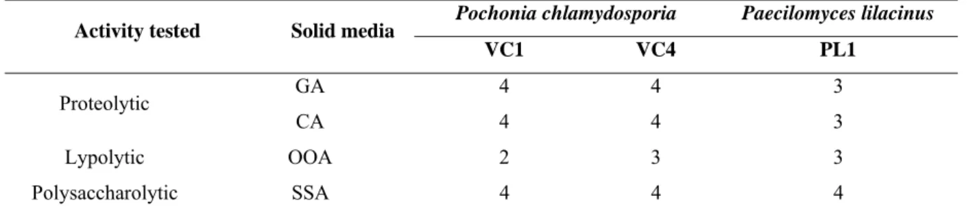
2.4. Growth and enzymatic production of P. chlamydosporia (VC1 and VC4) and P. lilacinus (PL1) on solid medium……… 103
2.5. Ovicidal effect of P. chlamydosporia and P. lilacinus on eggs of O. equi… 104 2.6. Assay B……….. 104
2.7. Proteolytic activity at different incubation time and protein content of the crude extract of P. chlamydosporia (VC1 and VC4)………. 105
3. Results……… 105
4. Discussion………. 108
References... 113
LISTA DE TABELAS
Página
CAPÍTULO 2
Table 1 - Percentages and standard deviations of the ovicidal activity of nematophagous fungi Duddingtoniaflagrans (AC001), Monacrosporium thaumasium (NF34a), Pochonia chlamydosporia (VC1 and VC4) and control group without fungi on eggs of Oxyuris equi
at five, 10 and 15 interaction days.
33
Table 2 - Percentages and standard deviations of the ovicidal activity of nematophagous fungi Duddingtonia flagrans (AC001), Monacrosporium thaumasium (NF34a), Pochonia chlamydosporia (VC1 and VC4) and control group without fungi on eggs of Austroxyuris finlaysoni at five, 10 and 15 interaction days.
33
CAPÍTULO 3
Table 1 – Percentages of ovicidal activity for the nematophagous fungus Pochonia chlamydosporia (VC4) and the control, without fungal treatment, against eggs of Oxyuris equi at faeces collection times 8, 12, 24, 36, 48 and 72 hours, after 30 days of interaction.
49
CAPÍTULO 4
Página
CAPÍTULO 6
Table 1 - High (+1) and low levels (-1) with the eight selected variables (g/l) : glucose (A), casein (B), K2HPO4 (C), MgSO4 (D), ZnSO4 (E), FeSO4 (G), CuSO4 (H) and temperature (ºC) in the Placket-Burman experimental design.
90
Table 2 - Matrix of the Plackett-Burman experimental design with different production levels (-1 and 1) of proteases (U/ml) by the nematophagous fungus Duddingtonia flagrans
(AC001).
90
Table 3 - Factors (g/l): glucose, casein, K2HPO4, MgSO4, ZnSO4, FeSO4, CuSO4 and temperature (ºC) in the Plackett-Burman experimental design and significance ranking.
90 Table 4 - Experiments used for response surface methodology (RSM) with three dependent variables (casein MgSO4 and CuSO4), with five levels each (-1, +1, 0, -1.68179 and 1.681793) and proteolytic activity values.
90
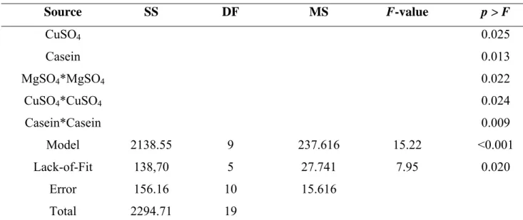
Table 5 - Analysis of variance (ANOVA) for the response equation developed for protease production by Duddingtonia flagrans (AC001).
90
CAPÍTULO 7
Table 1 - Enzymatic activity of Pochonia chlamydosporia (VC1 and VC4) and
Paecilomyces lilacinus (PL1) on solid agar medium supplemented with gelatin (GA), casein (CA), olive oil (OOA) and starch (SSA) independent of incubation time.
Página
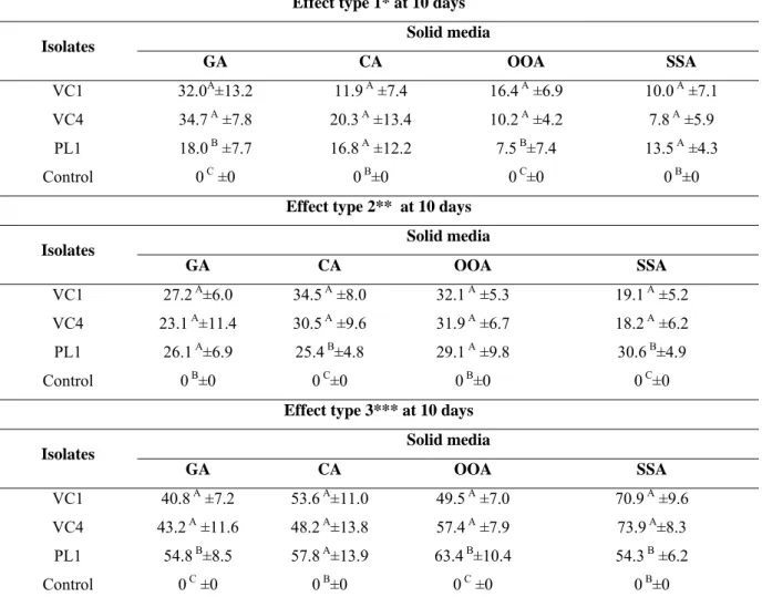
Table 2 - Percentages of ovicidal activity and standard deviations of Pochonia chlamydosporia (VC1 and VC4) and Paecilomyces lilacinus (PL1) on solid agar medium supplemented with gelatin (GA) casein (CA), olive oil (OOA) and starch (SSA) and control without fungi against eggs of Oxyuris equi at 10 days of interaction.
119
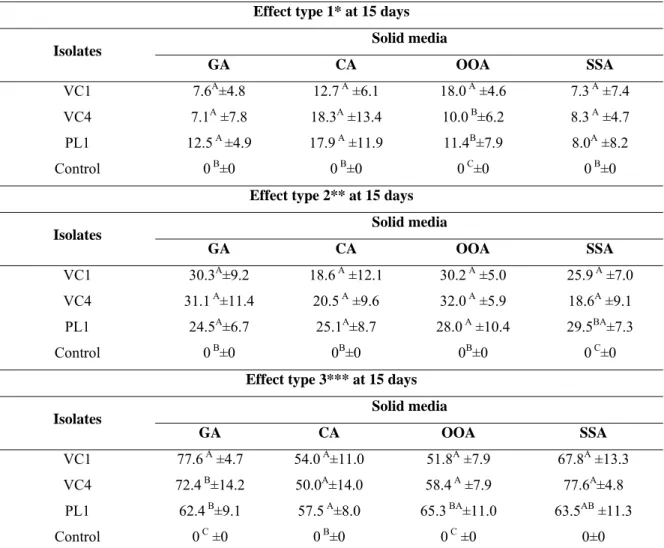
Table 3 – Percentages of ovicidal activity and standard deviations of Pochonia chlamydosporia (VC1 and VC4) and Paecilomyces lilacinus (PL1) on solid agar medium supplemented with gelatin (GA) casein (CA), olive oil (OOA) and starch (SSA) and control without fungi against eggs of Oxyuris equi at 15 days of interaction.
LISTA DE FIGURAS
Página
CAPÍTULO 1
Fig. 1 Monthly means of eggs per gram of feces (EPG) of fungus-treated and control animals collected from May to October 2007, Viçosa – MG – Brazil. Significant difference (p<0.01) between the treated group and the control denoted by asterisk- Tukey test.
20
Fig. 2 Mean monthly number of cyathostomin larvae recovered from coproculture of fungus-treated horses and control group collected from May to October 2007, Viçosa – MG – Brazil. Significant difference (p<0.01) between the treated group and the control denoted by asterisk- Tukey test.
20
Fig. 3 Monthly counts of number of infective nematode larvae per kilogram of dry matter recovered from pastures of fungus-treated horses and control collected in sampling distances up to 20 cm and 20-40 cm from fecal pats, from May to October 2007, Viçosa – MG- Brazil.
21
Fig.4 Averages of maximum, average and minimum monthly temperatures (ºC) and air relative humidity (%) recorded from May to October 2007, Viçosa – MG- Brazil.
21
Fig. 5 Monthly rainfall (mm3) recorded from May to October 2007, Viçosa – MG- Brazil.
Fig. 6 Monthly means of weight (kg) of fungus-treated horses and control from May to October 2007, Viçosa – MG- Brazil. Significant difference (p<0.01) between the treated group and the control denoted by asterisk- Tukey test.
22
CAPÍTULO 4
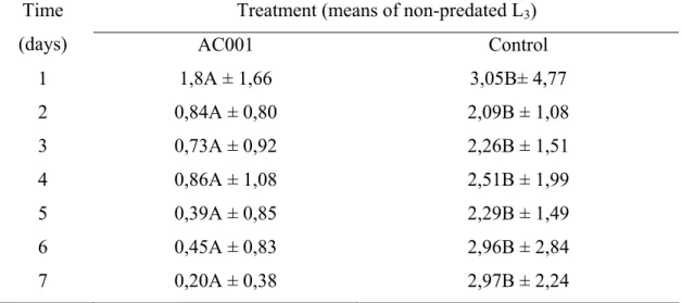
Fig. 1 Mean number of non-predated cyathostomin infective larvae (L3) recovered in 2% water-agar by the Baermann method at the seventh treatment day after interaction with the fungal isolate Duddingtonia flagrans (AC001) and group control . Lines on Bars represent standard deviation. Means followed by at least one common capital letter (A) in the row are not significantly different by the Tukey's test at a 1% probability level.
60
CAPÍTULO 5
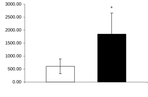
Fig. 1 Mean number and standard deviation (bar) of cyathostomin larvae recovered from plates of the treated group after 24 hours of interaction with the crude enzymatic extract of Pochonia chlamydosporia (VC4) and in the control group . Asterisk denotes significant difference (p<0.01) between the fungus-treated group and the control - Tukey's test at a 1% probability level.
75
Fig. 2 Mean number and standard deviation (bar) of cyathostomin larvae recovered from coproculture of the group treated with the crude enzymatic extract of
Pochonia chlamydosporia (VC4) and the control , after 8 days. Asterisk denotes significant difference (p <0.01) between the fungus-treated group and the control - Tukey's test at a 1% probability level.
Página
CAPÍTULO 6
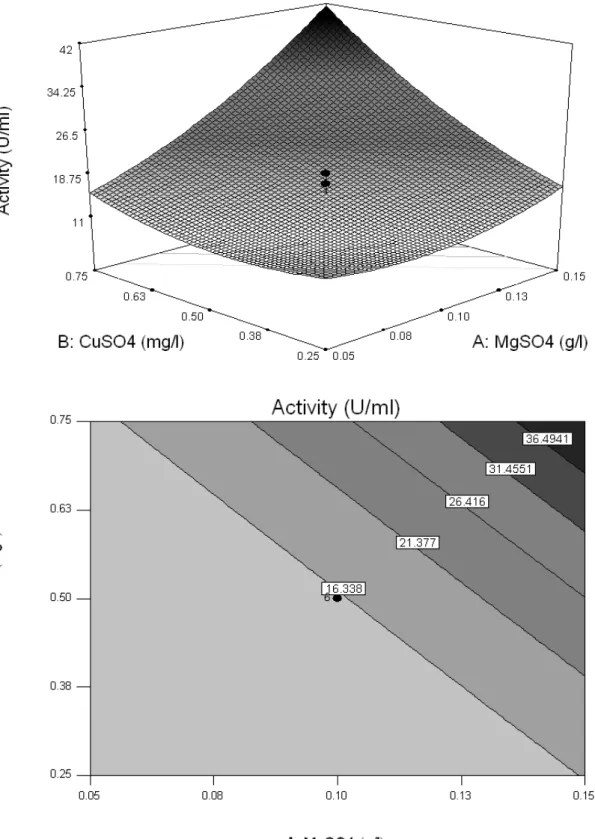
Fig. 1 Response surface curve (RSM) and contour map of the protease production by
Duddingtonia flagrans (AC001) and the interaction between the variables MgSO4 and casein (a); CuSO4 and casein (b); MgSO4 and CuSO4 (c).
97
Fig. 2 Mean number and standard deviation of L3 cyathostomins recovered after 24 hours of interaction in Petri dishes with optimized enzyme extract of
Duddingtonia flagrans (AC001) and control group . Significant difference (p <0.01) between treated group and control (*) – Tukey’s test.
98
CAPÍTULO 7
Fig. 1 Linear regression curves for effect type 3 on solid agar media supplemented with gelatin (GA) casein (CA), olive oil (OOA) and starch (SSA) against eggs of
Oxyuris equi at 10 and 15 days of interaction.
121
Fig. 2 Proteolytic activity of the fungal extracts of Pochonia chlamydosporia (VC1 and VC4) at different incubation times (15, 30, 45 and 60 minutes).
RESUMO
BRAGA, Fábio Ribeiro, D.Sc., Universidade Federal de Viçosa, fevereiro de 2011. Controle biológico de nematodioses intestinais de equinos por fungos nematófagos. Orientador: Jackson Victor de Araújo. Co-orientadores: José Humberto de Queiroz e Laércio dos Anjos Benjamin.
Com os objetivos de: testar uma formulação peletizada em matriz de alginato de sódio contendo massa miceliana do fungo Duddingtonia flagrans (isolado AC001) no controle biológico de ciatostomíneos de equinos criados a campo; avaliar a ação in vitro
de quatro isolados de fungos nematófagos dos gêneros D. flagrans (isolado AC001), Monacrosporium thaumasium (isolado NF34a) e Pochonia chlamydosporia (isolados VC1 e VC4) sobre ovos de Oxyuris equi e Austroxyuris finlaysoni; avaliar a atividade ovicida in vivo do fungo P. chlamydosporia (isolado VC4) sobre ovos de O. equi; avaliar a capacidade predatória in vitro do fungo nematófago D. flagrans sobre larvas infectantes (L3) de ciatostomíneos de equinos; testar a ação do extrato fúngico de P.
chlamydosporia (VC4) sobre a eclosão de ovos de ciatostomíneos; otimizar a produção de protease pelo fungo D. flagrans (isolado AC001) e sua ação larvicida sobre ciatostomíneos de equinos e; avaliar a produção enzimática do extrato bruto e a atividade ovicida dos fungos P. chlamydosporia (VC1 e VC4) e Paecilomyceslilacinus
produção de proteases pelo fungo D. flagrans (AC001) em meio líquido demonstrando também que o extrato otimizado teve ação larvicida sobre ciatostomíneos. Com a produção enzimática do extrato bruto e sua atividade ovicida dos fungos P. chlamydosporia (VC1 e VC4) e P. lilacinus (PL1) sobre ovos de O. equi, observou-se que os fungos P. chlamydosporia (VC1 e VC4) e P. lilacinus (PL1) cresceram e produziram atividade enzimática nos meios ágar suplementados com gelatina (GA), caseína (CA), óleo de oliva (OOA) e amido (SSA). Além disso, ao final do experimento (15 dias) os isolados testados de VC1, VC4 e PL1 demonstraram atividade ovicida (efeito do tipo 3) de (77,6%; 54,0%; 51,8% e 67,8%), (72,4%; 50,0%; 58,4% e 77,6%) e 62,4%; 57,5%; 65,3% e 63,5%), respectivamente, nos meios GA, CA, OOA e SSA. Os resultados demonstrados sugerem que os fungos nematófagos D. flagrans (AC001),
Monacrosporium thaumasium (NF34), P. chlamydosporia (VC1 e VC4) e P. lilacinus
ABSTRACT
BRAGA, Fábio Ribeiro, D.Sc., Universidade Federal de Viçosa, February 2011. Biological control of equine intestinal nematodiosis by nematophagous fungi. Adviser: Jackson Victor de Araújo. Co-advisores: José Humberto de Queiroz and Laércio dos Anjos Benjamin.
With the aim of: testing a pellet formulation in sodium alginate containing
Duddingtonia flagrans (isolate AC001) mycelial fungus for biological control cyathostomins of horses raised in field, evaluate in vitro effect of D. flagrans (isolate AC001), Monacrosporium thaumasium (isolate NF34a) and P. chlamydosporia (isolates VC1 and VC4) on Oxyuris equi and Austroxyuris finlaysoni eggs; evaluate the in vivo
ovicidal activity of P. chlamydosporia (isolate VC4) fungus on Oxyuris equi eggs and to evaluate the predatory capacity in vitro of D. flagrans nematophagous fungus on horses cyathostomin infective larvae (L3); test P. chlamydosporia (isolate VC4) fungal extract action on cyathostomins eggs eclosion; optimize the protease production by D. flagrans (isolate AC001) and its larvicidal action on horses cyathostomin and, to evaluate crude enzymatic extract production and ovicidal activity of P. chlamydosporia
(isolates VC1 and VC4) and Paecilomyces lilacinus (PL1) on O. equi eggs, tests were conducted in experimental laboratory and in field conditions. The animals in the group treated with D. flagrans (AC001) pellets showed a different (p<0.01) reduction in egg count per gram of feces and in fecal cultures compared to control animals. In vitro
laboratory tests results showed that P. chlamydosporia (VC1 and VC4) fungus influenced negatively O. equi and A. finlaysoni eggs. It was also observed that the isolate (VC4) remained viable and maintained its ovicidal activity (p<0.01) after passing through the gastrointestinal tract of horses when compared to control (without fungus). Moreover, we observed a significant reduction (p<0.01) of 93.64% in cyathostomin L3 recovered from Petri dishes in the group treated with D. flagrans
(AC001) comparing to the control group. However, in relation to P. chlamydosporia
1. INTRODUÇÃO GERAL
Criações de animais com fins produtivos, com destaque para os bovinos, ovinos, caprinos e equinos apresentam drásticas perdas econômicas associadas principalmente ao parasitismo por endoparasitos e ectoparasitos. No Brasil grande parte da criação ainda é feita em regime de pasto, o que leva as constantes infecções por parasitos presentes nas pastagens (Anualpec, 2003). Por outro lado, na criação comercial de animais silvestres em países com grandes diferenças regionais como o Brasil existem alguns entraves à produção comercial desses animais, e, dentre esses, podem-se mencionar as nematodioses gastrintestinais, que merecem destaque (Bonuti et al., 2002).
Nematóides gastrintestinais são comuns em equinos, representando um grupo de grande importância no Brasil, já que grande parte do rebanho encontra-se infectado. Além disso, esses animais apresentam uma grande variedade de parasitos em sua fauna helmíntica, e algumas espécies/gêneros são de relevada importância, como: Parascaris equorum, Anoplocephala perfoliata, Oxyuris equi, Cyathostomum spp., e Strongylus spp (Kaplan, 2002). O. equi relatado em cavalos no ano de 430 (DC) por Hipocrates é habitante comum principalmente do cólon menor desses animais, mas ocasionalmente pode ser encontrado também no cólon maior (Morgan & Hawkins, 1949). Segundo Urquhart et al. (1998) e Bowman et al. (2006), a maioria dos efeitos patogênicos desse parasito no intestino se deve aos hábitos alimentares das larvas infetantes de terceiro e de quarto estádios que resultam em pequenas erosões da mucosa. Contudo, um efeito mais importante é a irritação causada pelas fêmeas adultas durante a oviposição. Segundo Bowman et al. (2006) as fêmeas grávidas de O. equi migram para a parte inferior do cólon e reto e para fora do ânus para aderir massas de ovos pele do ânus e da região perianal. Essas massas de ovos consistem em um fluido pegajoso cinza-amarelado contendo de 8 mil a 60 mil ovos, causando intenso prurido anal.
Entre os nematóides parasitos gastrintestinais de animais silvestres está o
Austroxyuris finlaysoni, um oxyurídeo de marsupiais (Bowman et al., 1996). Gomes et al. (2003) mencionam que, alguns desses marsupiais, como o gambá de orelha branca (Didelphis albiventris) possui importância médico-veterinária por ser reservatório de nematóides, fungos como o Histoplasma capsulatum e também do Trypanosoma cruzi.
O controle das nematodioses gastrintestinais foi feito ao longo das décadas através do uso de antihelmínticos, muitas vezes de maneira indiscriminada e sem estratégias de controle adequadas, fato que conduziu a resistência destes parasitos a maior parte das classes de antihelmínticos disponíveis, principalmente aos benzimidazóis (Kaplan, 2002; Matthews et al., 2004). Nesse contexto, o controle biológico realizado com fungos nematófagos tem sido utilizado como uma alternativa de controle natural de formas infectantes (ovos e ou larvas) de nematóides, pois capturam e causam sua destruição. São divididos em três grupos: predadores, ovicidas e endoparasitas (Araújo et al., 2004). Braga et al. (2009a, b) mencionam que esses fungos nematófagos podem ser utilizados com sucesso no controle de nematóides parasitos gastrintestinais de animais domésticos e silvestres, e dentre esses, os pertencentes à subfamília Cyathostominae e o O. equi, que possuem grande prevalência em boa parte do território brasileiro.
No grupo dos predadores, destaca-se a espécie Duddingtonia flagrans considerada a mais promissora devido a sua grande produção de clamidósporos que são estruturas resistentes. Além disso, é classificada como predadora e produz uma série de enzimas, e dentre essas as proteases (Park et al., 2001; Meyer & Wiebe, 2003; Braga et al., 2010a). As espécies do gênero Monacrosporium são predadoras e caracterizadas por produzirem um único conídio em cada conidióforo e pela produção de redes adesivas, formando hifas septadas e ramificadas (Mota et al., 2003; Campos et al., 2008).
No grupo de fungos ovicidas destacam-se as espécies Pochonia chlamydosporia e
Paecilomyces lilacinus. O fungo P. chlamydosporia produz enzimas extracelulares do tipo serino proteases que desenvolvem um papel importante na infecção e na destruição (atividade ovicida) dos ovos de geohelmintos (Segers et al., 1994; Braga et al., 2008a, b, 2009a; 2010b). P. lilacinus tem sido utilizado com sucesso em condições laboratoriais e a campo no controle biológico de ovos de vários gêneros de helmintos parasitos gastrintestinais (Araujo et al., 2010).
(Joo and Chang, 2005). Por outro lado, as proteases produzidas por fungos nematófagos têm sido extensivamente estudadas, uma vez que tem relação na degradação da cutícula dos nematóides (Tunlid and Jansson, 1991). Gupta et al. (2002) mencionam que vários trabalhos têm objetivado a melhoria da produção de proteases por microorganismos por meio do uso de ferramentas estatísticas como o design Plackett–Burman e a metodologia
de superfície de resposta (RSM). De acordo com Djekrif-Dakhmouche et al. (2006) o uso
de ferramentas estatísticas como o design Plackett–Burman e a RSM têm sido utilizados para a otimização de meios de cultura. Por outro lado, segundo Hajji et al. (2008) tais ferramentas também são aplicadas para o entendimento das interações entre vários
parâmetros usando um número mínimo de experimentos.
A metodologia de superfície de resposta é uma técnica que avalia as relações existentes entre um grupo de fatores experimentais controlados e os resultados observados de um critério selecionado (Ambati & Ayyanna, 2001).Isso inclui design fatorial e análise de regressão, que ajudam na validação dos fatores efetivos e na construção de blocos para o estudo das suas interações, servindo também para a seleção das condições ótimas das variáveis para uma resposta escolhida (Sharma et al., 2007).
Além disso, cada fungo tem a sua exigência específica em condições especiais o que causaria a maximização da produção dessas enzimas. Ainda, segundo Araújo et al. (2004) o desenvolvimento de formulações fúngicas para uso no controle biológico é um dos principais passos para a produção comercial destes microorganismos. Sendo assim, este fato poderia contribuir para pesquisas futuras que visem a produção industrial em larga escala de fungos nematófagos.
2. OBJETIVOS
1. Testar uma formulação peletizada em matriz de alginato de sódio contendo massa miceliana do fungo Duddingtonia flagrans (isolado AC001) no controle biológico de ciatostomíneos de equinos criados a campo.
2. Avaliar a ação in vitro de fungos nematófagos das espécies D. flagrans (AC001),
Monacrosporium thaumasium (NF34a) e Pochonia chlamydosporia (VC1 e VC4) sobre ovos de Oxyuris equi e Austroxyuris finlaysoni por meio de dois ensaios experimentais (A e B) nos intervalos de cinco, 10 e 15 dias de interação.
3. Avaliar in vivo o fungo nematófago P. chlamydosporia (VC4) quanto a sua capacidade de passagem pelo aparelho gastrintestinal de equinos, sua resistência e viabilidade, observando sua capacidade predatória sobre ovos de O. equi.
4. Avaliar in vitro a capacidade predatória do fungo D. flagrans (AC001) sobre larvas infectantes de ciatostomíneos.
5. Testar a ação do extrato do fungo P. chlamydosporia (VC4) sobre ovos de ciatostomíneos e em culturas de fezes em dois ensaios experimentais (A e B).
6. Otimizar a produção de protease do fungo D. flagrans (AC001) pelo método de superfície de resposta e avaliar sua ação larvicida sobre ciatostomíneos de equinos.
CAPÍTULO 1
Biological control of horse cyathostomin (Nematoda: Cyathostominae) with the nematophagous fungus Duddingtonia flagrans in tropical southeast Brazil
Abstract
The viability of a fungal formulation using the nematode predator fungus Duddingtonia flagrans was evaluated for the biological control of horse cyathostomin. Two groups (fungus-treated and control), consisting of eight crossbred mares each, 3 to 18 years of age, were fed on Cynodon sp. pasture naturally infected with equine cyathostome larvae. Each animal of the treated group was orally administered sodium alginate mycelial pellets (1g/10 kg live weight/ week), during six months. Animals of the control group were not treated. Significant reduction (p<0.01) in the number of eggs per gram of feces and coprocultures was found for animals of the fungus treated group compared with the control group. There was difference (p<0.01) of 78.5% in pasture samples collected up to (0-20 cm) between the fungus treated group and the control group, during the experimental period (May to October). Difference of 82.5% (p <0.01) was found between the fungus treated group and the control group in the sampling distance (20-40 cm) from fecal pats. In the last three months of the experiment (August, September and October), animals treated with the fungus showed difference (P <0.01) for weight gain, 38 kg more than the control group. The treatment of horses with sodium alginate pellets containing the nematophagous fungus D. flagrans can be effective against cyathostomin in tropical southeastern Brazil.
1. Introduction
A large variety of helminths are known to parasite horses. Nematodes, mainly cyathostomin species, are the most common and important among them. Also known as small strongyles, cyathostomin infections are responsible for causing anemia, weight loss, intestinal colic, and death in horses (Assis and Araújo, 2003). They are the most prevalent parasites in horses, present throughout the year in the pasture, having however greatly varied distribution in different age groups (Barbosa et al., 2001; Quinelato et al., 2008).
Klei and Chapman (1999) reported field data suggesting that horses can acquire resistance to helminths with age, which is confirmed by the reduced parasite load and egg count in feces. This response is slow and inconsistent in most animals and unrelated to the intensity of previous contact with parasite.
Kaplan (2002) and Matthews et al. (2004) discussed that worm control in horses is usually carried out with antihelmintic drugs, which have not been totally effective for the control of these nematodes since their action is restricted to adult parasites and the occurrence of resistance.
However, the continued use of the same antihelmintic class, as well as the rapid rotation of compound groups, introduction of resistant worms and the use of doses lower than the recommendation should be avoided (Mota et al., 2003). Biological control using natural nematode antagonistic fungi is among the most viable alternatives. These organisms comprise different types of fungi divided into predators, endoparasites and opportunists, whose action is concentrated in the fecal environment and directed against free-living parasitic larvae. In the group of predators, the species Duddingtonia flagrans
stands out as the most promising for the control of gastrintestinal nematodiasis in domestic animals (Terril et al., 2004; Dias et al., 2007a). However, in order to be used as a biological control agent, these nematophagous fungi must have ability for nematode capture and survive passage through gastrointestinal tract (Waller et al., 1994).
Sodium alginate based formulations containing D. flagrans mycelial mass have been experimentally evaluated against parasitic nematodes of animals in laboratory and field conditions (Araújo and Sampaio, 2000; Araújo et al., 2000, Dias et al., 2007b). However, none these formulations has been developed to be used in the control of parasitic nematodes of horses in the field.
2. Material and methods
2.1. Organisms
D. flagrans isolate (AC001), nematode-trapping fungus belonging to the genus
Duddingtonia, was kept in test tubes at 4oC containing 2% corn-meal-agar (2% CMA) in the dark. This isolated originated from a Brazilian soil was obtained by the soil sprinkling technique (Duddington, 1955), modified by Santos et al. (1991).
For induction of fungal mycelia, culture disks, approximately 5 mm in diameter, from fungal isolates in 2% CMA were transferred to 250 mL Erlenmeyer flasks with 150 mL liquid potato-dextrose medium (Difco), pH 6.5, incubated under agitation (120 rpm), in the dark at 26oC, for ten days. Mycelia were then removed for preparation of pellets using sodium alginate as described by Walker and Connick (1983) and modified by Lackey et al. (1993).
2.2. In vivo experimental assay
The experiment was conducted at the horse experimental sector of the Federal University of Viçosa, Viçosa, MG-Brazil, latitude 20°45’20” S, longitude 42°52’40” W, from May to October 2007.
In the beginning of the experiment, 3-18 year old crossbred females were previously dewormed by oral administration of 200 µg/kg live weight Ivermectin 1% and 6.6mg /kg live weight Pyrantel Pamoate (Centurion Vallé®, Montes Claros-Minas Gerais, Brasil).
During the experiment the animals were fed daily with 2 kg of horse commercial ration with 14% soybean meal, 83.1% corn meal, 14.5% salt, 1.5% limestone and 14% protein.
Once a week, after the mares had been moved to the pastures, fresh feces were collected directly from the rectum, 72 h after the treatment, for egg count per gram of feces (EPG), according to Gordon and Withlock (1939) and modified by Lima (1989).
Coprocultures were established simultaneously with EPG counts; 20g of feces were mixed with ground, moistened and autoclaved industrial vermiculite (NS Barbosa Ind. Com. ®) and taken to an oven at 26oC, for 8 days, to obtain cyathostome larvae. Larvae were then identified to the genus level as described by Bevilaqua et al. (1993). EPG and larvae recovered from coprocultures of animals of both treated and control groups were recorded and percentage of larval reduction was determined according to Mendoza-de-Guives et al. (1999).
Reduction (%) = [Mean L3 recovered from control group – Mean L3 recovered from treated group]
_____________________________________________________________
Mean L3 recovered from control group
Every fifteen days, two pasture samples were collected from both treated and control groups, from each pasture, in a zigzag pattern from several and alternated points, 0-20 and 20-40 cm away from fecal pats, in each pasture of the different groups, according to Amarante et al. (1996). Pasture sample collections were always carried out in the morning at 8am. Then, a 500g pasture sample was weighed, and parasitic nematode larvae were recovered according to Lima (1989). The samples were incubated in a drying oven at 100oC, for three days, to determine dry matter. Data were transformed into larvae/kg of dry matter.
Climate data referring to averages of maximum, average and minimum monthly temperatures, air relative humidity and monthly rainfall were daily recorded in a meteorological station in the area.
The egg count curves (EPG) originated from the coprocultures, number of infective larvae recovered from pasture (L3), correlation between EPG and recovered L3 and animal weight were compared over the period of the experiment. Data were transformed in log (x+1) and then examined by analyses of variance (ANOVA), followed by Tukey’s multiple comparison test with 1% probability. The analyses were performed using the BioEstat 3.0 Software (Ayres et al., 2003).
3. Results
Figure 1 shows the monthly mean EPG counts. EPG of animals treated with D. flagrans was lower than the control group, especially in the last four months of the experiment, with significant difference (p<0.01), in which the EPG monthly mean of the treated group was 46.2 % lower than the control group. July, August, September and October showed smaller percentages of EPG reduction for fungus treated animals than the control group; 35.4%, 73.2%, 64.3% and 30.5%, respectively. Moreover, fungus-treated animals had EPG values lower than the control group throughout the experiment. Figure 2 shows coproculture data. There was significant difference (p<0.01) between the results of fungus-treated animals and the control group in the last four months of the experiment (July, August, September and October) with larval reduction of 57.2%, 59.4%, 68.5% and 51% respectively.
Weights of animals from both groups are shown in Figure 6. There was no significant difference (p>0.01) for animal weight during the three first months of the year (May, June and July) between the two groups. However, in the last three months of the experiment (August, September and October), significant differences (p<0.01) of 9.74%, 10.26% and 12.21%, respectively, were found for the weight between treated and non-treated animals.
4. Discussion
According to Amarante et al. (1996), EPG counts constitute a parameter that allows evaluation of infection levels in animals and pasture infestation levels by gastrointestinal nematode parasites. A number of studies on D. flagrans using horses and ruminants recorded average monthly EPG counts lower for treated animals than for the control group (Baudena et al. 2000b; Knox and Faedo, 2001; Fontenot et al., 2003; Araújo et al., 2006; Paraud et al., 2007). The efficacy of D. flagrans application on gastrointestinal of ruminants was also demonstrated in the work of Dimander et al. (2003). These findings are in agreement with results obtained in the present work, confirming that the fungus acts on the infective forms in the fecal environment, with consequently decrease in EPG. There is nevertheless a lack of studies involving nematophagous fungi and equine cyathostomin (Bird and Herd, 1985; Baudena et al., 2000b).
et al., 2006). Only the occurrence of small strongyles (Cyathostominae) was observed after the coprocultures, following the parameters described by Bevilaqua et al (1993). Silva et al. (1993) reported that the subfamily Cyathostominae has a high prevalence in a large part of the Brazilian territory, and Carvalho et al. (1998) identified nineteen species of small strongyles in necropsied horses from the state of Minas Gerais. The importance of these parasites for horses is directly related with larval cyathostomosis, a potentially fatal syndrome in most cases, and the high resistance of most gastrointestinal nematode parasites to routine antihelminthics (Reinemeyer, 1986, Reinemeyer et al., 1986).
The number of larvae recovered in the distances 0-20 cm and 20-40 cm from fecal pats (Figure 3) is likely to be directly related with the use of nematophagous fungi that act directly on the L3 present in pastures, making it clear that D. flagrans was responsible for the satisfactory reduction of environmental contamination (Araújo et al., 2004).
for recovering these larvae. The authors also argued that horses might be infected during the whole year in tropical climates, since L3 are always present in the pastures and, besides, the grass type can affect their recovery. Langrová et al. (2003), in central Europe, suggested that L3 respond to rain through dispersion within the vegetation, existing a moderate correlation between moisture and the L3 number in the pasture.
Courtney (1999) observed that during the dry period L3 development is slower, but they survive longer. Still, Fernández et al. (1997) and Baudena et al. (2000a) argued that possibly the survival of these parasites in the environment is strongly related with temperature and that few larvae would be found in feces in the summer. Baudena et al. (2000a) recorded field data in southern Louisiana, a region with subtropical climate in The United States, which suggested that months with milder temperature would have larger number of infective larvae present in the pasture. Such information is in accordance with the results found in this work in which the largest number of larvae recovered in pastures was found during months of milder temperature (Figure 3). Peña et al. (2002) and Chandrawathani et al. (2004) reported D. flagrans being able to reduce more than 90% of infective larvae present in fecal pats of ruminants.
Fontenot et al. (2003) also discussed that besides D. flagrans decreasing infectious forms of gastrointestinal nematode parasites in pastures, it would avoid contamination of new animals entering these sites.
The correlation coefficient between EPG and infective larvae recovered from pastures of group 1 within 0-20 cm from fecal pats was 0.0662; and for the distance 20-40 cm was 0.0416. For group 2, the correlation coefficient between EPG and infective larvae recovered within 0-20 cm from the fecal pats was –0.0394 and within 20-40 cm was 0.0401. These results showed weak, non-significant correlations, close to zero, nevertheless, as Dias et al. (2007b) pointed out, there might be dependence between EPG and infective larvae recovered from pastures even if the correlations are null. Besides, the availability of larvae on pasture may be determined by contamination from animals, as well as environmental factors, parasite and host (Lima et al., 1997).
In a work carried out with two fungal isolates of the genus Monacrosporium, Assis Araújo (2003) found fungal mycelia in horse feces up to 96 hours after passing through the gastrointestinal tract of horses. Therefore, in this work, for an efficient weekly coverage, D. flagrans was applied twice a week.
In Malaysia, the effectiveness of daily administration of D. flagrans to sheep was confirmed by Chandrawathani et al. (2003). Terrill et al. (2004) also reported reduction of larvae in feces of goats infected with predominantly H. contortus. They also found that the daily administration of fungi (D. flagrans) was more effective than every two or three days. The frequency of treatments in this work promoted reduction of pasture
contamination, mainly the weekly treatment.
The difference (p<0.01) observed in the weight gain of the treated animals compared to the control group was probably due to lower parasite load of animals that received pellets containing mycelial mass of the nematophagous fungus D. flagrans, which may have contributed to a better food conversion of treated animals. These results are similar to those reported by Dias et al. (2007a) on weight gain of cattle treated with pellets containing mycelial mass of D. flagrans.
The findings reported in this study suggest that the nematophagous fungus D. flagrans could be used in an integrated program to control horse cyathostomin in southeastern Brazil. It would be useful to carry out a previous anthelmintic treatment to decrease parasite load in animals and therefore the EPG, and starting from that to supply animal feed added with the fungus to control the larval forms present in the environment and thus prevent reinfection.
4. Conclusion
Treatment of horses with pellets containing mycelial mass of the nematophagous fungus D. flagrans can be effective to control cyathostomin in tropical southeastern Brazil.
Acknowledgements
The authors would like to thank Fapemig and CNPq for the financial support and grant concession.
References
Amarante, A.F.T., Padovani, C.R., Barbosa, M.A., 1996. Contaminação de larvas de nematóides gastrintestinais parasitos de bovinos e ovinos em Botucatu-SP. Rev. Bras. Parasitol. Vet. 5, 65-73.
Araújo, J.V., Freitas, B.W., Vieira, T.C., Campos, A.K., 2006. Avaliação do fungo predador de nematóides Duddingtonia flagrans sobre larvas infectantes de
Haemonchus contortus e Strongyloides papillosus de caprinos. Rev. Bras. Parasitol. Vet. 15, 76-79.
Araújo, J.V., Mota, M.A., Campos, A.K., 2004. Controle de helmintos de animais por fungos nematófagos. Rev. Bras. Parasitol. Vet. 13, 165-169.
Araújo, J.V., Sampaio, W.M., 2000. Effects of temperature, mineral salt and passage through gastrointestinal tract of calves on alginate formulation of Arthrobotrys robusta. Rev. Bras. Parasitol. Vet. 9, 55-59.
Araújo J.V., Sampaio, W.M., Vasconcelos, R.S., Campos, A.K., 2000. Effects of different temperatures and mineral salt on pellets of Monacrosporium thaumasium - a nematode-trapping fungus. Vet. Arhiv, 80, 181-190.
Assis, R.C.L., Araújo, J.V., 2003. Avaliação da viabilidade de duas espécies de fungos predadores do gênero Monacrosporium sobre ciatostomíneos após a passagem pelo trato gastrintestinal de eqüinos em formulação de alginato de sódio. Rev. Bras. Parasitol. Vet. 12, 109-113.
Ayres, M., Ayres, J.R.M., Ayres, D.L., Santos, A.S., 2003. Aplicações estatísticas nas áreas de ciências biológicas. Belém: Sociedade civil mamirauá: Brasília CNPq, p. 290.
Barbosa, O.F., Rocha, U.F., Silva, G.S., Soares, V.E., Veronez, V.A., Oliveira, G.P., Landim, V.J.C., Costa, A.J., 2001. A survey on Cyathostominae nematodes
Baudena, M.A., Chapman, M.R., Larsen, M., Klei, R.R., 2000a. Efficacy of the nematophagous fungus Duddingtonia flagrans in reducing equine cyathostome larvae on pasture in south Lousiana. Vet. Parasitol. 89, 219-230.
Baudena, M.A., Chapman, M.R., French, D.D., Klei, R.R., 2000b. Seasonal development and survival of equine cyathostome larvae on pasture in south Louisiana. Vet. Parasitol. 88, 51-60.
Bevilaqua, C.M.L., Rodrigues, M.L., Cocordet, D., 1993. Identification of infective larvae of some common Eqüinos strongylids of horses. Rev. Méd. Vét. 144, 989-995.
Bezerra, S.Q., Couto, M.C.M., Souza, T.M., Bevilaqua, C.M.L., Anjos, D.H.S., Sampaio, I.B.M., Rodrigues, M.L.A., 2007. Cyathostominae (strongylidae-cyathostominae) horse parasites: experimental ecology of free living stages on pasture tifton 85 (cynodon spp. cv. tifton 85) in baixada fluminense, RJ, Brazil. Parasitol. Latinoam. 62, 27-34.
Bird, J., Herd, R.P., 1985. In vitro assessment of two species of nematophagous fungi
(Arthrobotrys oligospora and Arthrobotrys flagrans) to control the development of infective cyathostome larvae from naturally infected horses. Vet. Parasitol. 56, 181-187.
Carvalho, R.O., Silva, A.V.M., Santos, H.A.; Costa, H.M.A., 1998. Nematodes Cyathostominae parasites of Equus caballus in the state of Minas Gerais, Brasil. Rev. Bras. Parasitol. Vet. 7, 165-168
Chandrawathani, P., Jamnah, O., Adnan, M., Waller, P.J., Larsen, M., Gillespie, A.T., 2004. Fields studies on the biological control the nematodes parasites of sheep in the topics, using microfungus Duddingtonia flagrans. Vet. Parasitol. 120, 177-187.
Courtney, C.H., 1999. Seasonal transmission of equine cyathostomes in warm
climates. Vet. Parasitol. 85, 173-80.
Dias, A.S., Araújo, J.V., Campos, A.K., Braga, F.R., Fonseca, T.A., 2007a. Application of a formulation of the nematophagous fungus Duddingtonia flagrans in the control of cattle gastrointestinal nematodioses. World. J. Microbiol. Biotechnol. 28, 10.1007.
Dias, A.S., Araújo, J.V., Campos, A.K., Braga, F.R., Fonseca, T.A., 2007b. Relação entre larvas recuperadas da pastagem e contagem de ovos por grama de fezes (OPG) de nematóides gastrintestinais de bovinos na microrregião de Viçosa, Minas Gerais. Rev. Bras. Parasitol. Vet. 16, 33-36.
Dimander, S.O., Höglund, J., Uggla, A., Spörndly, E., Waller, P.J., 2003. Evaluation of gastro-intestinal nematode parasite control strategies for first-season grazing cattle in Sweden. Vet. Parasitol. 111, 192-209.
Duddington, C.L., 1955. Notes on the thecnique of handling predaceous fungi. Trans. Brithis. Mycol. Soc. 38, 97-103.
Faedo, M., Larsen, M., Dimander, S.O., Yeates, G.W., Höglund, J., Waller, P.J., 2002. Growth of the Fungus Duddingtonia flagrans in soil surrounding feces deposited by cattle or sheep fed the fungus to control nematode parasites. Biol. Control. 23, 64-70.
Fernández, A.S., Larsen, M., Nansen, P., Gronvold, J., Henriksen, S.A., Wolstrup, J., 1997. Effect of the nematode-trapping fungus Duddingtonia flagrans on the free-living stages of horse parasitic nematodes: a plot study. Vet. Parasitol. 73, 257-266.
Fontenot, M.E., Miller, J.E., Peña, M.T., Larsen, M., Gillespie, A., 2003. Efficiency of feeding Duddingtonia flagrans chlamydospores to grazing ewes on reducing availability of parasitic nematode larvae on pasture. Vet. Parasitol. 118, 203-213.
Hasslinger, M.A., Bittner, G. 1984. Zur Saisondynamik gives larven von pferdestrongyliden und deren beziehung zoom infektiosrisko auf weid. Zentralbblatt fürVeterinärmedizin. 31, 25-31.
Kaplan, R.M., 2002. Antihelmintic resistance in nematodes of horses. Vet. Res. Comm. 33, 491-507.
Klei, T.K., Chapman, M.R., 1999. Immunity in equine cyathostome infections. Vet. Parasitol. 85, 123–136.
Knox, M.R., Faedo, M., 2001. Biological control of field infections of nematode parasites of young sheep with Duddingtonia flagrans and effects of spore in take on efficacy. Vet. Parasitol. 101, 155-160.
Lackey, B.A., Muldoon, A.E., Jaffe, B.A., 1993. Alginate pellet formulation of
Hirsutella rossiliensis for biological control of plant-parasitic nematodes. Biol. Cont. 3, 155-160.
Langrová, I., Jankovská, I., Borovský, M., Fiala, T., 2003. Effect of climatic influences on the migrations of infective larvae of Cyathostominae. Vet. Méd. 48, 18 – 24.
Lima, W., 1989. Dinâmica das populações de nematóides parasitos gastrintestinais em bovinos de corte, alguns aspectos da relação parasito-hospedeiro e do comportamento dos estádios de vida livre na região do Vale do Rio Doce, MG, Brasil. 78 p. Tese (Doutorado) - Instituto de Ciências Biológicas da Universidade Federal de Minas-Gerais, Belo Horizonte.
Lima, W.S., Fakuri, E., Guimarães, M.P., 1997. Dinâmica de helmintoses de bovinos de leite na região metalúrgica de Minas Gerais. Rev. Bras. Parasitol. Vet. 6, 97-103.
Mendoza-De-Guives, P., Davies, K.G., Clarck, S.J., Behnke, J.M. 1999. Predatory behaviour of trapping fungi against srf mutants od Caenorhabditis elegans and different plant and animal parasitic nematodes. Parasitol.119, 95-104.
Mota, M.A., Campos, A.K., Araújo, J.V., 2003. Controle biológico de helmintos parasitos de animais: estágio atual e perspectivas futuras. Pesq. Vet. Bras. 23, 93-100.
Paraud, C., Pors, I., Chartier, C., 2007. Efficiency of feeding Duddingtonia flagrans
chlamydospores to control nematode parasites of first-season grazing goats in France. Vet. Res. Comm. 31, 305-315.
Peña, M.T., Miller, J.E., Fontenot, M.E., Gillespie, A., Larsen, M., 2002. Evaluation of Duddingtonia flagrans in reducing infective larvae of Haemonchus contortus in feces of sheep. Vet. Parasitol.103, 259-265.
Quinelato, S., Couto, M.C.M., Ribeiro, B.C., Santos, C.N., Souza, L.S., Anjos, D.H.S., Sampaio, I.B.M., Rodrigues, L.M.A., 2008. The ecology cyathostomin infective larvae (Nematoda-Cyathostominae) in tropical southeast Brazil. Vet. Parasitol. (in press).
Reinemeyer, C.R., 1986. Small strongyles – recent advances. Vet. Clinics North America- Equine Pract. 2, 281-312.
Reinemeyer, C.R., Herd, R.P., 1986. Anatomic distribution of cyathostome larvae in the horse. Am. J. Vet. Res. 47, 510-513.
Santos, M., Ferraz, S., Muchovej, J., 1991. Detection and ecology of nematophagous fungi from Brazil soils. Nematol. Bras. 15, 121-134.
Silva, A.V.M., Costa, H.M.A., Santos, H.A., Carvalho, R.O., 1993. Cyathostominae (Nematoda) parasites of Equus caballus in some Brazilian states. Vet. Parasitol. 86, 15-21.
Terril, T.H., Larsen, M., Samples, O., Husted, S., Miller, J.E., Kaplan, R.M., Gelaye, S., 2004. Capability of the nematode-trapping fungus Duddingtonia flagrans to reduce infective larvae of gastrointestinal nematodes in goat feces in the southeastern United States: dose titration and dose time interval studies. Vet. Parasitol. 120, 285-296.
Waghorn, T.S., Leathwick, D.M., Chen, L.-Y., Skipp, R.A., 2003. Efficacy of the nematode-trapping fungus Duddingtonia flagrans against three species of gastro-intestinal nematodes in laboratory faecal cultures from sheep and goats. Vet. Parasitol. 118, 227-234.
Walker, H.L., Connick, W.J., 1983. Sodium alginate for production and formulation of mycoherbicides. Weed. Sci, 31, 333-338.
Waller, P.J., Larsen, M., Faedo, M., Henessy, D.R., 1994. The potential of nematophagous fungi to control the free-living stages of nematodes parasites of sheep:
0 500 1000 1500 2000 2500 3000
May/07 jun/07 jul/07 Aug/07 Sep/07 Oct/07
EP
G
fungus-treated control group
Fig.1 Monthly means of eggs per gram of feces (EPG) of fungus-treated and control animals collected from May to October 2007, Viçosa – MG – Brazil. Significant difference (p<0.01) between the treated group and the control denoted by asterisk- Tukey test.
0 20 40 60 80 100 120
May/07 jun/07 jul/07 Aug/07 Sep/07 Oct/07
cyat h o st o m in lar vae reco ver ed f ro m c op roc ul tur e s
fungus-treated control group
Fig.2 Mean monthly number of cyathostomin larvae recovered from coproculture of fungus-treated horses and control group collected from May to October 2007, Viçosa – MG – Brazil. Significant difference (p<0.01) between the treated group and the control denoted by asterisk- Tukey test.
0 50 100 150 200
May/07 jun/07 jul/07 Aug/07 Sep/07 Oct/07
La
rv
a
e
fungus-treated (0-20 cm) control group (0-20 cm) fungus-treated (20-40 cm) control group (20-40 cm)
Fig.3 Monthly counts of number of infective nematode larvae per kilogram of dry matter recovered from pastures of fungus-treated horses and control collected in sampling distances up to 20 cm and 20-40 cm from fecal pats, from May to October 2007, Viçosa – MG- Brazil.
0 20 40 60 80 100
may/07 jun/07 jul/07 aug/07 sep/07 oct/07
maximun temperature average temperature
minimum temperature relative humidity of air
0 5 10 15 20 25 30 35 40
May/07 jun/07 jul/07 Aug/07 Sep/07 Oct/07
mm
3
rainfall
Fig.5 Monthly rainfall (mm3) recorded from May to October 2007, Viçosa – MG- Brazil.
320 330 340 350 360 370 380 390 400
May/07 jun/07 jul/07 Aug/07 Sep/07 Oct/07
w e ight ( k g)
fungus-treated control group
Fig.6 Monthly means of weight (kg) of fungus-treated horses and control from May to October 2007, Viçosa – MG- Brazil. Significant difference (p<0.01) between the treated group and the control denoted by asterisk- Tukey test.
*
*
CAPÍTULO 2
Duddingtonia flagrans, Monacrosporium thaumasium and Pochonia
chlamydosporia as possible biological control agents of Oxyuris equi and Austroxyuris
finlaysoni
Abstract
The action of four fungal isolates of the species Duddingtonia flagrans (AC001), Monacrosporium thaumasium (NF34a) and Pochonia chlamydosporia (VC1 and VC4) on eggs of Oxyuris equi and Austroxyuris finlaysoni was evaluated in two assays (A and B). Eggs of O. equi (Test A) and A. finlaysoni (Test B) were plated on Petri dishes with 2% water-agar with grown fungal isolates and control without fungus. After five, 10 and 15 days, one hundred eggs were collected and classified according to the following parameters: type 1 effect, physiological and biochemical effect without morphological damage to the eggshell; type 2 effect, lytic effect with morphological alteration of the eggshell and embryo, and type 3 effect, lytic effect with morphological alteration of the eggshell and embryo, hyphal penetration and internal egg colonization. P. chlamydosporia
1. Introduction
O. equi is a common inhabitant of the lower colon or occasionally the large colon of horses. Urquhart et al. (1998) and Bowman et al. (2006) discussed that most pathogenic effects of this parasite in the intestine are caused by the feeding habits of third and fourth stage infective larvae, resulting in small erosions in the mucosa. However, a more important effect is the irritation caused by adult females during oviposition. Bowman et al. (2006) also stated that pregnant female parasites instead of eliminating their eggs in the fecal bulk migrate to the lower part of the colon and rectum and out of the anus to adhere egg masses (8 to 60 thousand eggs) in the anal skin and the perianal region, causing anal itching.
The commercial raising of wild animals in countries with large regional differences has been recognized as an important protein source for subsistence of poor populations. There are however some important barriers to the commercial production of these animals, particularly gastrointestinal nematodiosis (Bonuti et al., 2002).
Austroxyuris finlaysoni, a marsupial Oxyuridae, is a gastrointestinal nematode parasite of Petauroides volans (Petauridae, Marsupiala). Marsupials are primitive mammals only found in the Americas and Australia. Gomes et al. (2003) reported that some marsupials, for instance, the white-eared opossum (Didelphis albiventris), have great medical-veterinary importance as reservoir for gastrointestinal nematode parasites and definitive host of potentially zoonotic intracellular parasites with wide distribution in neo-tropical regions. Studies on marsupial endoparasites are scarce and generally aimed at helminth systematic (Hugot & Bougnoux, 1988).
Alternative treatments may help to reduce the continued use of the same class of antihelminths as well as doses above the recommendation. Biological control carried out with natural nematode antagonists, such as nematophagous fungi, is among these alternatives. These fungi are biologically very important in the environment, playing a role in recycling carbon, nitrogen and other elements derived from nematode degradation (Araújo et al., 2004).
important biological control agent of parasite eggs (Araújo et al., 2008; Braga et al., 2008).
This study evaluated the in vitro effect of the nematophagous fungi D. flagrans, M. thaumasium and P. chlamydosporia on eggs of O. equi and A. finlaysoni in two experimental assays (A and B) in intervals of five, 10 and 15 days of interaction.
2. Material and methods
Four nematophagous fungal isolates, (AC001) from D. flagrans, (NF34a) from M. thaumasium and (VC1 and VC4) from P. chlamydosporia, were kept in test tubes containing 2% corn-meal-agar (2% CMA), at 4oC in the dark for 10 days. The isolates were previously stored at the Laboratory of Parasitology in the Department of Veterinary Medicine, Federal University of Viçosa, Minas Gerais, Brazil.
Culture disks, 4mm in diameter, were extracted from the fungal cultures kept in test tubes containing 2% CMA and transferred to 9.0cm in diameter Petri dishes containing 20 ml of 2% potato-dextrose-agar (2% PDA), at 26oC, in the dark for 10 days. After fungal growth, new 4-mm culture disks were transferred to 9.0cm diameter Petri dishes containing 20 mL of 2% water-agar (2% WA) and kept at 26o C in the dark for 10 days.
Eggs of O. equi were recovered by dissection of adult female specimens from feces of an infected horse and identified according to Urquhart et al. (1998).
Eggs of A. finlaysoni were recovered by dissection of adult specimens obtained from the autopsy of a white-eared opossum (D. albiventris) that had died of natural causes. Adult parasites were identified according to Johnston & Mawson (1938) apud Hugot & Bougnoux (1988).
The work was divided into two experimental tests carried out in different stages during 15 days. The fungal isolates D. flagrans, M. thaumasium and P. chlamydosporia
adherence; type 2 - lytic effect with morphological changes in eggshells and embryos, without hyphal penetration; and type 3 - lytic effect with morphological change in eggshells and embryos, with hyphal penetration and internal egg colonization.
Data were examined by the nonparametric Friedman test at 1% probability level (Ayres et al., 2003).
3. Results and Discussion
Table 1 shows the percentage results of fungal effect on O. equi eggs at five, 10 and 15 interaction days. The fungal isolate D. flagrans AC001 showed 54.5%, 58.6% and 62.0% of type 1 effect on O. equi eggs at five, 10 and 15 days, respectively. Type 2 and 3 effects were not detected for this isolate in the studied periods. However, only AC001 hyphae were found adhered to O. equi eggshell, characterizing effect type 1. Isolate M. thaumasium (NF34a) had percentage results similar to AC001; 61.3%, 62.7% and 69.0% of only type 1 effect at five, 10 and 15 days, respectively.
No significant difference was found (p>0.01) for type 1 effect when comparing the effect of isolates AC001 and NF34a on O. equi eggs.
Isolates VC1 and VC4 of P. chlamydosporia showed type 3 effect on O. equi eggs in the three studied intervals. In this experiment, VC1 showed percentage results for this effect of 27.2%, 23.1% and 25.0% at five, 10 and 15 days respectively. Similarly, VC4 showed percentages of 13.6%, 25.4% and 21.8% for type-3 effect in the same intervals.
VC1 also showed results for type 1 and 2 effects, with percentages of 18.6%, 21.8% and 19.5% at five, 10 and 15 days for type 1 effect respectively; and 24.0%; 23.6% and 26.1% for type 2 effect. VC4 presented percentage results for type 1 effect of 25.0%, 24.5% and 25.1% and for type 2 effect of 26.3%, 24.5% and 28.6% at five, 10 and 15 days, respectively. No differences (p>0.01) were found between VC1 and VC4 for types-1, 2 and 3 effects on O. equi eggs during the interaction days.
Table 2 shows the percentage results for the effect of different fungi on
Austroxyuris finlaysoni eggs at five, 10 and 15 interaction days. D. flagrans AC001 showed percentages results of 55.9%, 60.0% and 59.0% for type 1 effect on A. finlaysoni
eggs at five, 10 and 15 days, respectively, but no results for type 2 and 3 effects, without egg damage. Therefore, D. flagrans was not classified as ovicidal. M. thaumasium
Comparison of AC001 and NF34a for type 1 effect on A. finlaysoni eggs showed no significant difference (p> 0.01), both had the same action.
The isolate VC1 showed type 1, 2 and 3 effects at five, 10 and 15 interaction days, respectively, on A. finlaysoni eggs. Ovicidal activity presupposes fungi have 3 effects of 16.8%, 27.0% and 21.0 % at five, 10 and 15 days, respectively; 25.9%, 29.5% and 28.6% for type 1 effect; and 27.2%, 22.2% and 23.9% for type 2 effect. Isolate VC4 also showed type 3 effect of 19.0%, 20.4% and 17.0%; type 1 effect of 16.3%, 30.0% and 34.0%; and type 2 effect of 26.3%, 20.4% and 20.8% at five, 10 and 15 days respectively.
No differences (p>0.01) were found between VC1 and VC4 for type 1, 2 and 3 effects on A. finlaysoni eggs, demonstrating that there was no difference between their forms of interaction with A. finlaysoni eggs.
This work confirmed the ovicidal activity of P. chlamydosporia isolates (VC1 and VC4) on O. equi and A. finlaysoni eggs, as well as the capacity of interaction of predatory fungal isolates AC001 and NF34a with parasite eggs.
Biological control using nematophagous fungi, biologically very important in the environment, is a promising strategy in the control of gastrointestinal nematode parasites in wild and domestic animals (Larsen, 1999; Sanyal et al., 2008). Nematophagous fungi have predatory capacity against L3 of gastrointestinal nematode parasites, specially the genera Duddingtonia and Monacrosporium (Araújo et al., 2004; 2007). Ovicidal nematophagous fungi have been successfully tested as biological control agents of gastrointestinal nematode parasites eggs, mainly the genus Pochonia (Braga et al., 2007; Araújo et al. (2008). Although some studies have shown the action of the nematophagous fungi Duddingtonia, Monacrosporium and Pochonia as biological control of gastrointestinal nematodiosis of domestic animals (Araújo et al., 2008), there are no reports demonstrating their action on eggs of O. equi and gastrointestinal nematodes of wild animals such as A. finlaysoni.
The appressorium was the first mechanical structure formed by ovicidal fungi, allowing adhesion to the host surface and providing mechanical strength for egg penetration (Lysek et al., 1982). According to Stirling & West (1991), the direct effect of parasitism of this fungus on embryo development is the enzymatic action on the eggshell increasing its permeability and favoring the passage of toxins. As a biological control agent, a fungus has to survive the passage through the gastrointestinal tract of animals, such as the chlamydospores producing species D. flagrans and P. chlamydosporia (Braga
Morgan-Jones et al. (1983) suggested that the ideal procedure would be to feed animals with eggs, after the prior contact of 15 days, to verify whether or not eggs and embryos become unviable.
As suggested by Araújo et al. (1995), the experimental tests were carried out with longer intervals of interaction between fungi and parasite eggs to observe a possible type 3 effect. After 15 days, nevertheless, hyphae and conidia of isolates AC001 and NF34a were found colonizing only the egg surface, characterizing type 1 effect. Isolates VC1 and VC4 produced hyphae adhering to the surface and colonizing inside the eggs, and, later, egg rupture was also observed, characterizing type 3 effect.
According to Lysek et al. (1982) the ovicidal conditions of a fungus is characterized by the type 3 effect during egg infection; and as pointed by Gams & Zare (2001) mechanical and enzymatic actions are involved in the process.
Differences in the interaction of P. chlamydosporia with eggs of some helminth genera have been attributed to peculiarities of these eggs. For instance, eggs of F. hepatica
and S. mansoni are large, with diameters between 150μm and 190μm and very resistant (Burger & Stoye, 1978). This could reduce the ovicidal activity of P. chlamydosporia
(Braga et al., 2008). Eggs of A. lumbricoides and A. sum have a thick chitin-protein capsule (Silva & Massara, 2005; Bowman et al., 2006). This is important information, since P. chlamydosporia has enzymatic and mechanical action on eggs during the interaction days, and eggs without capsule would undergo less damage when in contact with the fungus.
The penetration mechanism of ovicidal fungi on parasitized eggs is not yet fully understood. Still, several authors admit that the enzymatic activity is a key component in the attack and penetration of eggs by fungi (Lysek & Sterba, 1991).
References
Araújo, J.V., Braga, F.R., Araújo, J.M., Silva, A.R. & Tavela AO. (2008) In vitro
evaluation of the effect of the nematophagous fungi Duddingtonia flagrans,
Monacrosporium sinense and Pochonia chlamydosporia on Ascaris suum eggs.
Parasitology Research 102, 787-790.
Araújo, J.V., Mota, M.A. & Campos, A.K. (2004) Controle biológico de helmintos parasitos de animais por fungos nematófagos.
Revista Brasileira de Parasitologia Veterinária 13, 165- 170.
Araújo, J.V., Santos, M.A. & Ferraz, S. (1995) Efeito ovicida de fungos nematófagos sobre ovos embrionados de Toxocara canis.
Arquivo Brasileiro de Medicina Veterinária e Zootecnia 47, 32-42.
Araujo, J.M., Araújo, J.V., Braga, F.R., Carvalho, R.O., Silva, A.R. & Campos, A.K. (2009). Interaction and ovicidal activity of nematophagous fungus Pochonia chlamydosporia on Taenia saginata eggs.
Experimental Parasitology 121, 338–341.
Ayres, M., Ayres, J.R.M., Ayres, D.L. & Santos, A.S. (2003)
Aplicações estatísticas nas áreas de ciências biológicas. 290p. Brasília.
Bonuti, M.R., Nascimento, A.A., Mapeli, E.B. & Arantes, I.G. (2002) Gastrintestinal helminths of capybara (Hydrochoerus hydrochaeris) from the Paiaguás subregion, in the floodplain of Mato Grosso do sul, Brasil.
Ciências Agrárias 23, 57-62.
Bowman, D.D., Lynn, R.C., Eberhard, M. & Alacaraz, L. (2006)
Parasitologia Veterinária de Georgis, 8th edn. Editora Manole, Rio de Janeiro, pp 115–130.