Molecular and Functional Analyses of a
Maize Autoactive NB-LRR Protein Identify
Precise Structural Requirements for Activity
Guan-Feng Wang1*, Jiabing Ji2, Farid EI-Kasmi3, Jeffery L. Dangl4, Guri Johal2, Peter J. Balint-Kurti1,5*
1Department of Plant Pathology, North Carolina State University, Raleigh, North Carolina, United States of America,2Botany and Plant Pathology, Purdue University, West Lafayette, Indiana, United States of America,3Department of Biology, University of North Carolina, Chapel Hill, Chapel Hill, North Carolina, United States of America,4Department of Biology and Howard Hughes Medical Institute, Curriculum in Genetics and Molecular Biology, Department of Microbiology and Immunology, University of North Carolina, Chapel Hill, Chapel Hill, North Carolina, United States of America,5USDA-ARS Plant Science Research Unit, Raleigh, North Carolina, United States of America
*[email protected](GFW);[email protected](PJBK)
Abstract
Plant disease resistance is often mediated by nucleotide binding-leucine rich repeat (NLR) proteins which remain auto-inhibited until recognition of specific pathogen-derived mole-cules causes their activation, triggering a rapid, localized cell death called a hypersensitive response (HR). Three domains are recognized in one of the major classes of NLR proteins: a coiled-coil (CC), a nucleotide binding (NB-ARC) and a leucine rich repeat (LRR) domains. The maize NLR geneRp1-D21derives from an intergenic recombination event between two NLR genes,Rp1-DandRp1-dp2and confers an autoactive HR. We report systematic structural and functional analyses of Rp1 proteins in maize andN.benthamianato charac-terize the molecular mechanism of NLR activation/auto-inhibition. We derive a model com-prising the following three main features: Rp1 proteins appear to self-associate to become competent for activity. The CC domain is signaling-competent and is sufficient to induce HR. This can be suppressed by the NB-ARC domain through direct interaction. In autoac-tive proteins, the interaction of the LRR domain with the NB-ARC domain causes de-repres-sion and thus disrupts the inhibition of HR. Further, we identify specific amino acids and combinations thereof that are important for the auto-inhibition/activity of Rp1 proteins. We also provide evidence for the function of MHD2, a previously uncharacterized, though wide-ly conserved NLR motif. This work reports several novel insights into the precise structural requirement for NLR function and informs efforts towards utilizing these proteins for engi-neering disease resistance.
OPEN ACCESS
Citation:Wang G-F, Ji J, EI-Kasmi F, Dangl JL, Johal G, Balint-Kurti PJ (2015) Molecular and Functional Analyses of a Maize Autoactive NB-LRR Protein Identify Precise Structural Requirements for Activity. PLoS Pathog 11(2): e1004674. doi:10.1371/journal. ppat.1004674
Editor:David Mackey, Ohio State University, UNITED STATES
Received:November 10, 2014
Accepted:January 9, 2015
Published:February 26, 2015
Copyright:This is an open access article, free of all copyright, and may be freely reproduced, distributed, transmitted, modified, built upon, or otherwise used by anyone for any lawful purpose. The work is made available under theCreative Commons CC0public domain dedication.
Data Availability Statement:All sequence data from this article can be found in the EMBL/GenBank data libraries under the following accession numbers. Nucleotide sequences: D21 (KF951062), Rp1-dp2 (KF951063), Rp1-D (AF107293). Protein sequences: ZmRp1-D21 (AIW65617), ZmRp1-dp2 (AIW65618) and ZmRp1-D (AAD47197).
Author Summary
The plant hypersensitive defense response (HR) is a rapid, localized cell death, usually oc-curring upon the recognition of specific pathogen-encoded molecules and consequent acti-vation of nucleotide binding-leucine rich repeat (NLR) proteins. Rp1-D21, a naturally-occurring mutant caused by the recombination of two NLR genes, confers a‘lesion mimic’, HR-like phenotype in the absence of pathogen infection and provides a powerful tool to in-vestigate the molecular mechanisms of NLR regulation. Here we report the results of a ge-netic screen in maize that identified novel mutations abrogating Rp1-D21-induced HR. To characterize the function of Rp1-D21, we transiently expressed Rp1-D21 and various de-rivatives inNicotiana benthamianato observe the resulting levels of HR. We furthermore examined the protein-protein interactions between and within different Rp1-D21 deriva-tives. We report novel insights into the precise structural requirements for NLR function and determine the function of a previously undefined motif. These insights enable a better understanding of how NLRs regulate the switch between the resting and the active states.
Introduction
One of the main plant disease resistance mechanisms is mediated by dominant resistance (R) genes with a major effect [1]. ManyRgenes have been cloned from diverse plant species, most of which encode nucleotide binding leucine-rich-repeat (NB-LRR or NLR) proteins [2,3]. Based on the secondary structure of their N-termini, NLRs can largely be subdivided into two classes: one containing a Toll-interleukin 1 receptor (TIR) domain (TIR-NB-LRR, TNL hereaf-ter), and the other a putative coiled-coil (CC) domain (CC-NB-LRR, CNL hereafter). Both TNL and CNL proteins have been identified in dicots, while only CNL proteins have been found in monocots [4].
Each NLR is capable of directly or indirectly recognizing the presence of at least one effector protein, usually produced by a subset of isolates of a single pathogen species [5]. Recognition generally leads to activation of the NLR protein and initiation of signal transduction, resulting in the hypersensitive response (HR), which is often accompanied by the induction of a rapid lo-calized cell death at the point of pathogen penetration [6]. HR can contribute to the halting of pathogen growth. When the corresponding effector is not present, wild-type NLRs are held in an inactive state, largely through self-inhibitory intra-molecular interactions, which may also be facilitated or reinforced by additional host proteins [5,7,8].
In general, plant NLR proteins contain three separable structural domains, an N-terminal CC or TIR domain, a C-terminal LRR domain and a central NB-ARC (ARC: APAF1,Rgene prod-ucts and CED-4) domain. The ARC subdomain in plant NLRs is further divided into two sepa-rated structural units: ARC1 and ARC2 [9,10]. In the NB-ARC domain, several conserved motifs are recognized; in linear order: P-loop/Kinase-1a/Walker A (henceforce called the P-loop), RNBS (Resistance Nucleotide Binding Site)-A, Kinase-2/Walker B, RNBS-B, RNBS-C, GLPL, RNBS-D, MHD [11]. Among the different motifs, the P-loop and MHD motifs are known to be very im-portant for NLR function.
In their inactive form, NLRs likely have ADP bound to the NB-ARC domain. Activation of NLRs involves the exchange of ADP for ATP through a structural change that is thought to re-sult in an open structure of the proteins [12,13]. The P-loop motif within the NB-ARC domain is thought to be involved in binding ATP/ADP [13]. P-loop mutations in the NLR resistance genesI-2from tomato,Mfrom flax andRPM1from Arabidopsis impair ATP binding and abil-ity to confer HR-based resistance [14,15,16]. Mutations in the conserved MHD-motif lead to
Molecular Analysis of a Maize Autoactive NB-LRR Protein
Work in the lab of JLD was supported by NSF grant IOS—1257373 to JLD. JLD is an Investigator of the Howard Hughes Medical Institute and is supported by the HHMI and the Gordon and Betty Moore Foundation (GBMF3030). The funders had no role in study design, data collection and analysis, decision to publish, or preparation of the manuscript.
autoactivation of NLRs, which is thought to be due to weakened ADP-binding and resulting structural change into an open conformation, favoring nucleotide exchange [15,17].
Autoactivate NLRs, i.e. NLRs that can be activated without the need for a recognition event, can be generatedin vivoor experimentally via recombination between different NLRs, or through specific point mutations. One example of anin vivorecombination event leading to an autoactive NLR is from the maizeRp1locus.Rp1is a complex locus that carries multiple NLR paralogs. The number of these paralogs varies widely between haplotypes, some carrying more than 50 [18]. TheRp1-Dhaplotype consists of 9 NLR paralogs;Rp1-dp1toRp1-dp8which have no known function, andRp1-Ditself, which confers resistance against the biotrophic fungal pathogenPuccinia sorghi, the causal agent of maize common rust [19,20]. TheseRp1paralogs are more than 90% identical in nucleotide sequence, allowing them to undergo unequal cross-ing over and occasional intragenic recombination. An intragenic recombination between para-logsRp1-DandRp1-dp2produced the chimeric geneRp1-D21, which was identified on the basis of its‘lesion mimic’phenotype in the absence of pathogen infection [20,21].
We previously demonstrated that many of the hallmarks of pathogen-induced immune response, such as H2O2accumulation, increased expression of the defense-related genesPR1,
PRmsandWIP1, are associated withRp1-D21-mediated lesion phenotype in maize. We also demonstrated that theRp1-D21lesion phenotype is genetic background-, temperature- and light-dependent [22,23]. Recently, we deployedRp1-D21as a tool in a novel enhancer/suppres-sor screen to identify natural modifiers of the HR [23,24,25,26].
Different NLRR-genes appear to have a variety of different structural requirements for proper activation and functioning [27,28,29,30,31,32]. The autoactive nature of Rp1-D21 and the fact that its‘parental’proteins, Rp1-D and Rp1-dp2, are not autoactive makes it a very use-ful tool to explore the molecular requirements for NLR regulation, specifically the switch be-tween inactive and active states.
Here we report the characterization of the activity of Rp1 proteins in maize and inNicotiana benthamiana, a model system widely used for characterization ofR-gene mediated HR for both dicot and monocot NLRs [27,29,33,34]. Using a combination of genetic, molecular biolog-ical, biochemical and computational techniques we derive a model for Rp1 activity which pro-vides a better understanding of how NLRs regulate the switch between the resting and the activation states.
Results
Characterization of
Rp1-D21
sequence
Fig 1. Sequence alignment of Rp1-D, Rp1-dp2 and Rp1-D21.The CC (coiled-coil), NB-ARC (nucleotide binding), ARC1 (APAF1,Rgene products and CED-4), ARC2 and LRR (leucine-rich-repeat) domains were indicated by bars with cyan, green, dark blue, pink and red colors, respectively. The conserved motifs (EDVID, P-loop, GLPL, RNBS-D, MHD1 and MHD2) were indicated by orange bars and labeled below the sequences. The recombination point of Rp1-D21 was labeled by cyan bar. Patches 1 and 2 are the major difference regions between Rp1-D and Rp1-dp2 in the ARC2 domain. The positions of the mutations in the intragenic Rp1-D21 suppressor from maize were labeled by blue color. The landmark positions of recombination points in the constructs listed inFig. 6were labeled by numbers with red color. The black and light blue shaded regions represent 100% and above 50% similarity of the amino acids, respectively.
doi:10.1371/journal.ppat.1004674.g001
A targeted EMS-mutagenesis screen identifies intragenic suppressor
mutations in
Rp1-D21
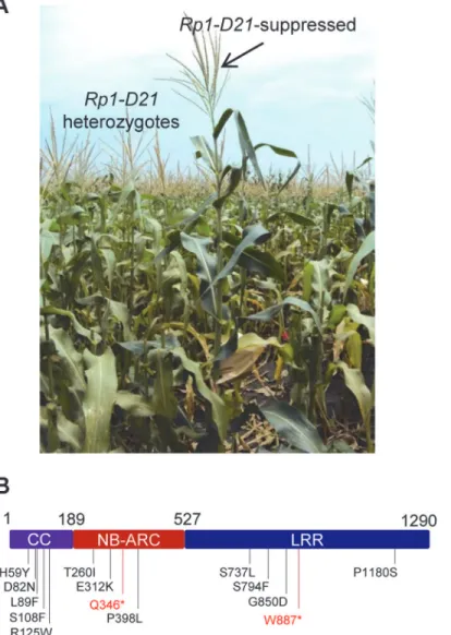
We performed a targeted ethyl methanesulfonate (EMS) mutagenesis screen in a maize line harboringRp1-D21to identify mutations that lost the autoactive HR phenotype conferred by Rp1-D21. Putative suppressor mutants were easily identified due to their robust growth com-pared to the stunted, lesion-mimic siblings heterozygous forRp1-D21(Fig. 2A). From about 23,000 EMS-mutated M1 plants, 12 missense and 2 nonsense intragenic mutants were identi-fied (Fig. 2B;Table 1). Among the 12 missense mutants, five had mutations in the CC domain, three in the NB-ARC domain and four in the LRR domain (Fig. 2B). Notably, one mutant (nucleotide G244A, thus D82N in amino acid) was in the conserved EDVID motif of the CC domain, and one (C1193T, P398L) in the conserved GLPL motif of the NB-ARC domain
Fig 2. EMS mutagenesis screening for mutants that lost the hypersensitive response (HR) induced by
Rp1-D21.(A) The growth phenotype ofRp1-D21heterozygotes in the field compared to theRp1-D21mutant in which the HR phenotype is suppressed (arrow). (B) Twelve missense (in black) and two nonsense (in red) point mutations that abolished the HR phenotype ofRp1-D21were identified in different domains (CC, NB-ARC and LRR) of Rp1-D21.
(Table 1; Figs.1and2B). The mutants T260I and E312K were adjacent to the conserved RNBS-A and Walker B motifs, respectively (Table 1; Figs.1and2B).
The HR phenotype conferred by Rp1-D21 in maize can be recapitulated
by transient expression in
N.
benthamiana
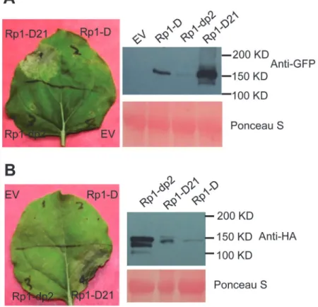
We used Agrobacterium-mediated transient expression inN.benthamianato investigate the structure/function of Rp1-D21. Rp1-D, Rp1-dp2 and Rp1-D21 were either fused to the N-ter-minus of enhanced green fluorescent protein (EGFP), a 3×HA (influenza hemagglutinin) tag or not tagged for subsequent functional analysis. When the three fusion proteins were tran-siently expressed using the cauliflower mosaic virus 35S promoter, a HR phenotype was ob-served 3 days post-infiltration (dpi) only with Rp1-D21, but not with Rp1-D, Rp1-dp2 or the empty vector (EV) control. The same phenotypes were obtained regardless of the tags used (EGFP, 3×HA or no tag) and regardless of the differing levels of protein accumulation observed for the three proteins (Fig. 3;S1A Fig.).
To further confirm that the phenotype conferred by Rp1-D21 in ourN.benthamiana sys-tem conformed to the phenotypes observed in maize, we constructed Rp1-D21 expression vec-tors carrying the missense Rp1-D21 suppressor mutations identified in maize. When
transiently expressed inN.benthamiana, Rp1-D21 produced a strong HR phenotype, rating 4 based on a 0–5 scale rating with 0 being no cell death or chlorosis at all and 5 being confluent cell death [36]. The HR rating is based on the average results from at least 10 individual leaves. All the mutants we identified as non-functional in maize, except for H59Y and G850D, Table 1. Mutations that abrogatedRp1-D21-induced HR identified in an EMS mutagenesis screen in maize.
Domain Nucleotide mutation
Amino acid change
HR rating of full lengh in
N.benthamiana
HR rating of the CC domain inN.benthamiana
Self-association of the CC domain
Conserved motif
Rp1-D21
4 4 +b
CC C175T H59Y 4 4 +
CC G244A D82N 0 5 +/- In EDVID
CC C265T L89F 2 2 +
CC C323T S108F 3 2 +
CC C373T R125W 0 0 +
NB-ARC
C779T T260I 1 Next to
RNBS-A
NB-ARC
G934A E312K 2 Next to walker
B
NB-ARC
C1036T Q346*a
NB-ARC
C1193T P398L 0.5 In GLPL motif
LRR C2210T S737L 2
LRR C2381T S794F 0
LRR G2549A G850D 4
LRR G2660A W887*a
LRR C3538T P1180S 2
a*indicates stop codon. b+: strong self-association; +/-: weak self-association. doi:10.1371/journal.ppat.1004674.t001
abolished or greatly reduced the level of Rp1-D21-induced HR (Table 1;S2A Fig.). All con-structs conferred high levels of protein expression (S2B Fig.). The concordance between the maize andN.benthamianasystems is very good and we expect that our conclusions based on the transient expressionN.benthamianasystem are generally relevant to the endogenous maize system. Thus, we used the transient expression of Rp1-D21 and its derivatives inN. benthamianato further analyze the molecular mechanism of activation/auto-inhibition of Rp1 proteins.
Rp1 protein activity depends on P-loop and MHD motifs
The MHD motif is a highly conserved region of the NB-ARC domain involved in nucleotide binding [15]. Mutations in the MHD domain confer autoactivity to a number of NLRs. Two MHD motifs, here termed MHD1 and MHD2 (actually LHD in Rp1-D and Rp1-dp2;Fig. 4A), separated by a single amino acid are apparent in Rp1-D, Rp1-dp2 and Rp1-D21, and in a num-ber of other CNLs [37]. To investigate whether mutations in either of the MHD motifs could Fig 3. Rp1-D21 triggers a hypersensitive response phenotype when transiently expressed inN.
benthamiana.(A) Rp1-D21, Rp1-D and Rp1-dp2 proteins fused with a C-terminal EGFP tag were agro-infiltrated intoN.benthamiana, with an empty vector (EV) as a negative control. A representative leaf was photographed at 3 days post infiltration (dpi, left). Total protein was extracted from agro-infiltrated leaves at 30 hours post infiltration (hpi), and anti-GFP antibody was used to detect the expression of the fused proteins (right). The sizes of the proteins were labeled on the right. Equal loading of protein samples was shown by Ponceau-S staining of the Rubisco subunit (below, right). (B) Rp1-D21, Rp1-D and Rp1-dp2 proteins fused with a C-terminal 3×HA tag were agro-infiltrated intoN.benthamiana, with empty vector (EV) as a negative control. A representative leaf was photographed at 3 dpi (left). Total protein was extracted from agro-infiltrated leaves at 30 hpi, and anti-HA antibody was used to detect the expression of the fused proteins. Equal loading of protein samples was shown by Ponceau-S staining of Rubisco subunit (below, right). These experiments were repeated three times with the same results.
cause autoactivity in the non-autoactive parental proteins, Rp1-D and Rp1-dp2, we generated the following mutations: Rp1-D(H517A), Rp1-D(D518V), Rp1-dp2(D513V) and Rp1-dp2 (H512A/D513V) in the MHD1 motif, and Rp1-D(H521A), Rp1-D(D522V), Rp1-dp2(H516A), Rp1-dp2(D517V) in the MHD2 motif. Transient expression of the Rp1-dp2(D517V) MHD2 mutant conferred an obvious HR, with symptoms appearing at 3.5 days, about 12 h later than that observed with Rp1-D21 (Fig. 4B). HR was not induced by transient expression of any of the other MHD mutants, including the Rp1-D(D522V) MHD2 mutation (S3 Fig.). However, Rp1-D(D522V) expression was not detectable by western blot analysis using anti-HA antibody. Fig 4. Functional characterization of the MHD2 motif and the P-loop motif.(A) Multiple sequence alignment of the MHD motifs from Rp1-D, Rp1-D21 and Rp1-dp2. Positions of the MHD1 and MHD2 motifs are indicated. The numbers indicated the positions of the aspartate (D) corresponding to Rp1-dp2. (B) The MHD1 and MHD2 point mutations in Rp1-dp2 background were generated and transiently expressed inN.benthamiana. (C) Investigating the MHD2 point mutations in V1 and V16, two recombinant constructs that were not autoactive as shown inFig. 6. (D) Point mutations in P-loop motif of Rp1-D21 and Rp1-dp2(D517V) caused the loss of the ability to induce HR. EV (empty vector) as a negative control. The picture of hypersensitive response (HR) phenotype was taken at 3 days post-infiltration and total protein was detected by anti-HA antibody from agro-infiltrated leaves at 30 hours post infiltration. Equal loading of protein samples was shown by Ponceau-S staining of Rubisco subunit. The experiments were repeated three times with the same results. doi:10.1371/journal.ppat.1004674.g004
Protein expressed from all the other constructs could be detected (Fig. 4B;S3 Fig.). We also generated the MHD2 mutations V1(D517V) and V16(D522V) in V1 and V16, two recombi-nant constructs that were not autoactive (see below) and found both of them induced HR (Fig. 4C).
The P-loop is a highly conserved motif in NLRs and mutations in this domain often result in loss of function and loss of autoactivity [14,15,27,34,38,39]. P-loop mutations (K225R) in-troduced into Rp1-D21 and the autoactive Rp1-dp2(D517V) mutant abrogated their ability to confer HR (Fig. 4D). This indicated that the Rp1-D21 autoactivity and presumably also the ac-tivity of the Rp1 proteins are P-loop dependent.
The CC domain is sufficient for inducing HR
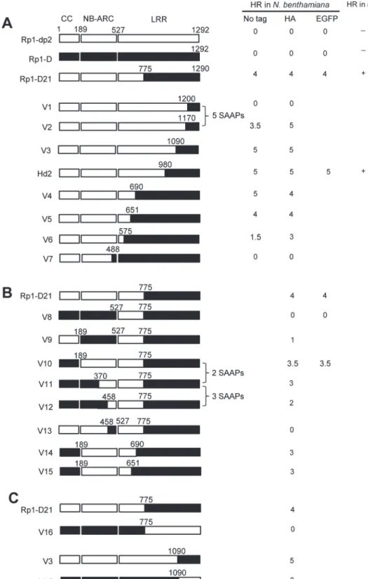
To examine which region of Rp1-D21 was required for triggering HR, different domains or do-main combinations of Rp1-D21, including CC, CC-NB-ARC, NB-ARC, NB-ARC-LRR and LRR domains (hereafter CCD21, CC-NB-ARCD21, NB-ARCD21, NB-ARC-LRRD21and LRRD21;
Fig. 5A), were fused to the N-terminus of EGFP. CCD21and NB-ARCD21were derived from (and are therefore identical to) the corresponding domains of Rp1-dp2 while LRRD21is recom-binant between the LRRs of Rp1-D and Rp1-dp2. The transient expression of CCD21, but of no other Rp1-D21 domains or domain combinations, conferred HR (Fig. 5B). CCD21, NB-ARCD21 and CC-NB-ARCD21produced higher protein accumulation than NB-ARC-LRRD21and LRRD21 (Fig. 5B), excluding the possibility that lack of HR phenotype seen with most of the domain con-structs was solely due to low protein accumulation. We also tested the HR phenotype conferred by different Rp1-D domains and found that the CC domain, but no others, induced HR when fused with EGFP (Fig. 5A). Surprisingly, no untagged or HA-tagged domains from either Rp1-D or Rp1-D21 induced HR (Fig. 5;S1B Fig.).
CCD21, but not CC-NB-ARCD21, induced HR, which suggested that NB-ARCD21can inhibit CCD21-induced HRin cis(when the two domains were fused in the same molecule). Consistent with this result, no HR was observed when EGFP-tagged CCD21and NB-ARCD21were tran-siently co-expressedin trans(when the two domains were co-expressed as separate molecules) inN.benthamiana(Table 2). LRRD21did not restore HR when co-expressed with CCD21and NB-ARCD21separately, or with CC-NB-ARCD21in trans. However, co-expressing CCD21with NB-ARC-LRRD21retained HRin trans, suggesting that LRRD21might interact with NB-ARCD21 to regulate CCD21–induced HRin cisbut notin trans(Table 2). However, since LRRD21or NB-ARC-LRRD21are expressed at substantially lower levels than CCD21or CC-NB-ARCD21 (Figs. 5B and 5C), we cannot exclude the possibility that the concentration of LRRD21or NB-ARC-LRRD21is simply too low to affect the NB-ARC suppression of CCD21–induced HR or CCD21–induced HR itself when co-expressedin trans. In contrast to thein cisresult of CC-NB-ARCD, we found that NB-ARCDdid not suppress CCD-induced HRin trans(Table 2), suggest-ing NB-ARCDmight interact with CCDin cisbut notin trans.
To further investigate the inhibition region of NB-ARC in CC-induced HR, we generated a series of deletion constructs from CC-NB-ARC (S4 Fig.). We found that extension including AAs 190–260 from the NB domain was sufficient to suppress CC-induced HR. Consistent with the data, the nonsense EMS mutant (Q346) suppressed the Rp1-D21 lesion-mimic and
stunted growth phenotype in maize (Fig. 2B;Table 1).
Fig 5. Investigating the functional domains of Rp1-D21 and Rp1-D to trigger a hypersensitive response (HR).(A) Schematic diagram of the Rp1-D21 and Rp1-D domain structure and the derived fragments used for agro-infiltration ofN.benthamiana. The different domains are indicated using different colors: CC (purple); NB-ARC (red); LRR (dark blue). The positions of the amino acids that form the domain boundaries are indicated on the top and the abilities to induce (+) or not induce (-) HR are listed on the right of each construct. (B) The HR phenotype of the constructs fused with a C-terminal EGFP tag and transiently expressed inN.benthamiana. Representative leaves were photographed at 3 days post infiltration (dpi, left). Total protein was extracted from agro-infiltrated leaves at 30 hours post infiltration (hpi), and anti-GFP antibody was used to detect the expression of fused proteins. Equal loading of protein samples was shown by Ponceau-S staining of Rubisco subunit (right, below). The results shown were from different domains of Rp1-D21, and similar results were observed from different domains of Rp1-D. (C) The phenotype of the constructs fused with a C-terminal 3×HA tag and transiently expressed inN.benthamiana. A representative leaf was photographed at 3 dpi (left), and the same leaf was cleared by ethanol (middle). Total protein was extracted from agro-infiltrated leaves at 30 hpi, and anti-HA antibody was used to detect the expression of the fused proteins. Equal loading of protein samples was shown by Ponceau-S staining of Rubisco subunit (right). The results shown were from different domains of Rp1-D21, and similar results were observed from different domains of Rp1-D. The experiments were repeated three times with the same results.
doi:10.1371/journal.ppat.1004674.g005
last 34 AAs of the acidic tail in C-terminus (S5 Fig.). This is despite the fact that all the con-structs conferred high levels of protein expression (S5B Fig.).
Domain swaps between Rp1-D and Rp1-dp2 delineate precise
requirements for self-inhibition
To delineate the functional regions that cause Rp1-D21 to be autoactive and keep its progenitor proteins auto-inhibited, we generated a series of chimeric constructs recombinant at different positions between Rp1-D and Rp1-dp2 (Fig. 6). These proteins, both with and without a 3×HA C-terminal tag, were tested in theN.benthamianatransient expression system. Previously, the chimeric construct Rp1-dp2-D2, with the N-terminus deriving from Rp1-dp2 and the C-termi-nus from Rp1-D, with a recombination point at 980 AA (Fig. 6A), had been shown to cause HR in transgenic maize [21]. The identical chimeric construct, hereafter named Hd2, induced a strong HR when transiently expressed inN.benthamiana(Fig. 6A), consistent with the trans-genic maize result.
The results of the transient expression of a series of chimeric proteins are summarized in
Fig. 6. The HR phenotype conferred by the constructs fused with 3×HA was largely similar to that conferred by the constructs without any tag (Fig. 6A). All the tagged proteins accumulated to high and broadly comparable levels (Figs.7and8;S6 Fig.).
Constructs V1 through V7 were generated with recombination points between Rp1-dp2 and Rp1-D ranging from AAs 1200 to 488 (Fig. 6A). V1 with a recombination point at AA 1200 did not confer HR while V2 with a recombination point just 30 additional AAs N-terminal to AA 1170 produced a strong HR, indicating that AAs 1170–1200 from Rp1-dp2 are important for auto-inhibition of V1. V4 and V5 (recombination point at 690 and 651, respectively) also pro-duced strong HR phenotype, while V6 and V7 (recombination point 575, 488 respectively) con-ferred very weak or no HR (rating 1.5 and 0, respectively,Fig. 6A). These results suggest that the AAs 488–651 from Rp1-dp2 are important for the autoactivity of Rp1-D21. Together these data suggest that proteins with a combination of AAs 488–651 from Rp1-dp2 and AAs 1170–1292 from Rp1-D replacing the corresponding sequences of either parental protein are unable to maintain a self-inhibited state.
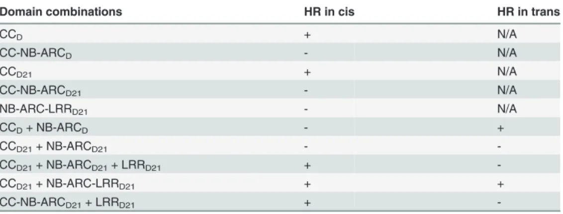
Table 2. Summary of the HR phenotype conferred by transient expression inNicotiana benthamiana
of specific domains or domain combinations from Rp1-D and Rp1-D21in cisandin trans(i.e the two or three listed domains either fused in the same molecule in the order presented here, or expressed as separate molecules, respectively).
Domain combinations HR in cis HR in trans
CCD + N/A
CC-NB-ARCD - N/A
CCD21 + N/A
CC-NB-ARCD21 - N/A
NB-ARC-LRRD21 - N/A
CCD+ NB-ARCD - +
CCD21+ NB-ARCD21 -
-CCD21+ NB-ARCD21+ LRRD21 +
-CCD21+ NB-ARC-LRRD21 + +
CC-NB-ARCD21+ LRRD21 +
Fig 6. Schematic diagram of domain swap constructs between Rp1-D and Rp1-dp2 used for transient expression inN.benthamiana.For ease of interpretation, the gene structure has been divided into CC (coiled coil), NB-ARC (nucleotide binding) and LRR (leucine rich repeat). The amino acid position of the recombination site of each construct was indicated above the construct. For each construct, either with no tag, 3×HA tag or EGFP tag, the strength of the hypersensitive response (HR) resulting from transient expression inN.benthamianais shown. HR was scored on a 0 (no HR) to 5 (strong HR) scale according to Slootweg et al. (2013). If there was no score recorded, the particular experiment was not performed. Where
To test this hypothesis, we investigated whether swapping the N-terminal region (CC, NB-ARC or AAs 1–488) in Rp1-D21 affected its autoactivity. We generated 8 more chimeric con-structs in which we exchanged the Rp1-dp2-derived N-terminal parts of Rp1-D21 with the cor-responding parts from Rp1-D (Fig. 6B). Construct V8, in which the CC-NB-ARC (AAs 1–527) of Rp1-dp2 was replaced with the corresponding Rp1-D sequence, did not confer an HR. Simi-lar results were obtained by exchanging the NB-ARC domain in V9. However, V10, exchang-ing the CC domain of Rp1-D into Rp1-D21 conferred a strong HR (Fig. 6B). These results suggest that the‘parental origin’of the CC domain (AAs 1–189) in Rp1-D21 plays only a minor or no role in its autoactive phenotype. V11 and V12 were generated by replacing AAs 190–370 or 190–458 (NB-ARC1 plus part of ARC2, respectively) of V10 with the correspond-ing Rp1-D sequence. They differed from V10 by only 2 and 5 scorrespond-ingle amino acid polymorphisms (SAAPs), respectively. V11 conferred a slightly weaker HR than V10 and the HR conferred by V12 was weaker still (Fig. 6B). Construct V13, of which the CC-NB-ARC domain was‘ recipro-cal’of V12, did not confer HR, confirming that AAs 458–527 (ARC2 region) from Rp1-dp2 were important for the autoactivity of Rp1-D21 (comparing V13, V12 and V8). Constructs V14 and V15 were generated by exchanging AAs regions 690–775 and 651–775 respectively of V10 with the corresponding Rp1-D sequence. Both constructs showed strong HR (Fig. 6B), similar with the corresponding AAs exchanges in Rp1-D21 (constructs V4 and V5 inFig. 6A). V16 and V17 were‘reciprocals’of Rp1-D21 and V3, respectively and did not confer an HR phenotype (Fig. 6C).
In summary, this set of experiments indicated that the combination of AAs 458–651 from Rp1-dp2 and AAs 1170–1200 from Rp1-D is central to the deficiency of self-inhibition of Rp1-D21.
Defining specific regions (amino acids) important for Rp1-D21
autoactivity
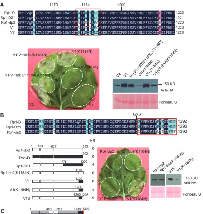
There are only 17 single amino acid polymorphisms (SAAPs) between AAs 1170–1292 of Rp1-D and Rp1-dp2, and only 5 SAAPs in the AAs 1170–1200 region that differentiate con-structs V1 and V2 (Figs.6Aand7A). However, V2 was autoactive, while V1 was not (Fig. 6). To analyze which SAAP is important for the self-inhibition of Rp1 proteins, we generated several mutants based on these 5 SAAPs in the autoinhibited construct V1, including V1 (V1181A), V1(K1184N), V1(V1181A/K1184N) and V1(V1186T/F1188L/C1189D). When transiently expressed inN.benthamiana, V1(V1181A) was not autoactive, while V1(K1184N) and V1(V1181A/K1184N) produced a strong HR (rating 4), and V1(V1186T/F1188L/C1189D) a weak HR (rating 2;Fig. 7A). The results indicated that residue K1184 from Rp1-dp2 plays an important role in self-inhibition, and the residues V1186/F1188/C1189 play a minor role. Inter-estingly, one of the Rp1-D21 suppressor EMS mutants had a mutation at residue 1180 (P to S), only 2 AAs away from N1182 in Rp1-D21 (equivalent to N1184 in Rp1-D), emphasizing the im-portance of this region for regulating the activity of the Rp1 proteins.
To investigate whether mutating K1184 was sufficient to convert Rp1-dp2 into an autoac-tive NLR, we generated Rp1-dp2(K1184N). However, this single point mutation was not able to‘activate’Rp1-dp2 (Fig. 7B). This result suggests that additional SAAPs in the region of AAs known, the abilities of constructs to induce (+) or not induce (-) HR in maize were indicated. In some cases the number of single amino acid polymorphisms (SAAP) between constructs is indicated. All experiments were performed three times with similar results. (A) Constructs with the N-terminus from Rp1-D and C-terminus from Rp1-dp2. (B) Constructs designed to delimit the domains of Rp1-D or Rp1-dp2 important for self-inhibition. (C) The reciprocal constructs: Rp1-D21 with V16, and V3 with V17.
Fig 7. Investigating the functional regions of amino acids (AAs) from 1170–1200 and the C-terminal end of Rp1-D21.(A) Investigating the point
mutations in the region of AAs 1170–1200 which are important for inducing hypersensitive response (HR). Top: Multiple sequence alignment of D, Rp1-D21, Rp1-dp2, V1 and V2. The black, red and light blue shaded regions represent 100%, 75% and above 50% similarity of the amino acids, respectively. The polymorphic amino acids are boxed, and the position of 1184 (corresponding to 1182 of Rp1-D21) is labeled above the sequences. Bottom left: The HR phenotype of V1, V2 and V1-derived point mutants as indicated. A representative leaf was photographed at 3 days post infiltration (dpi). Bottom right: Total protein was extracted from agro-infiltrated leaves at 30 hours post infiltration (hpi), and anti-HA antibody was used to detect the expression of the fused proteins. Equal loading of protein samples was shown by Ponceau-S staining of Rubisco subunit. (B) Investigating the phenotype conferred by various recombinants and point mutants at the C-terminal end of Rp1-D21. Top: Multiple sequence alignment of the C-terminus of Rp1-D, Rp1-D21 and Rp1-dp2. The polymorphic amino acids are boxed. Bottom left: Schematic diagram of the constructs used. The K1184N mutation is indicated by a red vertical bar in Rp1-dp2(K1184N), V1(K1184N) and V18. Bottom middle: The HR phenotype of the constructs transiently expressed inN.benthamiana. A representative leaf was photographed at 3 dpi. Bottom right: Total protein was extracted from agro-infiltrated leaves at 30 hpi, and anti-HA antibody was used to detect the expression of the fused proteins. Equal loading of protein samples was shown by Ponceau-S staining of Rubisco subunit. The experiments were repeated three times with the same results. (C) The final model presenting the functional regions important for autoactivity. In this model, white represents regions derived from Rp1-dp2 and black the region from Rp1-D that need to be present in the same construct to result in the autoactive HR inN.benthamiana. Regions in grey can be derived from either paralog. The combination of AA 458–651 from Rp1-dp2 and AA 1184–1292 (especially N1184 and the last 16 AAs in red vertical bars) from Rp1-D are important for HR induction.
doi:10.1371/journal.ppat.1004674.g007
1200–1292 are also important for self-inhibition of Rp1-dp2. There are 12 additional SAAPs in the last 92 AAs between Rp1-D and Rp1-dp2 and of these six are found in the last 16 AAs (Fig. 7B). Therefore, we constructed the chimeric protein V18 by replacing the C-terminal 16 AAs in V1(K1184N), the autoactive V1 protein, with the equivalent AAs from Rp1-dp2. Inter-estingly, this chimeric protein V18 lost the ability to induce HR (Fig. 7B), demonstrating that the very C-terminal 16 AAs are very important for the self-inhibition of the Rp1 proteins.
In conclusion, these experiments further defined the combination of AAs required for the autoactivity of Rp1-D21: a combination of AAs 458–651 from Rp1-dp2 and AAs 1184–1292 (especially N1184 and the last 16 AAs) from Rp1-D are central to the autoactivity (see model inFig. 7C).
Homology modeling of the NB-ARC from Rp1-D and Rp1-D21 reveals
novel structural elements required for HR
As shown inFig. 6B(construct V13), AAs 458–527 from Rp1-dp2 were very important for the autoactivity of Rp1-D21. There are two major regions of polymorphism, which we termed Patch 1 and 2, between Rp1-D and Rp1-D21 within the ARC2 sub-domain, from AAs 458–527 (Figs.1and8). Homology modeling based on the crystal structure of the NB-ARC domain of the ADP-bound human apoptosis regulator APAF1 (Protein data bank entry 1z6t; [40]) pre-dicted that Patch 1 and 2 were surface-exposed (Figs. 8A and 8B). Surface electropotential pre-dictions indicated that the surfaces of both patches in Rp1-D were mostly negatively charged, while for Rp1-D21 Patch 1 was slightly positively charged and Patch 2 was mostly positively charged (Fig. 8C). To investigate whether Patch 1 and 2 played roles in the autoactivity of Rp1-D21, we constructed another two chimeric proteins, V19 and V20, by replacing AAs 458–463 (Patch 1) and 488–527 (Patch 2) in Rp1-D21 with the corresponding AAs from Rp1-D, respectively (Fig. 8D). Both V19 and V20 conferred a greatly reduced HR compared to Rp1-D21, while V13 (containing Patch 1+2 from Rp1-D) did not cause HR (Fig. 8D), sug-gesting that these patches have an additive effect on the autoactivity of Rp1-D21.
Intra- and inter-molecular interactions of Rp1 proteins
Self-association has been observed in both CNL (eg. barley MLA, Arabidopsis RPS5) and TNL (eg. tobacco N, flax L6) proteins and is important for the activity of NLRs [38,41,42,43]. To test whether Rp1-D21, Rp1-D or Rp1-dp2 could self-associate, we transiently co-expressed EGFP-and 3×HA-tagged proteins inN.benthamianaand performed co-immunoprecipitation (Co-IP) analyses. We observed that full-length Rp1-D21, Rp1-D and Rp1-dp2 all self-associated (Fig. 9A). We further showed that all three of the domains (CCD21, NB-ARCD21and LRRD21) of Rp1-D21, were able to self-associate (Fig. 9B).
To further investigate whether Rp1-D or Rp1-dp2 could form heteromers with Rp1-D21, we transiently co-expressed Rp1-D21:EGFP and 3×HA-tagged Rp1-D or Rp1-dp2 proteins in N.benthamianaand performed Co-IP analyses. We found that dp2 interacted with Rp1-D21 (Fig. 9C). We did not detect interaction between Rp1-D and Rp1-D21, which might have been due to the low expression of Rp1-D (Fig. 3B). Consistent with the interaction data, we ob-served that Rp1-dp2 could partially suppress Rp1-D21-induced HR (Fig. 9C).
observe interactions between NB-ARCDand CCDor between NB-ARCDand LRRDunder our conditions (Fig. 10;S7A–S7B Fig.).
Since the CC and the NB-ARC domains in Rp1-D21 and Rp1-D have different interactions, we investigated the regions required for this interaction. As noted above, the ARC2 domain (including Patch 1 and 2) is the major difference in the NB-ARC domain between Rp1-D21 and Rp1-D. In order to test whether this region affects the interaction between the CC and NB-ARC domains, we performed a Co-IP assay with transiently co-expressed CCD21/CCD Fig 8. Investigating the function of Patch 1 and 2 from the ARC2 domain for Rp1-D21 autoactivity.(A) Sequence alignment of Rp1-D, Rp1-D21 and Rp1-dp2 in Patch 1 and 2 regions from the ARC domain. Positive (+), negative (-) and neutral (n) charges were labeled. RNBS-D, MHD1 and MHD2 motifs are indicated by orange bars and labeled below the sequences. (B) Homology modeling of NB-ARCD21based on the crystal structure of APAF1 (PDB: 1z6t). The P-loop, MHD, GLPL, RNBS-A, RNBS-D and Walker B motifs are color-coded in magenta, yellow, orange, pink, green and blue, respectively. Two boxed regions (Patch 1 and 2) are the major polymorphic regions between Rp1-D and Rp1-dp2 and are labeled in red. The positions of the suppressor mutations T260I and E312K identified by EMS mutagenesis are indicated in green. The N- and C-termini are indicated as N and C, respectively. (C) The surface electropotential of NB-ARCD21and NB-ARCD. The positive and negative charges are indicated in blue and red, respectively. The positions of Patch 1 and Patch 2 are indicated by pink dots in the yellow oval boxes. (D) Schematic diagram and the hypersensitive response phenotype of the constructs indicated. (E) Protein expression was detected by anti-HA antibody at 3 days post-infiltration. Ponceau-S staining of Rubisco subunit was used as loading control. The experiment was repeated three times with the same results.
doi:10.1371/journal.ppat.1004674.g008
and NB-ARCV12/NB-ARCV13, and observed that NB-ARCV12interacted with both CCDand CCD21, while NB-ARCV13did not interact with CCD21(Fig. 10;S7A Fig.). The results suggested that ARC2D21(AAs 458–527 from Rp1-dp2) is important for the interaction between CC and NB-ARC domains. In other words, ARC2Dcan suppress the interaction.
We also observed that LRRD21interacted with NB-ARCV12, but not NB-ARCV13(Fig. 10;
S7B Fig.), suggesting that NB-ARC1Dis required for the interaction between NB-ARC and Fig 9. Investigation of self-association of full length Rp1-D21, Rp1-D, Rp1-dp2, the separate domains of Rp1-D21, and the interaction of full length Rp1-D21 with Rp1-dp2.(A) Self-association of 3×HA- and EGFP-tagged Rp1-D21 (left panel), Rp1-D (middle panel) and Rp1-dp2 (right panel). Arrows indicate the target bands. (B) Self-association of CCD21, NBD21and LRRD21. (C) Left: Co-IP after co-expression of Rp1-D21 and Rp1-dp2. Right: HR induced by co-expression of Rp1-D21:EGFP with Rp1-D21:HA compared to HR induced by co-expression of Rp1-D21:EGFP with Rp1-dp2:HA. EGFP- and 3×HA-tagged constructs were transiently co-expressed inN.benthamianaand samples were collected at 30 hours post infiltration for Co-IP assay. Protein extract was immunoprecipitated by anti-GFP microbeads and detected by anti-GFP and anti-HA antibodies. The experiments were repeated three times with the same results.
Fig 10. Schematic diagram of the intra-molecular interactions and HR phenotype of the constructs indicated, summarizing the data shown inS7 Fig. in which the various domains were co-expressed and co-immunoprecipitatedin trans.The gene structure has been divided into CC, NB-ARC and LRR domains. The amino acid position of the recombination site of each construct was indicated above the construct. For each construct, the strength of the hypersensitive response (HR) resulting from transient expression of each full length molecule inN.benthamianawas scored on a 0 (no HR) to 5 (strong HR) scale. The ability to form or not to form intra-molecular interactions among different domains was indicated by arrows or arrows with crosses, respectively.
doi:10.1371/journal.ppat.1004674.g010
LRRD21, which is consistent with previous report that ARC1 is required for binding to LRR of potato CNL protein, Rx [44].
N1184 and the last 16 AAs from Rp1-D are required for the interaction
between NB-ARC
D21and LRR
V1(K1184N)To determine whether, as suggested by the recombination studies, N1184 and the C-terminal 16 AAs are involved in the intra-molecular interaction, we performed Co-IP assays with NB-ARCD21and LRRV1, LRRV1(K1184N)or LRRV18and observed that NB-ARCD21interacted with LRRV1(K1184N), but not with LRRV1and LRRV18(Fig. 10;S7C Fig.), indicating that N1184 and the last 16 AAs from Rp1-D are required for the interaction between NB-ARCD21 and LRRV1(K1184N).
The effects of CC domain EMS mutants on CC
D21-induced HR
We investigated the effect of the five CC domain missense EMS mutations on CCD21-induced HR. Consistent with the results from full length proteins, CCR125Wabolished HR induction and CCL89Fand CCS108Freduced HR, while CCH59Yhad no obvious effect compared to CCD21 (Table 1). Surprisingly, CCD82Ninduced stronger HR than CCD21, while CCD82Nin the full length Rp1-D21 did not induce HR (Table 1).
Co-IP experiments were performed to explore whether the CC domain mutations affect the self-association of CCD21. CCD82Nbut not the other four point mutants reduced the strength of CCD21self-association (S8A Fig.). To further test whether the mutations affect the interaction between CCD21and NB-ARCD21, we chose CCD82Nand CCR125W, two mutants that completely abolished HR in full length, to test the inter-domain interaction. We found that both CCD82N and CCR125Wgreatly reduced the interaction between CCD21and NB-ARCD21(S8B Fig.).
Discussion
A genetic screen identifies intragenic Rp1-D21 suppressor mutants
While 10 of the 12 Rp1-D21 loss of function EMS mutants isolated from maize also reduced or abolished the HR phenotype when transiently expressed inN.benthamiana, two, H59Y and G850D, did not. It is possible that these two mutations reduced the activity of Rp1-D21 to a level below the threshold for signaling in maize, but not inN.benthamianadue to the higher expression.Three of the missense mutations were within or next to the ADP binding pocket of the NB-ARC domain when the NB-NB-ARC was modeled onto the APAF-1 structure; T260I was close to the RNBS-A motif, E312K was next to Walker B motif and P398L was in the GLPL motif (Table 1; Figs.1and8B), suggesting that these mutations might change the ADP binding state. Consistent with our results, important roles for the RNBS-A, Walker B and GLPL motifs on modulating HR have been reported in other NLRs [14,42,44]. Mutations in the Walker B and GLPL motifs can also affect the intra-molecular interaction between CC and NB-ARC-LRR do-mains as evidenced in Rx [30]. Thus, we infer that the three mutations in the NB-ARC domain might also affect the interaction between CCD21and NB-ARCD21.
Finally, four of the suppressor mutations (S737L, S794F, G850D and P1180S) were in the LRR domain (Fig. 2B). Loss-of-function mutations in the LRR domain have been reported in several NLRs [16,50,51,52,53,54]. According to our model (see below), the mutations likely abolish the ability of the LRRD21domain to destabilize the interaction between NB-ARCD21 and CCD21.
Rp1 activity is P-loop- and MHD-dependent
Mutations from H (histidine) to A (alanine) or D (aspartate) to V (valine) in the highly con-served MHD domain result in constitutive activity of multiple NLRs from multiple species [17,27,37,41,55,56]. In the Flax NLR, M, ADP is bound to wild-type NB-ARC domain, while ATP is bound to the autoactive MHD mutant M(D555V) [15], providing direct evidence that the inactive“off”NLR binds ADP while the active“on”NLR binds ATP. It appears therefore that the MHD motif is important for maintaining the NLR protein in its appropriate state of activity.
Rp1-dp2 and Rp1-D as well as several other CNLs, but not TNLs, have two MHD motifs [17,37]. We have termed these motifs MHD1 and MHD2 (Fig. 4A). MHD1 is more conserved and is the functional MHD motif for many NLRs as defined in previous studies
[15,17,27,38,56]. The aspartate (D) in MHD2 is quite widely conserved throughout most CNLs [37]. Of the CNLs that contained two MHD motifs [37], the effects of mutations in the MHD1 domain only have been reported for tomato NLR Mi-1.2(H840A) and Mi-1.2(D841V). Both mutations activate these proteins [34]. The possible function of the MHD2 domain has recently been examined in rice CNL-RGA5, but no functional effect was observed [57]. Transient ex-pression of Rp1-dp2(D517V) and of V1(D517V) and V16(D522V), in which the MHD2 was mutated, conferred autoactive HR, while no MHD1 mutation had this effect (Figs. 4B and 4C;
S3 Fig.). It is possible that the MHD2 rather than the MHD1 domain is functional in Rp1-dp2, or that the MHD1 and MHD2 domains coordinate the activity. This is the first report showing the functional significance of the MHD2 domain of a CNL for its activity.
The P-loop motif in the NB-ARC domain regulates nucleotide binding. P-loop mutations abolish the ability to confer disease resistance or HR induction in multiple NLR proteins [14,15,27,34,38,39,56]. As expected, the HR induced by D21 and the MHD2 mutant Rp1-dp2(D517V) was abrogated by the introduction of a P-loop mutation (Fig. 4D), indicating that the activity of the Rp1 proteins is P-loop dependent.
Two minimum functional regions in the NB-ARC and the LRR domain
are important for regulating Rp1 protein activity
autoactivity [36,44]. Domain swaps between RPS5 and RPS2 [29] and between 1.1 and Mi-1.2 [58] also gave rise to genes conferring an autoactive phenotype. In contrast to these artifi-cially-constructed autoactive NLRs characterized in transient assays, Rp1-D21 is an autoactive protein that occurred via recombination and was identified in its endogenous genetic back-ground. To identify the precise structural requirement for its activity, we performed a systemat-ic structural analysis of Rp1-D21 using a set of artifsystemat-icial recombinants between the two
‘parental’alleles (Fig. 6) and showed that the combination of AAs 458–651 (the ARC2 and N-terminus of the LRR region) from Rp1-dp2 and the C-terminal LRR region (especially N1184 and the C-terminal 16 AAs) from Rp1-D was critical for the autoactivity of Rp1-D21. This combination either destabilized the Rp1-D21 intra-molecular interactions that cause the inac-tive resting state, or stabilized interactions resulting in the acinac-tive state (see below). In other words, these two regions appear to be important for maintaining the parental proteins in an in-active‘resting’state. Thus, in light of the“mousetrap”model [48], this region of the Rp1 family NB-ARC and LRR domains is involved in a precarious autoinhibiting conformation that is eas-ily broken by alterations including exchange of a very small C-terminal region of the LRR do-main, or presumably, in a wild-type context by the action of the cognate effector protein. It seems evident that the NB-ARC and LRR domains within each NLR must co-evolve to main-tain the NLRs in a suitably auto-inhibited resting state in the absence of pathogen infection while maintaining the ability to respond to the cognate effector via intra-molecular interactions [36,38,55], but that precise mechanisms of auto-inhibition and activation may vary between NLRs.
The role of Patch 1 and 2 in the ARC2 domain
The ARC2 domain is important for function in several NLRs [36]. We identified two major polymorphic regions localized in ARC2 that differentiated the NB-ARCD21and NB-ARCD do-mains, Patch 1 and Patch 2, and showed that they are important for the autoactivity of Rp1-D21 (Figs.1and8). Replacing either Patch 1 or Patch 2 of Rp1-D21 with the region from Rp1-D al-most completely suppressed the autoactive phenotype (constructs V13, V19 and V20;Fig. 8D). In agreement with our structural modeling, these two patches were surface-exposed and carried largely opposite charges in NB-ARCD21compared to NB-ARCD(Fig. 8B). The prevailing model for the activation of NLRs is that the off state binds ADP while the on state binds ATP [36,59]. Patch 2 is located three AAs N-terminal to the MHD1 motif and is localized in the exposed sur-face next to the nucleotide binding pocket [40], suggesting it might affect the state of nucleotide binding. Patch 1 is adjacent to a conserved RNBS-D motif in the ARC2 domain (Figs.1and3A). Mutations in or next to the RNBS-D motif of PM3, RPM1 and Rx affected their function [16,36,45,55]. Thus, the sequence differences of Patch 1 between NB-ARCD21and NB-ARCD might also affect the protein activity through disturbing the function of the RNBS-D motif.
to suppress CCD21-induced HR (S4 Fig.), suggesting that ARC2D21is not the only region which can regulate Rp1-D21 autoactivity.
The role of different Rp1-D21 domains in autoactivity
CCD21and CCDdomains alone were sufficient to induce HR when fused with EGFP, but not with 3×HA or on their own (Fig. 5;S1 Fig.). This phenomenon of“tag-dependent activity”has been observed in other NLR studies [30,61]. The functional domains required for HR induc-tion vary in different NLR proteins. The CC domain alone is sufficient for the HR phenotype triggered by MLA10, and also by NRG1 and ADR1, which contain atypical CC domains [28,41], while the NB-ARC domain of Rx can trigger HR [30], and for RPS5 the CC-NB-ARC is sufficient [29].
The fact that transient expression of CC-NB-ARCD21or CC-NB-ARCDdid not induce HR suggested that their respective NB-ARC domains repressed the signaling by the CC domain. Since full length Rp1-D21 induced HR and full length Rp1-D did not (Fig. 3), we inferred that the LRRD21or LRRDdomains are structurally incompatible with, respectively, repression of CCD21autoactivity or with CCDautoactivity (Fig. 5). While interaction between the LRR and NB-ARC domains was detected in some combinations (e.g., see proteins V12, V1(K1184N) in
Fig. 10), we did not detect any interaction between the LRR and NB-ARC domains of Rp1-D, Rp1-dp2 or Rp1-D21in trans(Fig. 10;S7 Fig.). It is possible though that these domains interact in the full length context, or that their interactions are weak or transient and cannot be detected in our Co-IP conditions. Consistent with this, LRRD21was unable to alter the suppression of CCD21autoactivity by NB-ARCD21in trans(Table 2). However, we cannot exclude the possibil-ity that the lack of suppressionin transwas due to the relatively low expression of LRRD21 (Table 2;Fig. 5B). A similar finding ofcisbut nottransinteraction/autoactivity was reported for potato Rx and autoactive MHD mutants of tomato Mi-1.2 [34,44,62]. It is notable also that the CC-NB-ARC fusion from MLA10 and Rx can still trigger HR [27,30], but not in Rp1-D and Rp1-D21.
LRR domains have multiple reported roles in NLR activation. They are essential for the acti-vation of the NLRs Rx and Mi-1.2 [34,62]. In the case of RPS5, the first four LRRs are the mini-mum region sufficient to inhibit the autoactive phenotype conferred by the CC-NB-ARC domain [29]. Conversely, the activation of RPS5 in response to disease requires the entire LRR domain [29]. Our analyses of Rp1-D21 C-terminal deletion constructs and LRR domain swap data (Fig. 7B;S5 Fig.) further confirmed the importance of the C-terminal LRR domain in reg-ulating the activity of the Rp1 proteins. Similarly, the importance of C-terminal LRR domain has also been observed in flax TNL, L proteins [37].
The interaction patterns observed between NB-ARC and LRR from V1, V1(K1184N) and V18 suggested that K1184N and the last 16 AAs from Rp1-D are required for the inter-domain interaction (Fig. 10;S7 Fig.). No interaction was detected between the NB-ARC and LRR do-mains from V1 or between those from V18 when they were expressedin trans, but atrans- in-teraction was detected between these domains using the V1(K1184N) construct. Assuming that these interactions are maintained in the full length proteins, this result implies that the ability of V1(K1184N) to induce HR is due to the interaction of the LRR with the NB-ARC, which apparently is then locked into an activated state allowing CC-dependent HR. This also requires the C-terminal 16 amino acids of the Rp1-D21 LRR domain (Fig. 10;S7 Fig.). A fur-ther inference is that the C-terminal 16 AAs and N1182 in Rp1-D21 (corresponding to N1184 in Rp1-D) are crucial for the inhibition of NB-ARC activity that is required for CC-dependent HR.
A model for the autoactivity of Rp1-D21
Alternate“on”or“off”states mediated through intra-molecular interactions are thought to be one of the major mechanisms regulating NLR activity [51,62]. For example, intra-molecular in-teractions were detected between CC and NB-ARC-LRR, and CC-NB-ARC and LRR domains of Rx in the absence but not in the presence of its cognate effector, the PVX coat protein [62]. Similarly the differentially-activated states of Rp1-D, Rp1-dp2, Rp1-D21 and our additional re-combinant proteins are likely due to the specific intra-molecular interactions found within each protein. While Rp1-D21 and V3 confer HR, their reciprocal constructs, V16 and V17, do not (Fig. 6C), indicating that the autoactivity of the recombinant protein is triggered by specific combination of sequences from Rp1-dp2 and Rp1-D. Our model (Fig. 11) explaining the intra-molecular interactions underlying the activity of the Rp1 proteins is as follows:
1. Self-association and heteromer formation between Rp1 proteins are important for their activity. Self-association of NLRs has been shown to be important for activity in other cases
[38,41,42,43]. While we have not been able to show that this is the case for Rp1 proteins, we have shown that Rp1-D21, Rp1-D, Rp1-dp2 and each of the three major domains, CCD21, NB-ARCD21, LRRD21can self-associate (Fig. 9). Based on these related studies, we propose that this self-association is important for function.
Heteromers formed by different NLRs have been shown to be important for NLR activity [57,63]. We found that Rp1-dp2 and Rp1-D21 physically interacted and that their co-expres-sion suppressed Rp1-D21-induced HR, presumably due to their physical interaction
(Fig. 9C). This is also consistent with our previous study in which we identified a quantitative trait locus mapping directly to Rp1 which regulated the severity of Rp1-D21-induced HR in maize [26].
2. The CC domain is the signaling competent region of the Rp1 proteins.
This is demonstrated by the ability of CC expressed alone to induce HR (Fig. 5).
3. The NB-ARC domain inhibits CC-induced HR in Rp1-D21 via intra-molecular interactions involving the ARC2 domain.
In Rp1-D, NB-ARCDcan inhibit CCD-induced HRin cis, but notin trans(Fig. 5A;Table 2). Though we did not detect direct physical interaction between NB-ARCDand CCD, the fact that NB-ARCDinhibits HR caused by CCDin cissuggests that there is inhibitory interaction between the CC and NB-ARC domains in both Rp1-D and Rp1-D21. Data from the recom-bination (Fig. 6) and Co-IP experiments (Fig. 10;S7 Fig.) indicate that the ARC2 domain is important for controlling both activity and intra-molecular interactions of Rp1 proteins. We propose that the intra-molecular CC/NB-ARC interactions in which ARC2 domain plays a critical role are important for setting up a‘primed’state as proposed under the “mousetrap model”for NLR activation [48,49].
4. InRp1-D21and the other autoactive recombinant proteins,the LRR destabilizes the CC/NB-ARC interaction,leading to auto-activation.
The best evidence for this comes from the comparison of the interaction patterns of V1, V1 (K1184N) and V18 inFig. 10. These proteins differ by only 1 and 6 AAs, respectively. In V1 (K1184N) the LRR and NB-ARC domains interactin trans. This is correlated with the abili-ty of the corresponding full length protein to confer HR in the transient expression system, since neither V1 nor V18 confer HR and their respective LRR domains do not interact with the shared NB-ARC. Consistent with these data, truncation of the C-terminal 34 AAs of Rp1-D21 eliminates autoactivity (see also #6, below). Thus, 7 AA changes from Rp1-D in Rp1-D21 in a protein otherwise derived from Rp1-dp2 are sufficient to de-stabilize the LRR’s ability to inhibit ectopic NB-ARC activity and consequent CC-dependent HR. Although we did not observe Co-IP between LRRD21and NB-ARCD21, we propose, for rea-sons detailed above, that there is an interaction between these two domainsin cisbut notin trans. This would be consistent with our data that NB-ARC-LRRD21is unable to inhibit CCD21signaling when presentin trans(Table 2).
5. Destabilization of the CC/NB-ARC inhibitory interaction allows for the exchange of bound ADP for ATP in a P-loop,RNBS-A,Walker B,GLPL and MHD2/ARC2-dependent manner. Mutants in or next to P-loop, RNBS-A, Walker B, GLPL and ARC2 lose the ability to confer HR (Table 1;Fig. 4;S2 Fig.).
6. The autoactivated state of Rp1-D21 and various other derived autoactive recombinants in which the CC/NB-ARC interaction is destabilized,depends upon the combined presence of AAs 458–651 (Patches 1 and 2 in ARC2 and the N-terminal LRR region) from Rp1-dp2 and of the C-terminal LRR region (especially N1184 and the last 16 AAs) from Rp1-D.
The data for this is discussed above and detailed in Figs.6and7.
While we have not identified a cognate effector of Rp1-D and do not know how it is de-tected, we hypothesize that its presence is necessary for activating Rp1-D via direct or indirect interaction with the LRR domain. This model is based both upon the data presented here and a review of the related literature. While plant NLR resistance genes share similar structures and appear to perform very similar functions, it is clear that the molecular mechanisms underlying their function and their appropriate activation, while sharing certain similarities, vary substan-tially. This variation likely reflects the intimate and unique co-evolutionary processes each NLR has undergone both with respect to the interactions among their different domains and with their cognate pathogen effector proteins, and/or the host targets or decoys of those effec-tors. The molecular mechanisms underlying the auto-inhibition of the maize NLR common rust resistance protein Rp1-D and its autoactive derivative Rp1-D21 show both broad similari-ties to and distinct differences from what is known of other NLRs. Furthermore, we have
identified several unique, or previously un-observed features, including: the AAs required for HR induction identified through EMS mutagenesis screening; the functional characterization of MHD2 motif; the involvement of the ARC2 domain in the interaction of the NB-ARC do-main with the CC dodo-main; the observation that N1184 and the C-terminal 16 AAs are involved both in the LRR/NB-ARC physical interaction and in regulating activity.
Recently, several NLR genes that have been transferred between plant species were demon-strated to still confer expected disease resistance specificities without obvious fitness effects, eg. ArabidopsisRPS4(Resistance to Pseudomonas syringae 4),RRS1(Resistance to Ralstonia sola-nacearum 1) and barleyMLA1[64,65]. Genetic manipulation ofRxindicates stepwise artificial evolution can be used to reduce the costs associated with disease resistance of NLRs [66]. Rp1-D21 is an autoactive mutant conferring nonspecific response to multiple maize rust species, in-cludingP.sorghiandP.triticina[19,21]. Presumably the resistance conferred byRp1-D21 might extend beyond rusts to other biotrophic and hemi-biotrophic pathogens, however, the severe growth penalties associated with the expression of this gene make its application in agri-cultural production impractical. In conjunction with suitable promoters, it may be possible to engineer maize or even other plants with some of the chimeric constructs characterized here that confer a weaker HR phenotype to achieve an elevated disease resistance with fewer fitness consequences.
Materials and Methods
Plant materials and growth conditions
Wild typeN.benthamianaplants were grown at 23°C with a cycle of 16 hrs light/8 hrs dark. Maize lineRp1-D21-H95 [25] was used to isolate the genomic DNA sequence ofRp1-D21. To generate material for EMS mutageneis,Rp1-D21was first introgressed into A632 by 7 back-crosses (BC7). A few of these BC7Rp1-D21heterozygous plants were self-pollinated to gener-ateRp1-D21homozygotes in the A632 background which were identified on the basis of their enhancedRp1-D21severity compared to the heterozygotes.
EMS mutagenesis screening
Pollen from the homozygousRp1-D21plants in an A632 background was collected and treated with EMS for 45 min before using it to fertilize the ears of an inbred line H95. The resulting M1 population was all heterozygous forRp1-D21and showed an HR phenotype with a relative-ly stunted growth stature. In this background,Rp1-D21-suppressed plants due to EMS muta-genesis were easily distinguished from the rest of the M1 siblings because of their non-lesioned and highly robust growth phenotype (Fig. 2A). About 23,000 M1 progenies were screened in field to identify intragenic suppressor mutants that had lost theRp1-D21phenotype. In total, 32 mutants which lacked the HR phenotype ofRp1-D21were identified and 14 of them were characterized in detail. Gene specific primers forRp1-D21(S1 Table) were used to sequence theRp1-D21gene in these mutants.
Plasmid construction
epitopic tag in the C-terminus) [67,68].Rp1-D21was also isolated from maize lineRp1-D21 -H95 using the primers listed inS1 Table.
The different domains (CC, CC-NB-ARC, NB-ARC, NB-ARC-LRR, and LRR) of Rp1-D21 or Rp1-D were amplified using primer pairs listed inS1 Table. The resulting PCR products were cloned into D-TOPO and sequenced, then transferred into pGWB2, pGWB14 or pSI-TEII-N1-EGFP by gateway LR reactions.
Site-directed mutagenesis
Overlapping extension PCR primers (S1 Table) were designed for generating the site-directed mutations: Rp1-D21(K225R), Rp1-D(H517A), Rp1-D(D518V), Rp1-D(H521A), Rp1-D (D522V), Rp1-dp2(D513V), Rp1-dp2(H512A/D513V), Rp1-dp2(H516A), Rp1-dp2(D517V), Rp1-dp2(K225R/D517V) and 12 missense EMS mutations listed inTable 2. The site-directed mutations were cloned into D-TOPO and verified by sequencing and sub-cloned into the gate-way vector pGWB14 by LR reaction.
Sequence alignment and homology structure modeling
NLR sequences were aligned by ClustalW (www.ebi.ac.uk), and edited by BioEdit software. Ho-mology modeling of the NB-ARC domain was performed with Phyre 2 [69] based on the crys-tal structure of human APAF1 (PDB: 1z6t). The three-dimensional structure and the surface electropotential were mapped using PyMOL (http://www.pymol.org/).
Agrobacterium tumefaciens-mediated transient expression
Agrobacterium tumefaciensstrain GV3101 (pMP90) transformed with binary vector constructs was grown at 28°C overnight in 5ml L-broth medium supplemented with appropriate antibiot-ics. The bacteria were collected at 4,000g by centrifugation and resuspended in 2 ml resuspen-sion buffer (10 mM MES pH5.6, 10 mM MgCl2and 200μM acetosyringone). The final concentration of the bacteria was diluted to the OD600of 0.5 using the same resuspension buff-er. To prevent the onset of post-transcriptional gene silencing and improve the efficiency of transient expression, a strain containing p19 protein was included at OD600of 0.2 to all the bac-teria strains [70]. The solution was left at room temperature for 3 hrs on bench before infiltra-tion into the abaxial side ofN.benthamianaleaves. After infiltration, plants were put at room temperature with 16h-light/8h-dark. At least 15 individual leaves were infiltrated by different constructs, and each experiment was repeated at least three times.
Protein analysis
For protein expression analysis, three leaf discs (1.2 cm diameter) from different single plants were collected at 30 hours post infiltration (hpi). The samples were ground with prechilled plastic pestles in liquid nitrogen, and total protein was extracted in 160μl extraction buffer [20 mM TrisHCl (pH 8.0), 150 mM NaCl, 1 mM EDTA (pH 8.0), 1% Triton X-100, 0.1% SDS, 10 mM DTT, 40μM MG132, and 1× plant protein protease inhibitor mixture (Sigma-Aldrich)]. Samples were centrifuged at 14,000 g for 15 min at 4°C, and 12μl supernatants were mixed with 2× Laemmli buffer and loaded for SDS-PAGE. For Co-IP assay, EGFP- and 3×HA-tagged constructs were transiently co-expressed inN.benthamiana. Agrobacterium carrying each construct were diluted to a final concentration of OD600= 0.4 plus p19 with OD600= 0.2. Samples were collected at 30 hpi, and proteins were extracted by grinding 0.8 g of leaf tissues in 2.4 ml extraction buffer [50 mM HEPES (pH 7.5), 50 mM NaCl, 10 mM EDTA (pH 8.0), 0.5% Triton 100, 4 mM DTT and 1× plant protein protease inhibitor mixture (Sigma-Aldrich)]. Extracts were centrifuged at
14,000 rpm for 20 min at 4°C, and 2 ml supernatant was mixed with 30μl anti-GFP microbeads (Miltenyi Biotec) and rotated for 3 hrs at 4°C. The samples were passed through pre-equilibrated MACS separation columns, and washed four times (1 ml, 1 ml, 500μl and 500μl) by washing buffer [50 mM HEPES (pH 7.5), 150 mM NaCl, 10 mM EDTA (pH 8.0), 0.2% Triton 100, 4 mM DTT and 1× plant protein protease inhibitor mixture (Sigma-Aldrich)]. The proteins were eluted by 100μl pre-heated elution buffer and separated by SDS-PAGE. Proteins were transferred to ni-trocellulose membrane (Fisher), and analyzed by western blot. HA detection was performed using a 1:350 dilution of anti-HA-HRP (horseradish peroxidase) (Cat# 12013819001, Roche). GFP detection was performed using a 1:8,000 dilution of primary mouse monoclonal anti-GFP (Cat# ab1218, Abcam), followed by hybridization with a 1:15,000 dilution of anti-mouse-HRP second antibody (Cat# A4426, Sigma). The HRP signal was detected by ECL substrate kit (Super-signal west femto chemiluminescent substrate, Thermo Scientific).
Supporting Information
S1 Fig. Transiently expressed of proteins without any tag fusion inN.benthamiana.(A) Rp1-D21, Rp1-D and Rp1-dp2 proteins without any tag fusion were agro-infiltrated intoN. benthamiana, with an empty vector (EV) as a negative control. A representative leaf was pho-tographed at 3 days post infiltration (dpi). (B) The different domains of Rp1-D21 without any tag fusion were transiently expressed inN.benthamiana. A representative leaf was photo-graphed at 3 dpi. These experiments were repeated three times with the same results. (TIF)
S2 Fig. The phenotype of the intragenic Rp1-D21 suppressor mutants transiently expressed inN.benthamiana.(A) HR phenotype of leaves transiently expressing D21 and Rp1-D21-derived intragenic suppressor mutations. All proteins indicated were fused with a C-ter-minal 3×HA tag and were agro-infiltrated intoN.benthamiana. A representative leaf was pho-tographed at 3 days post infiltration (dpi). (B) Detecting the protein expression of the
constructs used in (A). Total protein was extracted from agro-infiltrated leaves at 30 hours post infiltration, and anti-HA antibody was used to detect the expression of fused proteins. Equal loading of protein samples was shown by Ponceau-S staining of Rubisco subunit. These experiments were repeated three times with the same results.
(TIF)
S3 Fig. Investigating the function of the MHD motifs of Rp1-D.(A) Point mutations in MHD1 and MHD2 motifs from Rp1-D were fused with a C-terminal 3×HA tag and agro-infil-trated intoN.benthamiana. The representative leaves were photographed at 3 days post infil-tration. (B) Protein expression analysis of MHD mutations in Rp1-D. Total protein was extracted from agro-infiltrated leaves at 30 hours post infiltration, and anti-HA antibody was used to detect the expression of the fused proteins. Equal loading of protein samples was shown by Ponceau-S staining of Rubisco subunit. Experiments were repeated three times with the same results.
(TIF)
S4 Fig. Summary of results from transient expression inN.benthamianaof a series of
C-terminal deletion constructs of CC-NB-ARCD21.(A) Schematic diagram of a series of deletion
constructs from CC-NB-ARCD21and the derived fragments used for agro-infiltration ofN.
antibody was used to detect the expression of fused proteins. Equal loading of protein samples was shown by Ponceau-S staining of Rubisco subunit (below right). The experiments were re-peated three times with the same results.
(TIF)
S5 Fig. Investigating the ability of C-terminal deletions of Rp1-D21 to trigger hypersensi-tive response (HR) inN.benthamiana.(A) Schematic diagram of the C-terminal deletions of Rp1-D21 and the derived fragments used for agro-infiltration ofN.benthamiana. The posi-tions of the deletion breakpoints are indicated and their abilities to induce (+) or not induce (-) HR are indicated on the right of each construct. (B) The HR phenotype of the C-terminal dele-tions of Rp1-D21 observed at 3 dpi (left). All proteins were fused with a C-terminal 3×HA tag and anti-HA antibody was used for detecting the total proteins extracted from the samples in-dicated at 30 hours post infiltration (right). Equal loading of protein samples was shown by Ponceau-S staining of Rubisco subunit (below right). The experiments were repeated three times with the same results.
(TIF)
S6 Fig. Protein expression analysis of constructs fromFig. 6.Chimeric constructs fused with a 3×HA tag were infiltrated intoN.benthamiana. Total protein was extracted from agro-infil-trated leaves at 30 hours post infiltration, and anti-HA antibody was used to detect the expres-sion of the fused proteins. Equal loading of protein samples was shown by Ponceau-S staining of Rubisco subunit. The experiments were performed three times with similar results. (TIF)
S7 Fig. Intra-molecular interactions from different Rp1 proteins.(A) Inter-domain interac-tions between the CC and NB-ARC domains in different Rp1 proteins. EGFP- and 3×HA-tagged proteins were transiently co-expressed inN.benthamianaand samples were collected at 30 hours post infiltration for Co-IP assays. Protein extracts were immunoprecipitated by anti-GFP microbeads and detected by anti-anti-GFP and anti-HA antibodies. (B) Inter-domain interac-tions between NB-ARC and LRR in different Rp1 proteins. (C) Inter-domain interacinterac-tions be-tween NB-ARCD21and LRRV1, LRRV1(K1184N)or LRRV18. These experiments were performed three times with similar results.
(TIF)
S8 Fig. Investigating the function of the intragenic suppressor mutations in CCD21.(A)
Self-association of different CC domain variants. EGFP- and 3×HA-tagged proteins were tran-siently co-expressed inN.benthamianaand samples were collected at 30 hours post infiltration for Co-IP assay. Protein extracts were immunoprecipitated by anti-GFP microbeads and de-tected by anti-GFP and anti-HA antibodies. (B) Investigating the inter-domain interactions be-tween NB-ARCD21and the CC variants. These experiments were performed three times with similar results.
(TIF)
S1 Table. Primer sequences used in this study. (DOC)
Acknowledgments
We thank Dr. Scot H. Hulbert for providing the plasmids harboringRp1-DandRp1-D21and Dr. Michael Goodin for providing the vectors of pSITEII-N1-EGFP. We are grateful to Dr. Shannon Sermons for help with obtaining reagents. The Genomic Sciences Laboratory at