__________________________________________________________________________________ MORPHOLOGIA • 2015 • 9 • № 4 • О ФО О І
43
Y.Kuzenko
O.Diachenko
O.Kuzenko
A.Olishkevych
Sumy State University
Key words: periodontitis, cells microenvironment, collagen.
Наді шла: 24.11.2015
Пр нята: 18.12.2015
UDC 616.314.18-002.2/.4-092
CELLULAR MICROENVIRONMENT AND
COLLAGEN DESTRUCTION DURING
PE-RIODONT INFLAMMATION
ABSTRACT. Background. The association between periodontitis and collagen damage with immune cells is an actual problem. Periodontitis is a bacterially induced exacerbation of chronic process and chronic inflammatory disease that destroys teeth supporting tive tissue. Bacteria initiate periodontitis and destruction of the alveolar periodontal connec-tive tissue. Objecconnec-tives. Immune cells location between damaged collagen fibers remains obscure and this is the purpose of the current study. Results. We have determined five va-riants of immune cells microenvironment: nodular, trabecular, diffuse, mixed. We have ob-served five types of collagen structures destruction in exacerbation of chronic process and chronic periodontal inflammation. They are characterized by swelling, pulping and insigni-ficant necrosis. Conclusion. Connective tissue has signs of swelling and destruction during inflammation; edema is observed between collagen fibers. Collagen fiber damage during periodontitis is caused by neutrophils. Widespread edema of collagen fibers increasing of depth cells infiltration during chronic inflammation. Nodular type of immune cells microen-vironment is observed during outcome of chronic inflammation. Trabecular type of immune cells microenvironment is observed during exacerbation of chronic process. Diffuse type of immune cells microenvironment is observed during chronic inflammation process.
Morphologia. – 2015. – . 9, № 4. – . 43-48.
© Y.Kuzenko, O.Diachenko, O.Kuzenko, A.Olishkevych, 2015
Є., О., О., О і А. і і
-і .
. З ' , є
, . Н '
: , , , . В є
; - ;
- . П
-є , є .
і : , , .
Citation:
Kuzenko Y, Diachenko O, Kuzenko O, Olishkevych A. Cellular microenvironment and collagen destruction during periodont inflammation. Morphologia. 2015;9(4):43-8.
Introduction
Gingivitis and its treatment have been with mankind at least since the days of the Pharaohs [1]. Periodontitis is a group of inflammatory diseases that affect the connective tissue attachment and sup-porting bone around the teeth [2]. The periodontium includes the vasculature, epithelium, non-mineralized and non-mineralized connective tissues, as well as the inflammatory and immune cells that have infiltrated into the periodontal tissues. The cellular components of periodontitis are 70–80% granulo-cytes, 10–20% monocytes/macrophages, 5% mast cells and 5% T lymphocytes [3]. Pathogenesis of periodontal diseases has two major aspects: micro-organisms and host response. Interactions between microbial plaque and host immune system play a critical role in the initiation and progression of
peri-odontal diseases [4]. Monocytes, macrophages and other cells (including fibroblasts and endothelial cells) respond to the dental plaque microorganisms. The primary hallmark of periodontitis, the destruc-tion of periodontal tissue, is widely accepted to be a result of the host immune inflammatory response caused by periodontal microorganisms [5].
One important effector mechanism of the in-flammatory mediators present in periodontal tissue is stimulation of the monocytes, macrophages for-mation, this cells believed to be the major cell type responsible for cologne destruction. Destruction of the cologne may occur as a result of acute inflamma-tory, mechanical stimulation, or neoplastic processes.
__________________________________________________________________________________ MORPHOLOGIA • 2015 • 9 • № 4 • О ФО О І
44
bone may occur. In addition, seven major categories of destructive periodontal diseases were listed: Chronic periodontitis, Localized aggressive peri-odontitis, Generalized aggressive periperi-odontitis, Peri-odontitis as a manifestation of systemic disease, Ne-crotizing ulcerative gingivitis/periodontitis, Ab-scesses of the periodontium, Combined periodontic-endodontic lesions [6].
The aim of this study is to compare the levels of cologne destruction and variants of immune mi-croenvironment in gingival tissues on exacerbation of chronic process and chronic stages of periodontal disease.
Materials and Methods
Patient selection and of gingival tissues collec-tion
The study samples have included the periodon-tal and epulis tissues of the patients. Group consisted of 34 patients who had died in Sumy Regional Hos-pital. The patients had various somatic diagnoses (not atherosclerotic complications) and dental – pe-riodontitis. 17 males and 13 females, age range 43 to 69 years, mean age, 57.33 ± 8.31 years have been investigating. The specimens of overgrown gingiva were collected during jaws sawing procedures. After group tissue samples stained by hematoxylin eosin for, all samples were divided in two groups (acute and chronic). Group has 3 subgroups.
Exacerbation of chronic process - 13 (5 males and 3 females, age range 43 to 69 years, mean age, 54.38 ± 7.9 years).
Chronic subgroups - 17 (7 males and 15 fe-males, age range 44 to 68 years, mean age, 59.58 ± 8.11 years).
Normal periodontal tissues -4 (1 males and 3 females, age range 18 to 23 years, mean age, 20 ± 2.16 years)
Informed written consent was obtained from all study subjects in accordance with guidelines estab-lished by the Ukraine Health Council. The present study was approved by the Sumy State University (Protocol no. 5/ 2012).
Periodontal tissues samples have stained ac-cording to the methods specific for Hematoxylin and eosin (H&E) staining respectively.
Periodontal tissues were immersed in 10% pa-raformaldehyde pH 7.4 fixative at 18º C. These were then dehydrated with increasing concentration of ethanol before being embedded in paraffin.
Paraffin sections were prepared for acridine orange staining by mounting on the slides, dried on a hot plate, and then immersed into three sets of xy-lene for 2 minutes each followed by three sets of absolute ethanol for 5 minutes and finally rinsed with tap water. The aim was to remove the wax and dehydrate the sections. Slides (paraffin) were placed into acridine orange staining solution for 15 minutes, and rinsed with phosphate-buffered saline (PBS).
Then the slide was soaked in 0.1% calcium chloride solution for 3 minutes and was washed with PBS once again. Cover glass was mounted for observa-tion under a fluorescence microscope to observe and read the result.
Immunostainings
For MPO have been performed formalin fixed (pH 7,4) tissue. Paraffin-embedded tissue sections have been treated dy mouse monoclonal anti-myeloperoxidase (Thermo Fisher Scientific UK). Briefly, 4 μm thick tissue sections were dewaxed in xylene and were placed in to water through graded alcohols. Antigen retrieval has been performed by microwaving slides in 10 mM citrate buffer (pH 6.2) for 30 min at high power, according to the manufac-turer’s instructions. To remove the endogenous pe-roxidase activity, the sections have been treated with freshly prepared 1.0 % hydrogen peroxide in the dark for 30 min at 37 °C temperature. Non-specific antibody binding was blocked by means of blocking serum.
The sections were incubated for 30 min, at 37 °C temperature, with the primary antibodies against myeloperoxidase diluted 1:100 in phosphate buf-fered saline (PBS) pH 7.2 then a triple washing with PBS follows. Anti-(mouse IgG)–horseradish perox-idase conjugate (1:40 000 dilution) has been fulfilled for the detection of the MPO primariy antibodies, then the sections were incubated for 20 min, at 37 °C temperature. The colour was visualized by DAB. The appearance of the positive factors was detected semiquantitatively by counting of positive giant cells in visual field.
The data were analysed using STATISTICA 8.0 software, user version STA862D175437Q. The results have been presented as mean ± SD. The nor-malize test have been used before analysis of the data. Also, the nonparametric Student method was applied to perform a simple comparative analysis. The value of P < 0.05 have been considered to be a significant.
Results
The periodontal measurements of participants recruited for this study have been summarized in Table 1.
The normal periodontal tissues a shown in Fig. 1(1). Connective tissue gingival fibres support the gingival margin as a cuff around the tooth.
__________________________________________________________________________________ MORPHOLOGIA • 2015 • 9 • № 4 • О ФО О І
45
Fig. 1. Changes in collagen during inflammation.
1 - normal connective tissue: A – connective tissue, B – immune cells;
2 - exacerbation of chronic inflammation: A – swelled destruction of connective tissue B – immune cells;
3 - chronic inflammation: A – collagen fibers edema B – complex patterns of collogen destruction C - immune cells form the layers or trabeculae;
__________________________________________________________________________________ MORPHOLOGIA • 2015 • 9 • № 4 • О ФО О І
46
Table 1 Periodontal measurements of patients
Note: p value – *p < 0.05; **p < 0.01; ***p < 0.001.
The results of the chronic a shown in Fig. 1 (4). Consists of dilated thin-walled vessels in loose ede-matous stroma with superimposed edema. Wide-spread edema of collagen fibers increasing of depth cells infiltration (Fig. 1 (4A)). The results of the chronic exacerbation inflammation a shown in Fig. 1 (5). Destruction have progressed until tooth support becomes inadequate and more complex patterns of collagen fibers loss develop. Despite the deep exten-sion of inflammation it may have recovery of alveo-lar collagen fibers complex.
We have determined five variants of immune cells microenvironment:
1. Nodular – the leukocytes accumulate in fo-cuses, sometimes with the formation of pseudo-follicles. Fig 1(4A)
2. Trabecular – the immune cells form the layers or trabeculae Fig. 1(2)
3. Diffuse - the leukocytes are located around
the collagen fibers as single scattered cells Fig. 1(3C).
4. Mixed – a combination of different forms of inflammatory infiltration. Fig. 1(5A).
In the stroma of the control group tissue the immune cells were also presented, because they are a part of the fibrous tissue of any organs, including the oral cavity. But they are located singly and very rare.
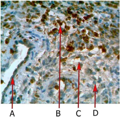
The immunoexpression of MPO have been con-firmed by the presence of brown stained cytoplasm in cell infiltration. Myeloperoxidase was expressed in 98,2 ± 5,9 % (P < 0.01) positive cells in exacerba-tion of chronic process. Figure 2 shows the results of the MPO expression in chronic and exacerbation of chronic process. Myeloperoxidase 72,8 ± 4,5 % P < 0.01 was expressed in chronic plasmatic cell infiltra-tion during inflammainfiltra-tion. Normal periodontal tis-sues single neutrophil have been detected.
Fig. 2. Immunoexpression of MPO in chronic inflammation. A – vessel; B – MPO positive cells; C – edema; D - MPO nega-tive cells. ×320.
Clinical parameters Exacerbation of
chron-ic process Chronic subgroups
Normal periodontal tissues
∑ N = 13 N = 17 N=4
probing pocket depth ≤4 mm 14.61 ± 0.58*** 3.51 ± 0.12*** probing pocket depth 4-6 mm 26.19 ± 8.55* 0.00 probing pocket depth≥ 7 mm 18.34 ± 3.62* 0.00 clinical attachment level 5.62 ± 0.56** 3.98±0.14*
bleeding on probing 4.86 ± 0.43* 15± 0.11
__________________________________________________________________________________ MORPHOLOGIA • 2015 • 9 • № 4 • О ФО О І
47 Discussion
Periodontal disease have caused by selected species of bacteria that colonize the tooth surface and invade the adjacent tissue, causing inflammation and, ultimately, connective tissue destruction and bone resorption. Most patients with plaque-induced periodontitis will have the chronic form [7]. In most cases the disease is slowly progressing [8], but short periods of rapid attachment loss can occur [9]. Al-though chronic periodontitis can occur in localized or generalized patterns, the two forms appear to be identical with regards to their etiology and pathoge-nesis. However, evidence that tissue loss caused by periodontal disease is reduced by inhibitors of pros-taglandins and inhibitors of matrix metalloproteinas-es indicatmetalloproteinas-es that much of the tissue damage is caused by up-regulation of the host response [10, 11] We have reported that MMP1 significantly simulat tis-sue destruction and inflammation [12]. MMPs are the main collagen-degrading enzymes in gingival and they are believed to be mainly responsible for collagen degradation in inflamed tissue during peri-odontitis. They produced by fibroblasts, macrophag-es and neutral metalloproteinase such as pro colla-genase. The cells participating in exacerbation of chronic and chronic inflammation are circulating leucocytes, plasma cells and tissue macrophages.
Myeloperoxidase (MPO) have catalyzed the synthesis of microbicidal hypochlorous acid enabl-ing the defence against bacteria [13]. Furthermore, plasma cells synthesize hypochlorous acid from H2O2 and NaCl. Hydroxyl radical (−OH) is mostly active in damaging important molecules such as
DNA proteins and lipids [14]. Hydrogen peroxide (H2O2) being a potent agent of oxygen species, is capable of crossing the nuclear membrane and da-maging the DNA [15]. There is growing support for the claim that inflammation induces DNA damage which leads to apoptosis in periodontal cells [16].
Inflammation is typically characterised by vas-cular and cellular events with emigration of neutro-phils leukocytes, not all examples of acute inflam-mation show infiltration by neutrophils. On the other hand, some chronic inflammatory conditions are characterised by neutrophilic infiltration. Osteomye-litis is an example of chronic inflammation but the cellular response in this condition is mainly neutro-philic. The morphological variation in inflammation depends upon a number of factors and processes. This testifies to the formation of different morpho-logical forms of infiltration which we first described in periodontal disease.
Conclusion
Connective tissue have swelled and destruction during inflammation. Edema have observed between collagen fibers. The collagen damage during peri-odontitis is the action of neutrophils. Widespread edema of collagen fibers increasing of depth cells infiltration during chronic inflammation. Nodular type cells microenvironment have observed during outcome of chronic inflammation. Trabecular – the immune cells form have observed during exacerba-tion of chronic process. Diffuse type cells microen-vironment have observed during chronic inflamma-tion process.
References
1. Nikiforuk G, Fraser D. The etiology of enamel hypoplasia: a unifying concept. J Pediatr. 1981 Jun;98(6):888-93.
2. de Queiroz AC, Taba M Jr, O'Connell PA, da Nóbrega PB, Costa PP, Kawata VK, Trevisan GL, Novaes AB Jr, de Souza SL, Palioto DB, Grisi MF. Inflammation markers in healthy and periodon-titis patients: a preliminary data screening. Braz Dent J. 2008;19(1):3-8.
3. Lamster IB. Evaluation of components of gingival crevicular fluid as diagnostic tests. Ann Periodontol. 1997 Mar;2(1):123-37.
4. Pinar Gumus, Nurcan Buduneli. Diabetes mellitus and periodontitis: signs of a bidirectional relationship. EMJ Diabet. 2013;1:30-6.
5. Darveau RP. Periodontitis: a polymicrobial disruption of host homeostasis. Nature Reviews Mi-crobiology. 2010 Jul; 8: 481-90. doi: 10.1038/nrmicro2337.
6. Armitage GC. Periodontal diagnoses and classification of periodontal diseases. Periodontol 2000. 2004;34:9-21.
7. Albandar JM. Periodontal diseases in North
America. Periodontol 2000. 2002;29:31-69.
8. Brown LJ, Löe H. Prevalence, extent, se-verity and progression of periodontal disease. Peri-odontol 2000. 1993 Jun;2:57-71.
9. Jordan RC. Diagnosis of periodontal ma-nifestations of systemic diseases. Periodontol 2000. 2004;34:217-29.
10. Williams RC, Jeffcoat MK, Kaplan ML, Goldhaber P, Johnson HG, Wechter WJ. Flurbipro-fen: a potent inhibitor of alveolar bone resorption in beagles. Science. 1985 Feb 8;227(4687):640-2.
11. Golub LM1, Ramamurthy NS, Llavaneras A, Ryan ME, Lee HM, Liu Y, Bain S, Sorsa T. A chemically modified nonantimicrobial tetracycline (CMT-8) inhibits gingival matrix metalloproteinas-es, periodontal breakdown, and extra-oral bone loss in ovariectomized rats. Ann N Y Acad Sci. 1999 Jun 30;878:290-310.
12. Kuzenko Y, Romaniuk A, Horobchenko D. Periodontitis and atherosclerosis – mechanisms of association through matrix metalloproteinase 1 ex-pression. Dent Med Probl. 2014;51(2):187-92.
Myeloperox-__________________________________________________________________________________ MORPHOLOGIA • 2015 • 9 • № 4 • О ФО О І
48
idase isoform activities released by human neutro-phils in response to dental and periodontal bacteria. Oral Microbiol Immunol. 1997 Feb;12(1):27-32.
14. Klebanoff SJ. Myeloperoxidase: friend and foe. J Leukoc Biol. 2005 May;77(5):598-625.
15. Takane M, Sugano N, Iwasaki H, Iwano Y, Shimizu N, Ito K. New biomarker evidence of oxid-ative DNA damage in whole saliva from clinically
healthy and periodontally diseased individuals. J Periodontol. 2002 May;73(5):551-4.
16. Gamonal J, Bascones A, Acevedo A, Blan-co E, Silva A. Apoptosis in chronic adult periodonti-tis analyzed by in situ DNA breaks, electron micro-scopy, and immunohistochemistry. J Periodontol. 2001 Apr;72(4):517-25.
., ., ., О А.
.
. С ,
, . Н
: , , ,
. У ;
– ; -
-. П
-, .