Interobserver reproducibility of vascular indices obtained
with three-dimensional power Doppler ultrasonography
of embryos at 7 to 10 weeks and 6 days of gestation*
Reprodutibilidade interobservador dos índices vasculares do Doppler de amplitude tridimensional do embrião entre 7 e 10 semanas e 6 dias de gestaçãoJoão Bortoletti Filho1, Luciano Marcondes Machado Nardozza2, Edward Araujo Júnior3, Liliam Cristine Rolo1, Paulo Martin Nowak1, Hélio Antonio Guimarães Filho4, Antonio Fernandes Moron5
OBJECTIVE: To evaluate the interobserver reproducibility of vascular indices obtained with three-dimensional power Doppler (3D power Doppler) ultrasonography at the first trimester of gestation. MATERIALS AND METHODS: The present reproducibility study involved 32 healthy pregnant women with 7 to 10 weeks and 6 days of gestation. The VOCAL (Virtual Organ Computer-aided AnaLysis) method was utilized to calculate embryos volume, with a 12° rotational angle. Subsequently, the software automatically displayed three 3D power Doppler vascular indices: vascularization index (VI), flow index (FI) and vascularization and flow index (VFI). In order to calculate the interobserver variability, an investigator performed a second blind measurement of the 32 embryos, and another investigator performed a third blind measurement of the same volumes. The interclass correlation coefficient (ICC) and Bland-Altman plots were utilized for statistical analysis. RESULTS: A good interobserver reproducibility was observed in relation to the three vascular indices. The VI presented ICC = 0.9 and mean difference between measurements = –1.1. For the FI, the ICC was 0.9 and mean difference = –0.5. The VFI presented ICC = 0.9 and mean difference = –1.1. CONCLUSION: The vascular indices, particularly the FI, obtained with 3D power Doppler ultrasonography at the first trimester of gestation demonstrated a high reproducibility.
Keywords: First trimester of gestation; Color Doppler ultrasonography; Reproducibility of results; Three-di-mensional imaging.
OBJETIVO: Avaliar a reprodutibilidade interobservador dos índices de vascularização do Doppler de ampli-tude tridimensional (Doppler de ampliampli-tude 3D) no primeiro trimestre de gestação. MATERIAIS E MÉTODOS: Realizou-se estudo de reprodutibilidade com 32 gestantes normais entre 7 e 10 semanas e 6 dias. Para o cálculo do volume tridimensional dos embriões utilizou-se o método VOCAL (Virtual Organ Computer-aided
AnaLysis) com ângulo de rotação de 12°. Em seguida obtiveram-se, automaticamente, os três índices
vas-culares do Doppler de amplitude 3D: índice de vascularização (VI), índice de fluxo (FI) e índice de vascula-rização e fluxo (VFI). Para o cálculo da variabilidade interobservador, um examinador realizou uma segunda medida dos 32 embriões e um segundo examinador realizou uma terceira medida dos mesmos volumes, ambos desconhecendo os resultados do outro. Utilizaram-se, para análise estatística, o coeficiente de correlação intraclasse (CCI) e gráficos de Bland-Altman. RESULTADOS: Observou-se boa reprodutibilidade interobser-vador dos três índices vasculares. O VI apresentou CCI = 0,9 e média da diferença = –1,1; o FI apresentou CCI = 0,9 e média da diferença = –0,5; e o VFI apresentou CCI = 0,9 e média da diferença = –1,1. CON-CLUSÃO: Os índices vasculares do Doppler de amplitude 3D do embrião no primeiro trimestre de gestação foram altamente reprodutíveis, em especial o FI.
Unitermos: Primeiro trimestre de gestação; Ultrassonografia color Doppler; Reprodutibilidade dos resulta-dos; Imagem tridimensional.
Abstract
Resumo
* Study developed at the Unit of Three-Dimensional Ultraso-nography, Department of Obstetrics – Universidade Federal de São Paulo (Unifesp), São Paulo, SP, Brazil.
1. Masters, MDs, Department of Obstetrics – Universidade Federal de São Paulo (Unifesp), São Paulo, SP, Brazil.
2. Private Docent, Associate Professor, Department of Obste-trics – Universidade Federal de São Paulo (Unifesp), São Paulo, SP, Brazil.
Bortoletti Filho J, Nardozza LMM, Araujo Júnior E, Rolo LC, Nowak PM, Guimarães Filho HA, Moron AF. Interobserver re-producibility of vascular indices obtained with three-dimensional power Doppler ultrasonography of embryos at 7 to 10 weeks and 6 days of gestation. Radiol Bras. 2010;43(3):143–148.
3. Post-Doctorate, Associate Professor, Department of Obs-tetrics – Universidade Federal de São Paulo (Unifesp), São Pau-lo, SP, Brazil.
4. PhD, MD, Department of Obstetrics – Universidade Fede-ral de São Paulo (Unifesp), São Paulo, SP, Brazil.
5. Private Docent, Titular Professor, Department of Obstetrics – Universidade Federal de São Paulo (Unifesp), São Paulo, SP, Brazil.
Mailing address: Dr. Edward Araujo Júnior. Departamento de Obstetrícia da Universidade Federal de São Paulo (Unifesp). Rua Napoleão de Barros, 875, Vila Clementino. São Paulo, SP, Bra-zil, 04024-002. E-mail: [email protected]
INTRODUCTION
The embryonic vasculogenesis starts at the 18th day, separately from the extra-embryonic vasculogenesis, in the parietal mesoderm of the intraembryonic coelom wall. The aggregates of endothelial cells form networks of angioblastic cords that accrete, grow and transform into cavities (angiocysts). These invade other tissues to form the embryonic vasculature that ini-tially does not have blood vessels. As the embryo develops, the primary vessels net-work grows more by a process of vasculo-genesis than by a process of angiogen-esis(1).
Power Doppler ultrasonography, utiliz-ing the signal received (echo) by the num-ber of blood cells in motion, allows the detection of very slow flows as those in the small intraplacental vessels, and is not sub-ject to the same limitations presented by conventional color Doppler technique, namely angle dependence and the aliasing effect(2).
Three-dimensional power Doppler (3D power Doppler) allows the mapping of the embryonic vessels architecture, besides a quantitative evaluation by means of vascu-lar indices (vascuvascu-larization index – VI; flow index – FI; and vascularization and flow index – VFI)(1). The 3D power
Dop-pler vascular indices have not demon-strated any correlation with the crown-rump length (CRL) at the first gestational trimester(1,3).
The 3D power Doppler vascular indices have been applied in the evaluation of the placental vascularization at the second and third gestational trimesters, presenting a proven intra- and interobserver reproduc-ibility(4,5). Recently, a good intraobserver
reproducibility was also demonstrated for vascular indices of the intervillous and uteroplacental circulation between the 5th and 12th gestational weeks(6); however, no
description is found about their reproduc-ibility in the evaluation of the embryo’s vascularization at the first gestational tri-mester.
The present study was aimed at evalu-ating the interobserver reproducibility of 3D power Doppler vascular indices in embryos in the period between 7 and 10 weeks and 6 days of gestation.
MATERIALS AND METHODS
A prospective reproducibility study was developed with 36 healthy pregnant women at their 7th–10th gestational weeks. The present study was approved by the Committee for Ethics in Research of Uni-versidade Federal de São Paulo (Unifesp), and all the patients signed a term of free and informed consent.
The following inclusion criteria were considered: single gestation with live em-bryo; gestational age determined by the date of last menstruation and confirmed by ultrasonography performed up to the 10th gestational week using the CRL as a param-eter; absence of vaginal bleeding, thermal index between 0.6 and 0.7; and maximum mechanical index of 0.3. Exclusion crite-ria were the following: pregnant women with chronic diseases (diabetes mellitus, arterial hypertension and collagenosis); smokers; use of illicit drug in the current pregnancy and low quality of volumes not allowing the appropriate evaluation of vas-cular indices. Such thermal and mechani-cal indices are within the safety limits for Doppler effects established by the Euro-pean Committee for Radiation Safety(7) and
by the American Institute of Ultrasound in Medicine(8). The patients were submitted to
first- and second-trimester morphological ultrasonography, and underwent follow-up until their 22th gestational week in order to verify the occurrence of miscarriage which would lead to their exclusion.
All the patients were selected at the Unit of Low-Risk Prenatal Care, Department of Obstetrics of Unifesp. The sonographic studies were performed with a Sonoace 8000 Live unit (Medison; Seoul, Korea) equipped with a volumetric and multifre-quency endocavitary transducer (3D4-7EK). All the studies were performed by a single observer, with two-year experience in 3D ultrasonography in Obstetrics, with all the patients undergoing evaluation one single time.
Initially, a real-time 2D evaluation was performed in order to measure the CRL, gestational sac diameter (arithmetic mean of the three largest diameters), and to evalu-ate embryonic heart revalu-ate. The following 3D power Doppler settings were standardized: frame rate (10), penetration frequency
(low), enhance (1), reject (8), color Dop-pler gain (50), frame average (2), sensitiv-ity (15), denssensitiv-ity (activates), balance (16),
scala (1.5 kHz) and filter (1). An aperture angle of 30° was utilized, the scan veloc-ity was normal and qualveloc-ity was high. Sub-sequently, the 3D key was activated, the three-dimensional window was displayed and positioned in such a way to comprise only the embryo (region of interest – ROI). After the three-dimensional scanning, the image was displayed on the screen in three orthogonal planes: axial, sagittal and coro-nal. The sagittal plane was selected as a reference, and it was magnified by 50%. Then, the embryo’s image was moved to the center of the plane and rotated around the “z” axis, so that the embryo was hori-zontally visualized. In the sequence, the
vocal key was activated with an angle of 12° and manual plane delimitation mode. The measurement calibrators were posi-tioned at the embryo poles, and after the delimitation of 15 sequential planes, the equipment processed a reconstructed im-age with its volume expressed in cm3.
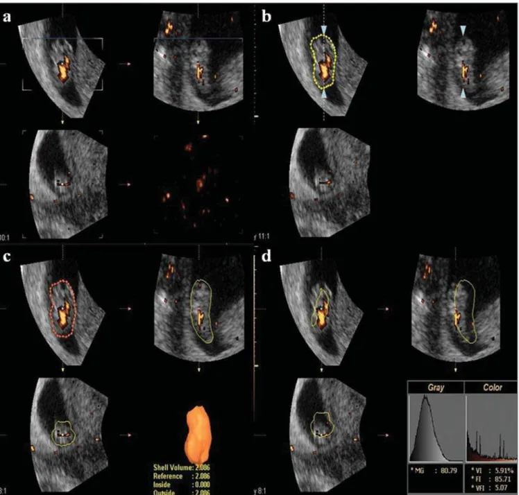
Af-ter adjustments in the areas constituting the three-dimensional image, such image was accepted, and the vocal shell histogram key was activated, and the 3D power Doppler indices of the embryo (VI, FI and VFI) (Fig-ure 1) were automatically calculated, VI corresponding to the number of color voxels and representing the percentage of the Doppler signal (vascularization) de-tected within the ROI; FI corresponding to the mean color value of all the color voxels, representing the mean intensity of the Dop-pler power signal within the ROI; and VFI, comprising the mean color value of all the color voxels and of the gray scale, consti-tuting a combination of the other indices (originated from their multiplication and division of the result by 100) and has been suggested to be representative of the vas-cularization and flow intensity(9). At the
were acquired from each patient, being such data stored in the equipment’s memory. The volume presenting the high-est vascular density was chosen to obtain the vascular indices. The volumetric and 3D power Doppler indices analyses were made in the absence of the patients by means of the software SonoView Pro ver-sion 1.03 (Medison; Seoul, Korea), being such analyses carried out by a single ob-server.
The data were stored on an Excel 2003 (Microsoft; Redmond, WA, USA) work-sheet and then analyzed by the statistical software SPSS for Windows version 13.0 (SPSS Inc.; Chicago, IL, USA). In order to evaluate the interobserver reproducibility of the 3D power Doppler vascular indices, a second observer, with a three-year expe-rience in obstetric three-dimensional ultra-sonography, performed blind measure-ments of the same 36 pregnant women. For
the reproducibility calculation, the paired Student t-test, the intraclass correlation coefficient (ICC) and Bland-Altman plots were utilized. An ICC < 0.40 is considered as poor; between 0.40 and 0.75 is satisfac-tory and a correlation ≥ 0.75 is excellent(10). The Bland-Altman plots demonstrate the mean value of the measurements per-formed by two observers against the differ-ence of their measurements with agreement limits of 95% and 1.96 standard
devia-Figure 1. Measurement of 3D power Doppler vascularization indices (VI, FI and VFI) after 3D reconstruction of embryonic volume utilizing the VOCAL method with a 12° rotation angle. a: positioning of the measurement calibrators on the embryo poles; b: Manual delimitation of the external surface of the embryo; c:
tion(11). In all the analyses, the significance
level (p) of 0.05 was utilized.
RESULTS
Initially, 36 healthy pregnant women were selected. However one case was ex-cluded because of a thermal index > 1.0 and three others were also excluded because of the low quality of obtained volumes as a consequence of transmission artifacts not allowing an appropriate evaluation of vas-cular indices. The 32 remaining pregnant women were followed-up until the 22nd gestational week, without any miscarriage, thus comprising the final sample.
A good interobserver reproducibility was observed for the three analyzed vascu-lar indices of 3D power Doppler. For VI,
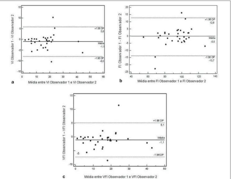
the mean difference between measure-ments was –1.1 (confidence interval [CI] 95%: –2.3 - 0.1) with the paired t-Student test demonstrating no significant difference among them (p = 0.09); the ICC was 0.9 (CI 95%: 0.8 - 0.9), while on the Bland-Altman plot the mean difference between measures was –1.1 (limits of agreement 95%: –8.0 -5.8) (Figure 2a). For FI, the mean difference between measurements was –0.5 (CI 95%: –2.9 - 1.1), with the paired Student t-test showing no significant difference among them (p = 0.65); the ICC was 0.9 (CI 95%: 0.8 - 0.9), while in the Bland-Altman plot the mean difference between measure-ments was –1.1 (limits of agreement 95%: –8.0 - 5.8) (Figure 2b). For VFI, the mean difference between measurements was –1.1 (CI 95%: –2.2 - 0.4), with the paired
Stu-dent t-test demonstrating no significant difference among them (p = 0.05); the ICC was 0.9 (CI 95%: 0.8 - 0.9), while on the Bland-Altman plot the mean difference between measurements was –1.1 (limits of agreement 95%: –5.0 - 5.1) (Figure 2c).
Such results confirm the applicability of 3D power Doppler in the study of the em-bryonic vascularization at the first trimes-ter of gestation, particularly with regards to FI, which presented the smallest mean dif-ference between the measurements.
DISCUSSION
The vascular indices of 3D power Dop-pler are not capable of quantifying the ac-tual blood flow or perfusion, as the flow is the quantity of blood that passes through a
Figure 2. Mean of the values obtained by observers 1 and 2 plotted against the difference of their measurements with limits of agreement 95% for VI (a), FI (b) and VFI (c).
c
vessel in a unit of time (generally per minute), while perfusion is defined as a quantity of flow in a volume of tissue per unit of time, and is generally measured as milliliters per minute per gram of tissue. As the three-dimensional vascular indices are not calculated as a function of time, their quantification cannot, consequently, match the actual perfusion or the flow of the stud-ied tissue(12,13). Such indices would be,
ac-tually, a representation of power Doppler data contained within the three-dimen-sional volume, with VI being the percent-age of power Doppler data contained within the volume, FI the mean intensity of the Doppler signal within the volume, and VFI a combination of both indices. How-ever, it is suggested that such indices rep-resent a semi-quantification of vasculariza-tion (VI) and flow intensity (FI)(13).
In the present study, the VOCAL method was utilized for the volumetric as-sessment of the embryo. Such technique is relatively simple and is commercially avail-able in several ultrasonography apparatuses Additionally, it presents a proven reproduc-ibility in the evaluation of placental power Doppler vascular indices (4,5,14). A rotation
angle of 12° was selected for allowing the determination of a higher number of planes (15) and, consequently, attempting to ob-tain higher accuracy in the measurements. In a study in vitro, measurements per-formed with an angle of 6° (30 planes) proved to be more reliable than those ob-tained with other rotation angles or multiplanar method, with the exception of the 9° angle (20 planes), and significantly more valid than those performed with a 30° angle or by the multiplanar method(15). In
another study using an endometrium-like experimental model, the measurements performed with the VOCAL method with rotation at 15° (12 planes) demonstrated to be valid and reproducible(16).
There are only two studies on the quan-titative evaluation of embryonic vascular-ization at the first trimester of gestation(1,3),
and in both cases no correlation between the vascular indices of the 3D power Dop-pler and CRL was observed. A possible explanation would be a state of homeosta-sis between demand of the tissues and the supply of blood from the vascular network in the first trimester of gestation(1).
The interobserver reproducibility was tested with two volumes previously re-corded by a single examiner. Therefore the reproducibility of the whole procedure, that is, from the volume acquisition, was not tested. There are two ways of analyzing reliability involving the use of the three-dimensional technique: by 3D volume ac-quisition and by volume calculation after definition of the contour by VOCAL method(17). The reliability of
three-dimen-sional volume acquisition is directly related to the level of confidence in accurately performing the contour definition on which the volume will be calaculated18).
Addition-ally, the data acquisition is much more sub-ject to uncertainties than a series of mea-surements of any given data, particularly in Dopplerfluxometric studies(15).
A good interobserver reproducibility was observed for the three 3D power Dop-pler vascular indices, with FI being the one with the lowest variability, with a mean difference between measurements of only –0.5. Previous studies in placentas have shown that FI is the index that best corre-lates with gestational age and the one pre-senting the lowest correlation coefficient in relation to the various measurements in the same placenta(14,19). Recently, the
intervil-lous vascularization was evaluated in a gestational period similar to the one in the present study, 5 to 12 weeks and 6 days, observing that FI was the 3D power Dop-pler vascular index with highest ICC(6).
This is explained by the increase of placen-tal vascularization as the gestation progresses, which is consistent with histo-logical data(20). Additionally, the increase in
the number of vessels is followed by a pro-gressive decrease in the vascular resistance, which contributes to the increase in perfu-sion and intraplacental flow(21). The results
of the present study are in agreement with those obtained by a Spanish group that has utilized the placental biopsy technique, in which the authors obtained ICC above 0.85 for the three indices, with best intraob-server reproducibility for the flow indices (FI and VFI)(4). Recently, researchers
evalu-ated the placental vascularization between the 12th and 40th gestational weeks, using the VOCAL method with manual delimi-tation of the whole external surface, and observed good intra- and interobserver
re-producibility for the three indices, with FI presenting the highest ICC and the lowest difference between measurements(5). It
should be highlighted that there are no studies on the reproducibility of 3D power Doppler vascular indices of embryos at the first trimester of gestation.
In the present study, some FI measure-ment presented values above 100, which contradicts the definition of such index(6).
The majority of the studies on 3D power Doppler have evaluated the placental vas-cularization at the first(6,22) as well as at the
second and third gestational trimes-ters(14,19,23), observing that the FI values
were within the normal variation range between 0 and 100. In previous studies the authors of the present study evaluated the vascularization in other structures such as the region of the middle cerebral artery of the fetus(24) and the embryo itself(3),
observ-ing that for these structures the maximum values of FI were above 100. A possible explanation for such fact would be the di-verse vascular development of the placenta in relation to other fetal organs. The authors do not believe that the apparatus or train-ing of the observers may have contributed for such result, considering that in a recent study developed by this same group, evalu-ating the placental vascularization at the second and third trimesters, using an appa-ratus from the same manufacturer with the same settings utilized in the present study, the mean values of FI ranged from 35.73 to 39.98(25). Further studies evaluating the
vascularization of other structures or fetal organs are necessary to prove the actual validity of the 3D power Doppler vascular indices.
CONCLUSION
The vascular indices, particularly the flow índex, obtained with 3D power Dop-pler ultrasonography at the first trimester of gestation demonstrated a high interob-server reproducibility.
REFERENCES
ultrasound. Ultrasound Med Biol. 2003;29:19– 23.
3. Bortoletti Filho J, Nardozza LM, Araujo Júnior E, et al. Embryo vascularization by three-dimen-sional power Doppler ultrasonography at 7-10 weeks of pregnancy. J Perinat Med. 2009;37:380– 5.
4. Mercé LT, Barco MJ, Bau S. Reproducibility of the study of placental vascularization by three-di-mensional power Doppler. J Perinat Med. 2004; 32:228–33.
5. de Paula CF, Ruano R, Campos JA, et al. Quan-titative analysis of placental vasculature by three-dimensional power Doppler ultrasonography in normal pregnancies from 12 to 40 weeks of ges-tation. Placenta. 2009;30:142–8.
6. Mercé LT, Barco MJ, Alcázar JL, et al. Intervil-lous and uteroplacental circulation in normal early pregnancy and early pregnancy loss assessed by 3-dimensional power Doppler angiography. Am J Obstet Gynecol. 2009;200:315.e1–8. 7. Duck FA. Is it safe to use diagnostic ultrasound
during the first trimester? Ultrasound Obstet Gynecol. 1999;13:385–8.
8. American Institute of Ultrasound in Medicine/Na-tional Electrical Manufactures Association. Stan-dards for real-time display of thermal and me-chanical acoustic output indices on diagnostic equipment. 2nd ed. Rockville, MD: American Institute of Ultrasound in Medicine; 1998.
9. Pairleitner H, Steiner H, Hasenoehrl G, et al. Three-dimensional power Doppler sonography: imaging and quantifying blood flow and vascu-larization. Ultrasound Obstet Gynecol. 1999;14: 139–43.
10. Shrout PE, Fleiss JL. Intraclass correlation: uses in assessing rater reliability. Psychol Bull. 1979; 86:420–8.
11. Bland JM, Altman DG. Statistical methods for assessing agreement between two methods of clinical measurement. Lancet. 1986;1:307–10. 12. Cosgrove D, Eckersley R, Blomley M, et al.
Quan-tification of blood flow. Eur Radiol. 2001;11: 1338–44.
13. Raine-Fenning NJ, Welsh AW, Jones NW, et al. Methodological considerations for the correct application of quantitative three-dimensional power Doppler angiography. Ultrasound Obstet Gynecol. 2008;32:115–7.
14. Guiot C, Gaglioti P, Oberto M, et al. Is three-di-mensional power Doppler ultrasound useful in the assessment of placental perfusion in normal and growth-restricted pregnancies? Ultrasound Obstet Gynecol. 2008;31:171–6.
15. Raine-Fenning NJ, Clewes JS, Kendall NR, et al. The interobserver reliability and validity of vol-ume calculation from three-dimensional ultra-sound datasets in the in vitro setting. Ultraultra-sound Obstet Gynecol. 2003;21:283–91.
16. Martins WP, Ferriani RA, Barra DA, et al. Reli-ability and validity of tissue volume measurement by three-dimensional ultrasound: an experimen-tal model. Ultrasound Obstet Gynecol. 2007;29: 210–4.
17. Järvelä IY, Sladkevicius P, Tekay AH, et al. Intraobserver and interobserver variability of ovarian volume, gray-scale and color flow indi-ces obtained using transvaginal three-dimen-sional power Doppler ultrasonography. Ultra-sound Obstet Gynecol. 2003;21:277–82.
18. Duin LK, Willekes C, Vossen M, et al. Reproduc-ibility of fetal renal pelvis volume measurement using three-dimensional ultrasound. Ultrasound Obstet Gynecol. 2008;31:657–61.
19. Mercé LT, Barco MJ, Bau S, et al. Assessment of placental vascularization by three-dimensional power Doppler “vascular biopsy” in normal preg-nancies. Croat Med J. 2005;46:765–71. 20. Jauniaux E, Jurkovic D, Campbell S. In vivo
in-vestigations of the anatomy and physiology of early human placental circulations. Ultrasound Obstet Gynecol. 1991;1:435–45.
21. Reynolds LP, Redmer DA. Angiogenesis in the placenta. Biol Reprod. 2001;64:1033–40. 22. Rizzo G, Capponi A, Cavicchioni O, et al.
Placen-tal vascularization measured by three-dimen-sional power Doppler ultrasound at 11 to 13 + 6 weeks’ gestation in normal and aneuploid fetuses. Ultrasound Obstet Gynecol. 2007;30:259–62.
23. Zalud I, Shaha S. Evaluation of the utero-placen-tal circulation by three-dimensional Doppler ul-trasound in the second trimester of normal preg-nancy. J Matern Fetal Neonatal Med. 2007;20: 299–305.
24. Nardozza LM, Araujo Júnior E, Simioni C, et al. Evolution of 3-D power Doppler indices of fetal brain in normal pregnancy. Ultrasound Med Biol. 2009;35:545–9.