Rev Bras Reumatol, v. 43, n. 5, p. IX-XII, set./out., 2003
Editorial
IX
E
DITORIALE
DITORIALThe Role of Ultrasonography in Rheumatology
(*)Kok Ooi Kong, MRCP(1), Damien Loeuille, MD PhD(2), Yohei Seto, MD(3),
Richard J. Wakefield, MRCP(4), Paul Emery, FRCP(5)
sculoskeletal ultrasound (US) is rapidly evolving into an important method for confirming the primary diagnosis and monitoring therapeutic response in many rheumatic conditions. Diagnosis US has achieved great advances since the reported detection of Baker’s cyst with B-mode scans in the early 1970s(1). Me-dical US was well developed in the 1980s when linear, sector, and Doppler US became available. However, the value of musculoskeletal US and its contribution in rheu-matology were not widely recognized until 1990s when high-resolution US (HRUS) became available. HRUS is non-invasive, painless, biologically safe, rapid to perform, and considerably less expensive then CT and MRI. This imaging technique is now being performed by rheumato-logists, particularly in Europe, as part of the standard clini-cal assessment of patients. The application of HRUS to the early diagnosis and evaluation of treatment response he-ralds an era where rheumatologists will be able to better target and manage various rheumatologic conditions, in particular inflammatory arthritis.
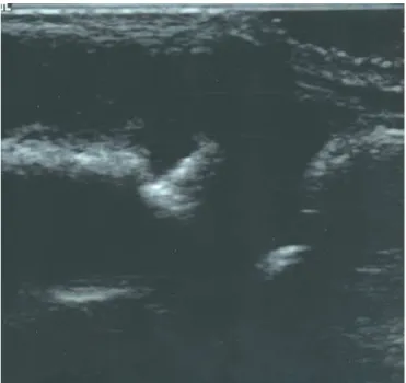
Effusion and synovitis
US allows visualization of inflamed sinovial tissue as a hypo-echoic structure while effusion as an anhypo-echoic structure (Figure 1). Many studies have highlighted the ability of US in detecting early sinovial disease in both large and small joints and its superiority over clinic examination(2-4). The-re have been several studies validating US against arthros-copy, MRI and scintigraphy. Backhaus et al.(4) found more synovitis in the joints of the hand and wrist with US when compared to radiography and clinical examination and it was comparable with MRI. Brown et al.(5) showed the
relevance of US in the detection of sub-clinical synovitis in their study of patients with RA, who were in clinical remission as defined by the ACR criteria(6). Almost half of the patients had US features of sub-clinical synovits in joints not thought to have any clinical synovitis. In a re-cent study of 80 patients with oligoarthritis(7), two third of the patients had sub-clinical synovitis detected on US and one third could be reclassified as having polyarticu-lar disease. Among those who were rheumatoid factor positive at baseline, 83% had evidence of sub-clinical
sy-* Academic Department of Musculoskeletal Medicine, First Floor, Old Nurse's Home, The General Infirmary at Leeds, Great George Street, Leeds, LS1 3EX. United Kingdom.
1. Research Fellow in Rheumatology.
2. Maitre de Conference Universitaire, Praticien hospitalier in Rheumatology. 3. Research Fellow in Rheumatology.
4. Senior Lecturer in Rheumatology. 5. ARC Professor of Rheumatology.
Correspondence to: Prof. Paul Emery, MD FRCP, ARC Professor of Rheumatology, Academic Department of Musculoskeletal Medicine, First Floor, Old Nurse's Home, The General Infirmary at Leeds, Great George Street, Leeds, LS1 3EX. United Kingdom. Tel: +44(0)113 392 5068, Fax: +44(0)113 392 2896, e-mail: [email protected].
X Rev Bras Reumatol, v. 43, n. 5, p. IX-XII, set./out., 2003 Editorial
novitis on US imaging. Of note, only 9% patients fulfil-led the ACR criteria for RA at baseline but, the addiction of US findings (synovitis and erosions) increased this per-centage to 50%. This finding demonstrates a potential roe for US in assisting diagnosis of early RA and highli-ghts an advantage over MRI (i.e., an ability to scan seve-ral joints at one time point). Clinical features of joint inflammation may not be present during this early stage as a result of subtleness af inflammation(8) or a marked response to non-steroidal anti-inflammatory drugs ( NSAI-Ds). In such situations, HRUS helps in identifying sub-clinical synovitis and allows reclassification of sub-clinical oli-goarthritis into polyarthritis(7).
Bony erosion
Furthermore, HRUS may detect bony erosions in affected joints when conventional radiography remains normal(9). Erosion is defined as a cortical “break” or defect with an irregular floor seen in longitudinal and transverse planes (Figure 2). Although MRI is as good as HRUS, if not better, in detecting synovitis and bony erosions(10), HRUS is chea-per and easily accessible in outpatient setting. The use of HRUS at early stage in identifying the presence of sub-cli-nical bony damages provides strong basis for early initiation of DMARD therapy. A study by Wakefield et al.(9) found that US was a reproducible technique and detected 3.5 ti-mes as many erosions as radiography; this difference was even grater with early disease. The superiority of US over
conventional radiography (CR) is explained by the multi-planar capability of US and the fact that US can detect smal-ler erosions. MRI was also used to assess the radial aspect of the 2nd MCP heads in 25 patients with early disease. All 10 MRI erosions corresponded exactly with US erosions. It is of interest to note that US detected 3 additional erosions. These can be explained by the superior spatial resolutions of US compared to MRI. A more recent study by Alarcon et al.(11) confirmed other diagnosis findings.
Reports on the value of HRUS in osteoarthritis suggest that may aid in diagnosis and assessment of severity of the disease. It allows assessment of joint space narrowing, os-teophytes, Baker’s cyst formation and its complication, and hyaline cartilage thickness of the trochlear groove (since the other knee articular surface are not directly ac-cessible to the probe). High prevalence of synovitis in the symptomatic osteoarthritis knees confirmed previous cli-nical findings(12). Use of US in other rheumatologic con-ditions is also developing. Its ability in assessing the skin thickness(13-15) and vascularity changes(16) in systemic scle-rosis had been reported. There has also been suggestion of its use in the management of myosistis(17).
HRUS does not involve ionizing radiation and is safe to be repeated as often as necessary on a number of different joints making it ideal for patient follow-up. HRUS also allows clinicians to distinguish inflammatory and non-in-flammatory diseases with more confidence, predict which patients have a poorer prognosis and require more aggres-sive early therapy and develop a more effective and appro-priate treatment plan for the patients. A study by Karim et al. demonstrated 50% of clinical decisions are changed with
the addition of the information from HRUS among those who were referred for HRUS examination(18). In our unit,
HRUS has been offered to rheumatology patients since
1997. Approximately 2000 patients a year have been be-nefited from it. Patients were seen by rheumatologists and had HRUS scanning of the joints done at the same clinic visit, if indicated. This not only provides a higher quality of care to patients with rheumatologic conditions, it also reduces the number of hospital visits for patients.
Power Doppler Sonography
Power Doppler Sonography (PDS) is playing an increasing role in the assessment of disease activity of inflammatory arthritis. Because PDS provides higher sensitivity to low-volume, low velocity blood flow at microvascular level, it is particularly useful for detecting changes of blood volu-me in sinovial issue and enthesitis, reflecting the degree of inflammation (Figure 3) and thus allowing the evaluation
Rev Bras Reumatol, v. 43, n. 5, p. IX-XII, set./out., 2003
Editorial
XI of the response to therapy. In addition, PDS signal is
inde-pendent of the transducer angle and thus does not have the problem of aliasing, an artifact as a result of inadequate signal sampling, and also much reduced background noi-se(19). PDS also improved of vessel characteristics that are not well visualized with colour-flow Doppler imaging. With the ability to identify low-velocity blood flow, PDS increases the specificity of HRUS in differentiating sinovial hypertrophy form fibrobtic sinovial tissues, blood clots, and complex effusion. The sensitivity of PDS can be fur-ther enhanced with the use of intravenous micro-bubble echo contrast agents(20). Validation of PDS against histopa-thology(21, 22) and MRI(23) had been published and it corre-lates well with the degree of the synovitis. Use of PDS in monitoring disease activity(24, 25) and treatment response(26-28) in rheumatoid arthritis has also been reported. The value of PDS has also been assessed in enthesitis(29), spondyloar-thorthopathy(30), myositis(31).
US-guided procedures
The use of US-guided procedures during daily clinical prac-tice is also of relevance to rheumatologists. US can visuali-ze deep joints that are not accessible to clinical examinati-on, and help in establishing the diagnosis of effusion. Moreover HRUS offers the opportunity for aspiration and biopsies in the same setting especially when sepsis in sus-pected. US can be useful for diagnostic and therapeutic
procedures. Steroid injection influences the efficacy(32, 33). Without imaging-guidance, the placement of needle was correct in only 50% of the time(32). It has been used to direct biopsy needles in erosions and enthesial sites. In addi-tion to the imaging guidance provided, the portability of the machine and lack of radiation allow more flexibility in aspiration, injection or biopsy.
It is particularly important for young rheumatologists to have a good understanding of functional limb anatomy in order to be able to manage regional pain syndromes effi-ciently and be experts in soft tissue rheumatology. The introduction of HRUS in academic rheumatology enhan-ces the teaching of functional anatomy to rheumatology trainees. Dynamic HRUS provides detailed real-time se-quential knowledge of the live functional anatomy that cannot easily be obtained otherwise. Trainees can observe the movement of muscles and tendons in relation to each other during the movement of interest. Many world-re-nown rheumatologic training canters have included HRUS as part of the core curriculum of training for young rheu-matologists. In fact, some even consider it a requirement for rheumatologists(34) (since it may be regarded) as an ex-tension of physical examination.
In order to use HRUS in daily practice of rheumatolo-gy, its limitations must be well understood. The interacti-ve and dynamic process of HRUS imaging means that such procedures are observer-dependent. This emphasizes the need for ultrasonographers to have adequate training at established rheumatology centers before service is provi-ded. The characteristic of US also means that is impossible to visualize structures behind or within bones. Some struc-tures, such as sacroiliac joints and hips, cannot be assessed adequately with the available transducers. The presence of artifacts, such as aniosotropy, must not be overlooked and at time may assist in identification of various structures, such as tendons or ligaments.
Worldwide, the use of HRUS in assisting the manage-ment of rheumatologic conditions, especially inflamma-tory arthritis, is growing rapidly. The availability of new, efficacious, expensive biologic therapies for inflammatory arthritis means the accurate diagnosis and monitoring of these patients has become even more important, particu-larly if there is a potential for stopping or reducing the use of these therapies among patients who are in remission. It is possible that HRUS in rheumatology may achieve a sta-tus similar to echocardiography in cardiology in future. With the advances in the technologies, the growth of HRUS in rheumatology must not be overlooked, especially when improved patient care and better disease outcome are the goals for all rheumatologists.
XII Rev Bras Reumatol, v. 43, n. 5, p. IX-XII, set./out., 2003 Editorial
REFERENCES
1. McDonald DG, Leopold GR. Ultrasound B-scanning differentia-tion of Baker’s cyst and thrombophlebitis. Br J Radiol 1972; 45(538):729-32.
2. Koski JM. Ultrasonographic evidence of hip synovitis in patients with rheumatoid arthritis. Scand J Rheumatol 1989;18(3):127-31. 3. Conaghan, Wakefield R, O’Connor P, Gibbon W, Emery P. The metacarpophalangeal joints in early rheumatoid arthritis: a comparison of clinical, radiographic, MRI and ultrasonographic findings, Ann Rheum Dis 1999;58(Suppl).
4. Backhaus M, Kamradt T, Sandrock D, et al. Arthritis of the finger joints- a comprhensive approach comparing conventional radiogra-phy, scintigraradiogra-phy, ultrasound and contrast-enhnaced magnetic re-sonance imageing. Arthritis Rheum 1999;42:1232-45.
5. Brown AK, Quinn MA, Karim Z, et al. Neither the ACR remission criteria nor the disease activity score accurately define true remission in rheumatoid arthritis. Arthritis Rheum 2002;46 Suppl 9:243. 6. Pinals RS, Mais AT, Larsen RA. Preliminary criteria for clinical
remission in rheumatoid arthritis. Arthritis Rheum 1981;24(10): 1308-15.
7. Wakefield RJ, Green MJ, Helena MO, et al. Should oligoarthritis be reclassified? – Ultrassound reveals high prevalence sub-clinical disease. Ann Rheum Dis 2003;(in press).
8. Wakefield RJ, Green M, Gibbon WW, et al. High-resolution ultrasound defined sub-clinical synovitis – predictor of outcome in early oligo arthritis? Arthritis Rheum 1998;41 Suppl 9:246. 9. Wakefield RJ, Gibbon WW, Conaghan PG, et al. The value of
sonography in the detection of bone erosions in patients with rrheumatoid arthritis – a comparison with conventional radiography. Arthritis Rheum 2000;43:2762-70.
10. McGonagle D, Conaghan PG, O’Connor P, et al. The relationship between synovitis and bone changes in early untreated rheumatoid arthritis: a controlled magnetic resonance imaging study. Arthritis Rheum 1999;42(8):1706-11.
11. Alarcon GS, Lopez-Ben R, Moreland LW. High-resolution ultrasound for the study of target joints in rheumatoid arthritis. Arthritis Rheum 2002;46(7):1969-70; author reply 1970-1. 12. Hukisson EC, Dieppe PA, Tucker AK, Cannel LB. Another look
at osteoarthritis. Ann Rheum Dis 1979;38(5):423-8.
13. Cosnes A, Anglade MC, Revuz J, Radier C. Thieteen-megahertz ultrasound probe: its role in diagnosing localized scleroderma. Br J Dermatol 2003;148(4):724-9.
14. Hesselstrand R, Westergreen-Thorsson G, Scheja A, Wildt M, Akesson A. The association between changes in skin echogenicity and the fibroblast production of biglycan and versican in systemic sclerosis. Clin Exp Rheumatol 2002;20(3)301-8.
15. Scheja A, Akesson A. Comparisson of high frequency (20 MHz) ultrasound and palpation for the assessment of skin involvement in systemic sclerosis (scleroderma). Clin Exp Rheumatol 1997; 15(3):283-8.
16. Keberle M, Tony HP, Jahns R, Hau M, Haerten R, Jenett M. As-sessment of microvascular changes in Raynaud’s phenomenon and connective tissue disease using color Doppler ultrasound. Rheuma-tology (Oxford) 2000;39(11):1206-13.
17. Fleckenstein JL, Reimers CD. Inflammatory myopathies: radiologic evaluation. Radiol Clin North Am 1996;34(2):427-38, XII.
18. Karim Z, Wakefield RJ, Conaghan PG, et al. The impact of ultrasonography on diagnosis and management of patients with musculoskeletal conditions. Arthritis Rheum 2001;44(12):2932-3. 19. Rubin JM, Bude RO, Carson PL, Bree RL, Adler RS. Power
Doppler US: a potentially useful alternative to mean frequency-based color Dopler US. Radiology 1994;190(3):853-6.
20. Blomley MJ, Cooke JC, Unger EC, Monaghan MJ, Cosgrove DO. Microbubble contrast agents: a nem era in ultrasound. BMJ 2001; 322(7296):1222-5.
21. Schmidt WA, Volker L, Zacher J, Schlafke M, Ruhnke M, Gromnica-ihle E. Colour Doppler ultrasonography to detect pannus in kenee joint synovitis. Clin Exp Rheumatol 2000;18(4):439-44. 22. Walther M, Harns H, Krenn V, Radke S, Faehndrich TP, Gohlke
F. Correlation of power Doppler sonography with vascularity of the sinovial tissue of the knee joint in patients with osteoarthritis and rheumatoid arthritis. Arthritis Rheum 2001;44(2):331-8. 23. Szkudlarek M, Court-payen M, Strandberg C, Klarlund M,
Klausen T, Ostergaard M. Power Doppler ultrasonography for assessment of synovitis in the metacarpophalangeal joints of patients with rheumatoid arthritis: a comparison with dynamic magnetic resonance imaging. Arthritis Rheum 2001;44(9):2018-23. 24. Qvistgaard E, Rogind H, Torp-pedersen S, Terslev L,
Danneskiold-Samsoe B, Bliddal H. Quantitative ultrasonography in rheumatoid arthritis: evaluation of inflammation by Doppler technique. Ann Rheum Dis 2001;60(7):690-3.
25. Carotti M, Slaffi F, Manganelli P, Salera D, Simonetti B, Grassi W. Power Doppler sonography in the assessment of synovial tissue of the knee joint in rheumatoid arthritis: a preliminary experience. Ann Rheum Dis 2002;61(10):877-82.
26. Hau M, Kneitz C, Tony HP, Keberle M, Jahns R, Jenett M. High resolution ultrasound detects a decrease in pannus vascularisation of small finger joints in patients with rheumatoid arthritis receiving treatment with soluble tumour necrosis factor alpha receptor (etanercept). Ann Rheum Dis 2002;61(1):55-8.
27. Newman JS, Laing TJ, McCarthy CJ, Adler RS. Power Doppler sonography of synovitis: assessment of therapeutic response-preliminary observations. Radiology 1996;198(2):582-4.
28. Stone M , Bergin D, Whelan B, Maher M, Murray J, McCarthy C. Power Doppler ultrasound assessment of rheumatoid hand synovitis. J Rheumatol 2001;28(9):1979-82.
29. D’Agostino MA, Said Nahal R, Hacquard-Bouder C, Brasseur JL, Dougados M, Breban M. Assessment of peripheral enthesitis in the spondylarthropathies by ultrassonography combined with power Doppler: a cross-sectional study. Arthritis Rheum 2003;48(2):523-33. 30. D’Agostino MA, Breban M, Said-Nahal R, Dougados M. Refractory inflammatory heel pain in spondylarthropathy: a significant response to infliximab documented by ultrasound. Arthritis Rheum 2002;46(3):840-1, author reply 841-3.
31. Meng C, Adler R, Pterson M, Kagen L. Combined use of power Doppler and gray-scale sonography: a new technique for the assessment of inflammatory myopaathy. J Rheumatol 2001;28(6): 1271-82.
32. Jones A, Regan M, Ledingham J, Pattrick M, Manhira A, Doherty M. Importance of placement of intra-articular steroid injections. Bmj 1993;307(6915):1329-30.
33. Eustace JA, Brophy DP, Gibney RP, Bresnihan B, FitzGerald O. Comparison of the accuracy of steroid placement with clinical outcome in patients with symptoms. Ann Rheum Dis 1997;56(1):59-63. 34. Canoso JJ. Ultrasound imaging-a requirement for rheumatologists.