Rev Bras M ed Esporte _ Vol. 11, Nº 5 – Set/Out , 2005
259e
1. PhD, Laboratory of Kinesiologic Electromyography, Department ofPhys-iotherapy, Londrina State University (UEL) – PR, Brazil.
2. Department of Physiotherapy, Londrina State University (UEL) – PR, Bra-zil.
3. Physiotherapist, Center of Sportive Traumato-Orthopedics, Sao Paulo Federal University (Unifesp-EPM ).
4. Academic of the Course of Physiotherapy, Londrina State University (UEL) – PR, Brazil.
5. Academic of the Course of Physical Education, Londrina State Univer-sity (UEL) – PR, Brazil.
Received in 4/8/04. 2nd version received in 10/4/05. Approved in 12/6/05. Correspondence to: Prof. Dr. Jefferson Rosa Cardoso, Rua Osaka 33, ap. 32 – 86050-330 – Londrina, PR. Tel.: (43) 3371-2649. E-mail: jeffcar@ sercomtel. com.br
Use of ankle bracing for volleyball activities
Jefferson Rosa Cardoso1, M s. Christiane de Souza M acedo Guerino2, M arcelo Bannw art Santos3,
Thiago André Del Arco M ustafá4, Anália Rosário Lopes4 and M arcelo Costa de Paula5
O
RIGINALA
RTICLEKeyw ords: Ankle. Electromyography. Orthoptic devices. ENGLISH VERSION
ABSTRACT
The ankle sprain is the most frequently found acute injury in volleyball. Aiming to prevent the occurrence of ankle injuries, pro-phylactic equipment as the bracing had been developed. This study had the purpose to evaluate the performance of the ankle mus-cles (tibialis anterior, peroneous longus, and medial and lateral gas-trocnemius) by measuring the electrical activity in different volley-ball activities (vertical jumping and lateral shuffling) w ith and w ithout using ankle bracing. Nine young female volleyball athletes w ith ages ranging from 14 to 17 years (x: 15.8 ± 1.3) w ere evaluated, all of them w ithout previous injuries in the dominant member. M aximal voluntary isometric contractions (M VIC) of each muscle w as collected, and after that, the electromyographic activity in dif-ferent situations w ith and w ithout using the bracing, randomly. The bracing used had tw o lateral supports. The electromyograph-ic signal w as quantified by the root mean square (RM S), and nor-malized by the M VIC. Analysis of the variance w ith repeated mea-surement w as used to verify the difference of the electric activity of the muscles involved in each activity, w ith and w ithout using the bracing, w ith 5% (p < 0.05) significance level. It w as identified a statistically significant difference in phase I of the jumping in favor of the tibialis anterior (p < 0.001) and in phase II in favor of the three flexors muscles (p < 0.001; p = 0.01; p = 0.003) in both situations, w ith and w ithout using the bracing. As to the lateral jump activity, a significant difference w as observed in the phase of braked in favor of the tibialis anterior and the lateral gastrocne-mius (p = 0.013) in both situations. It w as found no statistical dif-ference among muscles of the tw o groups. Results suggest that using the ankle bracing cannot influence the electrical activity of the muscles studied during the vertical jumping and the lateral shuffling.
INTRODUCTION
To participate in sports activities has been stimulated due to the benefits to the health. How ever, such practice predisposes the individual to specific injuries that may cause the removal of his daily activities, demanding a specialized treatment. Volleyball is a modality characterized by a great amount of repeated jump-ings, both during defense movements (blockage) w hile arming pitch movements (lifting), and attack movements (some types of attack and game finalizations). Goodw in et al.(1) observed that 63% of
the lesions in volleyball w ere related to the jumping.
The ankle sprain is the more frequent acute lesion found in this sports, w ith the incidence varying from 15 to 60% . The majority of the ankle sprains occur during the jump landing after a blockage or attack(2). A review in the literature describes the means to prevent
the ankle sprain: types of shoes, bandage, ankle bracing, and sen-sor-motor training.
The ankle bracing has the main purpose to promote an addition-al externaddition-al support to the stabilizing ligaments and muscles of the joint. In a biomechanical investigation on the ankle stability using brace, it w as verified an increasing in the torque of the joint of the ankle, thus neutralizing the inversion movement and keeping the joint in an appropriate anatomic position, w ith a better contact be-tw een the joint surfaces(4). The ankle bracing can be classified as:
lace-up (made by a flexible material, such as leather and strings to allow a better fixation), stirrup (w ith tw o lateral supports constitut-ed by plastic material) and elastic(5). Surve et al.(6) studied the
effi-ciency of the bracing aiming to prevent the ankle sprains in soccer players. In such study, the author observed a low er incidence of sprains in the group of athletes w ith previous ankle sprain and using the bracing compared to the group that did not use it. Silter et al.(7) found similar results in a study w ith 1601 basketball
ath-letes during tw o seasons. Some athath-letes w ith previous ankle sprain w ho used the bracing also obtained a low er occurrence of sprains in the joint.
M any studies have observed the effect of the bracings in the ankle muscles during sportive activities(8-10). Hopper et al.(8)
inves-tigated the electromyographic activity of the gastrocnemius, the tibial anterior and the peroneus longus muscles during the jump using adhesive tape, and jump using bracing. In those individuals using bracing, there w as a significant decreasing of the electromyo-graphic activity of the gastrocnemius and the peroneus longus muscles. Cordova et al.(9) verified the effect of different bracings
during stress in the inversion of the ankle by means of electromyo-graphic signals in the gastrocnemius (medial portion), the pero-neus longus, and the tibialis anterior muscles. Results confirmed the great importance of the peroneus longus as lateral stabilizer.
The aim of this study w as to compare the electrical activity of the tibialis anterior, the peroneus longus, and the gastrocnemius muscles in volleyball athletes w ith and w ithout using ankle brac-ing durbrac-ing tw o activities related to the volleyball (jumpbrac-ing and lat-eral displacement).
M ETHODS
Sampling
This study w as composed by nine childish-juvenile female vol-leyball athletes of the Londrina Sports Foundation, aging from 14 to 17 years old (x: 15.8 ± 1.3), w ith body mass index varying from 17.1 and 23.5 kg/m2 (x: 20.5 ± 2.4), and duration of training from
260e
Rev Bras M ed Esporte _ Vol. 11, Nº 5 – Set/Out, 2005Equipment
It w as used a sixteen channel electromyograph (EM G System do Brasil) composed by a tw elve bits A-D converter (analog-digital). Each channel w as coupled to tw o active electrodes and a reference one. The circular silver/silver chloride electrodes w ere connected to a high impedance preamp (1.0 x 10¹² Ohm) w ith common-mode
rejection ratio 120 dB. Signals w ere fitted for 2000 sampling/sec, and the filter w as adjusted in a 20 Hz to 500 Hz bandw ith. These data w ere analyzed in data acquisition softw are (AqDados, 5.0).
Procedures
The electrodes (simple differential active of surface) w ere put on the muscle-tendon junction located by means of palpation of the muscular belly, and parallel to the muscular fibers, according to positioning described by Basmajian and Deluca(11). The site w as
prepared w ith trichotomy and cleaned w ith alcohol to decrease the impedance. The reference electrode w as placed on the w rist. The method used to perform the quantitative analysis of the elec-tric potential amplitude throughout activities w as the root mean square (RM S) expressed in microvolts (µV).
The procedure to collect the electrical signals started w ith the maximum voluntary isometric contractions (M VIC), w ith the pur-pose to normalize such signals. M VIC of the tibialis anterior and the peroneus longus w ere acquired by means of the muscular function assessment, according to Kendall and M cCreary(12), w ith
the manual resistance performed by the same appraiser in every athlete. As to the gastrocnemius (both portions), M VIC w as ac-complished having the participant stand up under a transversal bar fixed to the w all, and applying strength in such a direction as to rise the bar w ith the shoulders. From the M VIC values, the per-centage of the electrical activation performed by the muscles w as calculated in every activity proposed.
Protocol
After collecting the M VIC, all individuals w ere analyzed in tw o different activities: vertical jumping (w ith a preparation movement – countermovement), and lateral displacement (defense move-ment) w ith and w ithout bracing. Such bracing (stirrup type, Air-cast Sports-Stirrup, Summit, NJ) has a contact base to the foot in w hich tw o lateral extensions are lifted around 15 cm up to the middle of the leg. They are fixed by means of tw o double stripes (velcro).
Firstly, the jumping w as performed having the athlete in bipedal support (w ith hands fixed on the w aist), and the feet parallel and separated approximately at the height of the shoulders.
After a verbal sign, the participant performed the movement inflecting the hip and knee joints, and extending the ankle joint (phase 1 – descending or pre-impulsion). In the next phase (2 – ascending or impulsion) the participant performed a continuous movement in w hich the hip and knee’s joints w ere extended and the ankle’s w ere inflected.
The movement should be performed as fast as possible. The third phase analyzed w as considered w hen the participant touched the ground (landing). The lateral displacement w as performed by means of tw o continuous lateral shuffling tow ards the predomi-nant side of the athlete: initially, the athlete started from the rest, stood up, extending the low er limbs, and next, performing the trunk flexion concomitant to a flexion of the knee, thus producing a passive extension of the ankle (closed chain) (phase 1 – begin-ning of the displacement). After that, the athlete propelled herself to the side w ith tw o jumps in a row performing a finalization as observed in the volley’s defense movement (phase 2 – brake). The order of the activities and use of bracings (before or after) w as randomized in each participant. For the data collection, all participants performed three repetitions of each activity. The tw o activities proposed (vertical jumping and side displacement) w ere
recorded by an S-VHS camera (JVC) w ith the purpose to synchro-nize the interpretation of the electromyographic signal w ith each phase of the activities, thus allow ing a better description of each muscle. The athletes w ere allow ed to train the activities, as to w arm and adapt themselves.
Statistical analysis
First, it w as accomplished a descriptive analysis of the results (presented in M VIC percentages – average and standard error). In order to verify the differences of the electrical activity in the mus-cles involved in each activity w ith and w ithout bracing, it w as used the analysis of variance w ith repeated measures. It w as applied the test of the M auchly W.’s sphericity test, and w henever the test w as violated, it w as performed the necessary technical cor-rections through the Huynh-Feldt test. Whenever the F test w as significant, the analysis w as complemented by means of the Bon-ferroni’s multiple comparison test. The statistical significance w as adopted in 5% (p < 0.05). For the data analysis, it w as used the Statistical Package for Social Sciences (SPSS) softw are version 11.5 for Window s.
RESULTS
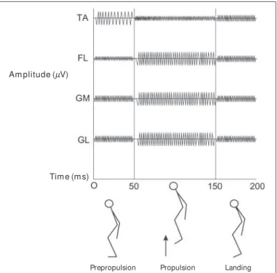
The electrical activities of the muscles involved (tibialis anterior, long fibular, and gastrocnemius – medial and lateral portions) in each phase of the vertical jumping as w ell as the lateral displace-ment are exemplified in figures 1 and 2. The tibialis anterior mus-cle in phase 1 of the jumping has an electrical activity higher than the peroneus longus and the tw o portions of the gastrocnemius muscles in both situations – w ithout and using the bracing (p < 0.001). In phase 2 of the jumping, the three flexor muscles w ith electrical activity higher than the tibialis anterior also in both situa-tions – w ithout and using the bracing (p < 0.001; p = 0.01; p = 0.003). In the third phase of the vertical jumping (landing) it w as not identified a statistically significant difference betw een the elec-trical activities of muscles in both situations.
Fig. 1 – Schematic figure of the electromyographic activity of the follow -ing muscles: tibialis anterior (TA), fibular longus (FL), and gastrocnemius – medium portion (GM ) and lateral (GL) during the three phases of the jump.
Am plitude (µV)
Tim e (m s)
Rev Bras M ed Esporte _ Vol. 11, Nº 5 – Set/Out , 2005
261e
DISCUSSION
The vertical jumping and the lateral displacement are quite com-mon sportive movements in volleyball. The jump is a frequent ac-tivity related to more incident muscle-skeletal sprains in such sports, and among them, the ankle sprain. Aiming to prevent the occurrence of this type of lesion, prophylactic equipments w ere developed such as the ankle bracing. This study has proposed to investigate the electrical activity of three muscles of the leg (tibia-lis anterior, peroneus longus, and gastrocnemius) in female vol-leyball athletes during tw o different situations, w ith and w ithout an ankle bracing.
It w as found no statistically significant difference in the electri-cal activity of the three muscles studied w ith or w ithout using the bracing during bipedal vertical jumping.
This result is different from other results found in a studied per-formed by Hopper et al.(8), w here the author verified a significantly
decreased electrical activity of the peroneus longus and the medi-al gastrocnemius muscles w henever the individumedi-als of the research used the bracing during one-leg landing movement.
In this research, it w as studied the bipedal jump in three differ-ent phases: pre-impulsion, impulsion and landing. Hopper et al.(8)
studied only the landing and in one-leg support w ith a different bracing than the one used in this study. Another difference of methods w as that in our study, the athletes performed the jump-ing only in vertical direction, w hile in the previous study the ath-letes performed a vertical jumping w ith horizontal component.
Tillman et al.(13) studied the jumping and landing techniques in
elite athletes of female volleyball. In this study, the author verified that 55% of attack landings and 57% of the defense landings are performed w ith symmetrical support from the tw o low er limbs (bipedal), as used in our research. How ever, the landing using one-leg support is described as a potential mechanism of sprain in the knees and ankle for volleyball athletes(14). This aspect suggests
the need to evaluate the electrical activity of the muscles studied also during the jump and the landing in one-leg support, and such aspect w as not mentioned in this study.
Compared to the electrical activities of the muscles studied dur-ing the three phases of the vertical jumpdur-ing described, it w as ob-served in the phase 1 (impulsion) that the tibialis anterior pre-sents a higher electrical activity than other muscles. This result may be understood by the fact that the individual performs a hip As to the electrical activity of the lateral displacement, it w as
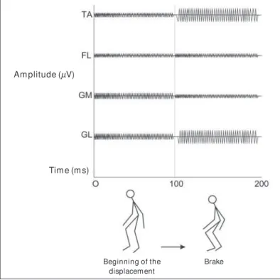
observed only one statistically significant difference betw een the tibialis anterior muscle and the medial portion of the gastrocne-mius muscle in the brake phase in both situations using the brac-ing (p = 0.013). The M VIC percentages for each muscle in differ-ent phases, and the comparison using or not the bracing are presented in table 1 and 2. It w as found no statistically significant differences betw een the tw o groups.
CHART 1
M ean (xxxxx ) and Standard Error (SE) of the CIVM ’s % of each muscle, phases I, II and III of the vertical jumping (prepropulsion, propulsion, and landing)
of groups w ithout and w ith using ankle bracing (n = 9)
G. w / out bracing G. w ith bracing
x xx x
x (SE) xxxxx (SE)
Vertical Jump
Phase I – Prepropulsion
Tibialis Anterior 78.9 0(4.7) 90.1 (10.3) Fibular Longus 36.2 0(4.9) 44.2 0(6.6) Gastrocnemius (P. M edium) 30.4 0(5.1) 29,00(5.1) Gastrocnemius (P. Lateral) 56.2 (21.3) 45.8 (13.2)
Phase II – Impulsion
Tibialis Anterior 043.6 0(5.7) 048.6 (7.6) Fibular Longus 102,00(3.6) 102.5 (3.6) Gastrocnemius (P. M edium) 089.9 0(6.1) 093,0 (8)0,
Gastrocnemius (P. Lateral) 091.7 (97.1) 097.1 (7.5)
Phase III – Landing
Tibialis Anterior 72.7 0(8.7) 81,00(9.9) Fibular Longus 62.9 0(4.1) 67.7 0(5.1) Gastrocnemius (P. M edium) 89.9 0(6.1) 93,00(8),0
Gastrocnemius (P. Lateral) 94.2 (14.5) 93.1 (14),0
Groups w ere compared through the ANOVA w ith repeated measures.
Phase 1 (Huynh-Feldt Test for sphericity of p = 0.677; F Test = 0.047; p = 0.832). Phase II (M auchly W Test for sphericity of p = 0.225; F Test = 0.111; p = 0.953). Phase III (M auchly W Test for sphericity of p = 0.096; F Test = 0.100; p = 0.960).
CHART 2
M ean (xxxxx ) and Standard Error (SE) of the CIVM ’s % of each muscle, phases I and II of the lateral displacement (beginning and brake)
of groups w ithout and w ith using ankle bracing (n = 9)
G. w / out bracing G. w ith bracing
x xx x
x (SE) xxxxx (SE)
Lateral Displacement
Phase I – Beginning
Tibialis Anterior 47.9 0(6.7) 53.8 0(8.6) Fibular Longus 49.9 0(8.4) 51.3 0(8.3) Gastrocnemius (P. M edium) 70,00(9.2) 70.2 0(9.8) Gastrocnemius (P. Lateral) 99.1 (14)0, 90.5 (10.6)
Phase II – Brake
Tibialis Anterior 48.8 0(6.1) 49.1 (8.8) Fibular Longus 38.6 0(6.1) 300, (4)0,
Gastrocnemius (P. M edium) 29,0 0(2.5) 30.6 (4.8) Gastrocnemius (P. Lateral) 48.1 (16.7) 42.9 (7.6)
Groups w ere compared through the ANOVA w ith repeated measures.
Phase 1 (Huynh-Feldt sphericity Test of p = 0.547; F Test = 0.031; p = 0.863). Phase II (Huynh-Feldt sphericity Test p = 0.632. F Test = 0.195; p = 0.664).
Fig. 2 – Schematic figure of the electromyographic figure of the tibialis anterior (TA), fibular Longus (FL) e Gastrocnemius – medium portion (GM ) and lateral (GL) during the tw o phases of the lateral displacement.
Am plitude (µV)
Tim e (m s)
Beginning of the displacem ent
262e
Rev Bras M ed Esporte _ Vol. 11, Nº 5 – Set/Out, 2005 and knee flexion associated w ith a passive extension of the anklein closed kinetic chain, demanding a higher activity of the ankle extensor studied (tibialis anterior).
In phase 2 of the vertical jumping, or impulsion, the flexor mus-cles of the ankle are more requested due to an active flexion move-ment of that joint. So, it w as observed a higher electrical activity in the tw o portions of the gastrocnemius and the peroneus longus. Finally, in phase 3 of the jumping it w as observed a higher elec-trical activity of the ankle flexors due to the eccentric contraction in closed kinetic chain after touching the ground, but it had no statistical significance.
Cordova et al.(9) studied the electric activity in the peroneus
lon-gus, the tibialis anterior, and the medial gastrocnemius during the lateral displacement. The author found a significant reduction in the electrical activity of the peroneus longus upon the use of both types of the studied bracing. In this study, it w as found no statis-tically significant difference in the electrical activity of the mus-cles evaluated during the lateral displacement compared to the tw o situations studied (w ith and w ithout bracing).
In the Cordova et al.(9) study, individuals w ere asked to perform
five to seven consecutive high speed displacements, and as soon as their foot got in touch w ith a platform, they should change the direction of the displacement quickly, coming back to their original position. In this study, the request did not include high speed move-ments nor changing in the direction, w hat w ould explain different results found. The fact that there w as no study on sudden decel-eration during the side displacement performed in this research must be taken into account, since the peroneus longus muscle could be more requested during activities that stimulates the in-version stress.
While the athletes w ere performing the lateral displacement, it w as observed that some of them w ere afraid to perform the activ-ity due to the presence of w ires of the electromyography chan-nels connected to their legs. This aspect may have influenced the speed and the biomechanics of the movement performed by them. Such difficult w as not detected during the execution of the verti-cal jump.
Compared to the electrical activity of the muscles studied dur-ing both phases of the lateral displacement described, it w as ob-served that only in phase 2 (the brake) it has occurred a statistical-ly significant difference in the electrical activity of the tibialis anterior and the medial portion of the gastrocnemius.
Another factor to be approached is the size of the sampling used in this study. The reduced number of individuals in the re-search may influenced the results obtained, causing the error type II. In this study, it w as possible to note that the use of ankle brac-ing did not influence the electrical activity of the muscles studied during the tw o proposed activities.
CONCLUSION
This research found no statistically significant difference in the electrical activity in the muscle w hile accomplishing the tw o activ-ities studied w ith and w ithout using the ankle bracing. These re-sults suggest that the use of ankle bracing may not influence the electrical activity of the three muscles studied during the vertical jumping and the lateral shuffling.
In order to better observe the activities studied, the authors suggest to perform new researches approaching sportive move-ments performed in one-leg support and high speed associated w ith changing in the direction w ith the purpose to evaluate the electrical activity of the muscles studied (w ith and w ithout brac-ing) during situations closest to the sportive activity.
ACKNOWLEDGEM ENT
The authors w ish thank Prof. Roger Burgo de Souza of the Physiother-apy Division of the University Hospital of the Londrina State University for his support in supplying all the necessary material to the data collection (electromyography), Daniel Jose de Carvalho, Instructional Production and Scientific Division, Daniel from the University Hospital of the Londrina State University for manufacturing the figures, coaches Ney Inácio da Sil-va, and Carlos Alberto Hipólito of the childish-juvenile team of the female volleyball of the Londrina Sports Foundation, and the athletes w ho partic-ipated of this study. Without their collaboration, it w ould be impossible to accomplish this w ork.
All the authors declared there is not any potential conflict of inter-ests regarding this article.
REFERENCES
1. Goodw in-Gerberich SG, Luhmann S, Finke C. Analysis of severe injuries associ-ated w ith volleyball activities. Phys Sportsmed 1987;15:75-9.
2. Briner WW, Benjamin HJ. Volleyball injuries: managing acute and overuse disor-ders. Phys Sportsmed 1999;27:48-60.
3. Thacker SB, Stroup DF, Branche CM , Gilchrist J, Goodman RA, Weitman EA. The prevention of ankle sprains in sports. A systematic review of the literature. Am J Sports M ed 1999;27:753-60.
4. Thonnard JL, Bragard D, Willems PA, Plaghki, L. Stability of the braced ankle. A biomechanical investigation. Am J Sports M ed 1996;24:356-61.
5. M attacola CG, Dw yer M K. Rehabilitation of the ankle after acute sprain or chronic instability. J Athl Train 2002;37:413-29.
6. Surve I, Schw ellnus M P, Noakes T, Lombard C. A fivefold reduction in the inci-dence of recurrent ankle sprains in soccer players using the Sport-Stirrup ortho-sis. Am J Sports M ed 1994;22:601-6.
7. Sitler M , Ryan J, Wheeler B, M cBride J, Arciero R, Anderson J, et al. The efficacy of a semirigid ankle stabilizer to reduce acute ankle injuries in basketball: a ran-domized clinical study at West Point. Am J Sports M ed 1994;22:454-61. 8. Hopper DM , M cnair P, Elliott BC. Landing in netball: effects of taping and bracing
the ankle. Br J Sports M ed 1999;33:409-13.
9. Cordova M L, Armstrong CW, Rankin JM , Yeasting R. Ground reaction forces and EM G activity w ith ankle bracing during inversion stress. M ed Sci Sports Exerc 1998;30:1363-70.
10. Pienkow ski D, M cM orrow M , Shapiro R, Caborn DN, Stayton J. The effect of ankle stabilizers on athletic performance. A randomized prospective study. Am J Sports M ed 1995;23:757-62.
11. Basmajian JV, Deluca CJ. M uscles alive. 5th ed. Williams & Wilkins, Baltimore.
12. Kendall FP, M cCreary EK. M úsculos. Provas e funções. 3a ed. São Paulo: M anole,
1987.
13. Tillman M D, Hass CJ, Brunt D, Bennett GR. Jumping and landing techniques in elite w omen’s volleyball. J Sports Sci M ed 2004;3:30-6.