1. Physiotherapist for Clube Grêmio Náutico União – Porto Alegre/RS, Physiotherapist for Clinic SOS ESPORTES, M aster in Human M ove-ment Sciences – UFRGS, Professor of the Physiotherapy Course, PU-CRS, Doctorship in Sciences of the Human M ovement – UFRGS. 2. Post-doctorship Calgary University, Canada, Doctorship by the Calgary
University, Canada, Professor of the Physical Education Course, UFRGS, Professor of the Post-graduation Program in Sciences of the Human M ovement, ESEF/UFRGS.
3. Orthopedist and Sports Traumatologist for Clinic SOS ESPORTES, Or-thopedist and Sports Traumatologist, and Coordinator for the M edicine and Rehabilitation Center of the Clube Grêmio Náutico União – Porto Alegre/RS, Physician Director of the Federação Gaúcha de Futevôlei, and Physician Director for the Federação Gaúcha de Futebol, M aster in Sciences of the Human M ovement – UFRGS, Doctorship in Sciences of the Human M ovement, UFRGS.
Received in 26/9/04. Final version received in 15/6/05. Approved in 17/7/ 05.
Correspondence to: Adriana M oré Pacheco, Rua Comendador Rheinga-ntz, 362, apto. 601, B. Auxiliadora – Porto Alegre, RS. E-mail: adrimpacheco @terra.com.br
Evaluation of the time for the electromyographic
response in volleyball athletes and
non-athletes w ho had ankle sprain
Adriana M oré Pacheco1, M arco Aurélio Vaz2 and Ivan Pacheco3
O
RIGINALA
RTICLEKeyw ords: Time for the electromyographic response. Sprain. Ankle.
ENGLISH VERSION
ABSTRACT
The purpose of this study w as to examine the time for the elec-tromyographic response of the fibular muscles in the sudden foot inversion in sprained and healthy ankles. Three groups of athletes w ere tested: one composed by healthy athletes (group 1), one group of athletes w ith recent history of ankle sprain (group 2), and another group composed by non-athletes w ith recent history of ankle sprain (group 3). For each individual from the three groups both ankles w ere tested. Individuals w ith ankle sprain (groups 2 and 3) w ere asymptomatic for the last tw o months prior to the
test. A platform able to produce a sudden 20o side inversion of the
ankle in the frontal plane simulated an ankle sprain event. Surface electromyography electrodes w ere placed over the fibular cles. Times for the electromyographic response of the fibular mus-cles w ere obtained and compared betw een groups. In group 1, the mean electromyographic response times w ere: 71 ms for the right leg, and 69 ms for the left leg. In group 2, the mean elec-tromyographic response times w ere: 72 ms for non-sprained an-kle, and 74 ms for sprained ankle. In group 3, the mean electromyo-graphic response times w ere: 72 ms for non-sprained ankle and 73 ms for sprained ankle. Results indicated no statistically signifi-cant difference betw een the right and left legs in group 1, and betw een non-sprained and sprained ankle in groups 2 and 3 for the fibular muscles. The findings in the present study suggest that the time for the electromyographic response of the fibular mus-cles to sudden angular displacement of the ankle w as not influ-enced by the ankle sprain.
INTRODUCTION
The sprain is considered the most common occurrence in the ankle articulation(1). The high incidence of this type of injury occurs
w hile practicing both contact(2,3) and non-contact sports such as
volleyball(1,4-6). This fact is mainly due to the sportive gesture
per-formed w hile practicing these sports, and the jumps, runs and ground falls after a jump are the main responsible causes for the
ankle sprains (21 to 25% )(3,7). Sprains account for 75% of these
injuries, w hile the mechanism by inversion reaches 85 or 90%(3,5,8).
Such injury is characterized by: the tensing up and/or the com-plete or incomcom-plete rupture of the ligaments of the 2 and 3 grades,
respectively, capsular loosening, and muscular instability(9-12). The
“ deformations” produced by an excessive tensing up of the tis-sues occur in the side portion of the leg, mainly reaching the short and long fibular muscles. With such tensing up, it can occur situa-tions such as changes in the proprioceptive ability caused by the injury and the articular instability(13-16).
The sprains can be classified according to the intensity of the trauma: a) Grade I or mild – the integrity of almost every ligamen-tous fibers is kept. There is a small vasomotor reaction generally characterized by an edema. During the acute phase, there is a mild pain. There is a ready reestablishment of the support and march; b) Grade II or moderate – there is hematoma and a larger dimension edema due to a w ider vascular injury. Through the prior draw er test, it is noted a small instability w henever the stressed articulation is submitted to the examination. There is a higher al-getic and inflammatory picture than grade I, and consequently, the support and march are difficult, only returning after the regres-sion of the symptoms. At such grade, it appears a partial rupture of the ligaments; c) Grade III or severe – there is intense pain, a w ide area w ith rupture of vases show ing an important edema, a w idely extent hematoma, and a tumefaction in the ankle articula-tion. It appears a radiological instability by a large aperture stress, and it may have bone avulsions. Also, upon the prior draw er test, it is possible to verify a great instability.
There is a complete rupture of the capsule-ligamentous struc-tures, and this is proved through the arthrography, due to the ex-travasation of liquids in regions w here they normally are not sup-posed to be found. In such a grade, generally it is required a surgical treatment(9,12).
One of the w ays to detect the reduction in the proprioceptive capability due to the ankle sprain is determining the time for the electromyographic response of the everter muscles of the foot
through the surface electromyography(17-21). So, the time for the
electromyographic response is defined as the value of the differ-ence w hich can be obtained betw een the muscle stimulus, and this can be supplied through a sudden movement to activate the distension of the muscles up to the reaction of the muscle to the stimulus. In the ankle sprain by inversion, it must be activated the everter muscles of the side portion of the leg simulating a sprain mechanism. Such electromyographic response is measured in milliseconds, and it can be obtained by the surface electromyo-graphy.
confirmed that the reduction in the proprioceptive capability after a sprain that affected the ankle articulation resulted in a delay in the time for the electromyographic response of the fibular long
and anterior tibialis muscles recorded by the electromyography(13).
Nevertheless, this idea has been debated by other authors w ho did not find any changes in the electromyographic response time of the everter muscles of the feet among healthy individuals, and individuals w ith a history of ankle sprain(8,20,22).
Having in mind the increasing number of volleyball players both in Brazil and around the w orld, as w ell as the high incidence of ankle sprain occurred in such sports(1,4,6,23), the solution for contro-versies related to the use of the electromyographic response time as a w ay to investigate the reduction in the proprioceptive capabil-ity in athletes, it w ould be very helpful for physicians and physio-therapists to determine the moment w hen an athlete can return to his regular training activities. Nevertheless, it w as found in the literature no systematic study evaluating the electrical response of the muscles in healthy volleyball athletes w ith recent history of ankle sprain.
Therefore, based on the investigation as to the existence or not of a delay in the time for the electromyographic response using a
20o falling platform simulating the inversion movements of the
ankle articulation, as w ell as the surface electromyography to record the electrical activity of the muscles, the purpose of this study w as to compare the times for the electromyographic response in the everter (short and long fibular) muscles from the athletes’ foot w ith healthy ankles, and athletes and non-athletes w ith recent his-tory of inversion sprain of the ankle.
M ATERIAL AND M ETHODS
The sampling w as constituted by sixteen 14 to 25 years old professional volleyball players (mean 17.5 years; standard devia-tion 2.12, and mode = 16), and fifteen 19 to 40 years old non-athlete individuals of both genders (mean 25.06 years; standard deviation 5.82, and mode = 22). The sampling used in this study w as intentional. Three groups w ere formed to the data collection. Group 1 (controlling group) w as composed by athletes practicing volleyball for the last three years, w ith a five days per w eek and three hours per day training frequency, w ho w ere considered pro-fessional players, and presenting healthy ankles.
Group 2 w as also composed by volleyball players w ith the same characteristics found in group 1, but presenting a recent history of
unilateral grade II ankle sprain(9-12) by foot inversion. Group 3 w as
composed by non-athlete individuals also w ith a recent history of grade II ankle sprain by inversion(9-12).
Individuals w ho had ankle sprain (groups 2 and 3) w ere asymp-tomatic as to the injury for the last tw o months prior to the test. Therefore, the inclusion criteria to the study w ere: volleyball ath-letes w ith three years practice in the modality, w ith grade II ankle
sprain(9,12), and others presenting sprained ankles, and
non-sprained ankles and non-athlete individuals w ith healthy ankles. The exclusion criteria included volleyball athletes w ho practiced the modality for less than three years, presenting injuries in the hips and knees or other diagnosed injury than second grade sprain. It w as also excluded from the study non-athletes injured in the hips and knees or w ho had other diagnosis than second grade sprained ankle.
Data collection procedure
It w as used as recent history of ankle sprain the period com-prised betw een the fourth and tenth w eeks after the injury, w hen every individual of the study w as analyzed. Group 2 individuals w ere evaluated by the mean sprain time of 6.8 w eeks (standard deviation, 2.70), and group 3 by mean sprain time of 5.8 w eeks (standard deviation, 2.20). That period w as related to the fact that the collagen fibers w ould already support almost normal loads,
and that is the phase in w hich individuals are released to return to their sportive practice(24,25).
In order to make the diagnosis of the second grade ankle sprain, it w as performed a physical examination through the previously mentioned draw er test, alw ays performed by the same Trauma-tologist, and using the classification according to the intensity of the trauma presented in the literature as grade I or mild, grade II or moderate, and grade III or severe(9-12).
From that diagnosis, athletes and patients w ere treated w ith the Air Cast immobilizer w hich w as performed by the same Trau-matologist, and they w ere guided to the physiotherapeutic treat-ment. Whenever they w ere nearby the end of the fourth w eek of the treatment, they w ere guided to the Exercise Laboratory (LA-PEX) of the Rio Grande do Sul Federal University (UFRGS) to start the data collection. At that moment, all individuals presented no pain signal or instability complaints.
An eight channel electromyography (Bortec Electronics Incor-poration, Canada) w as used to acquire the electromyographic sig-nals (EM G). Surface disposable adhesive electrodes in bipolar con-figuration (1 centimeter diameter each) w ere placed on the fibular muscles of the abdomen (right and left leg) tow ards their longitu-dinal axle (1/3 below the fibula’s head). The distance betw een elec-trodes w as approximately of three centimeters. An adhesive and disposable grounding electrode w as placed on the anterior tuber-osity of the tibia (left), parallel to the position of the electrodes in the fibular muscles. The skin under the electrodes w as shaved using a disposable razor, and it suffered abrasion w ith a cotton soaked in alcohol to remove dead cells in order to reduce the elec-trical impedance and oily from the spot w here the electrodes w ould
be placed(26), to prepared it to catch the electromyographic
sig-nals. Next, electrodes w ere fixed on the skin, and a mild pressure w as applied on the electrodes to increase the contact betw een
the electrode’s gel and the skin(27). The impedance betw een
elec-trodes w as measured through a voltmeter, and it w as kept below 5 KOhms. Both legs w ere prepared in the same w ay, once the one presenting the healthy ankle w as considered the control one. The electromyographic signals w ere caught along w ith the plat-form signal (synchronism). These signals w ere sampled at a 4,000 Hz frequency. The electromyographic signal w as filtered w ith the optimum filter w ith 20 Hz minimum frequency, and 700 Hz maxi-mum.
The Laboratory of M echanical M easurements from the UFRGS in Porto Alegre/RS School of Engineering developed an inversion platform, w hose board allow ed a foot inversion (at the frontal plane),
simulating a 20o subtalar inversion movement. Such platform w as
similar to the one used by Karlsson et al. (1992)(19). A manual
syn-chronism system w as installed in order to generate an electrical signal that w ould indicate the beginning of the inversion move-ment in the ankle. This w as an individual system for each of the sides of the platform, and it w as used to synchronize the inver-sion events in the ankle w ith the electrical activation of the mus-cle. The signal w as activated by the researcher by means of inde-pendently pulled strings for each of the platform’s sides (figure 1). When one of the sides of the platform w ould fall w hen the rope w as pulled, the synchronism w as turned off, thus generating a signal on the computer’s monitor together w ith the electrical sig-nal of the muscle that came from the electromyography. The dif-ference betw een both signals corresponded to the time for the electromyographic response of the muscle studied.
An extra care w as adopted so that every inversion w as sudden and unexpected. To use such surprise factor, mechanisms such as conversations betw een the researcher and the research assis-tant w ere adopted, for the individual to be distracted and he w ould not be aw are of w hen and w hich side the inversion movement of the ankle w ould happen. No signal of pain w as mentioned by indi-viduals during the tests. The electrom yographic signals w ere caught along w ith the platform signal (synchronism).
Every procedure in this study w as approved by the Human Re-search Ethics Committee from Hospital de Clínicas de Porto Alegre/ RS in M arch 28, 2001 under the number 01-074, and approved through an informed consent for each individual.
Statistical analysis
The paired t test w as used in each group, in order to compare the times for the electromyographic response betw een individu-als w ith eyes open and closed for the right and left ankles, and making a comparison betw een the right and left ankles (group 1), and betw een non-sprained and sprained ankles (groups 2 and 3) as to the times for the electromyographic response.
The significance level used w as p < 0.05 for every analysis.
RESULTS
The below graphics present as subtitle for group 1, right and left ankles, since it comprises the analysis of a non-injured group,
and therefore having healthy ankles. For groups 2 and 3, it w as used as subtitle the terms non-injured and injured, since they are groups of individuals w ith grade II ankle sprain.
Times for the electromyographic response
Results related to the time for the electromyographic response obtained in the three groups can be seen in figure 3. It w as found no statistical significance betw een right and left ankles in the group of healthy athletes (group 1), and betw een non-sprained ankles and the sprained ones from groups 2 and 3.
Fig. 1 – Inversion platform: show ing the 20o angular displacement on the right side of the board to w hich individuals w ere submitted during the experiment. The left side of the board show s the position w here the left ankle of the individual remained w hile the displacement of the right side occurred.
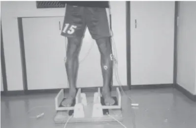
Fig. 2 – Anterior view of the individual on the platform
Fig. 3 – Time for the electromyographic (EM G) response obtained in the three groups of the sampling
Also, it w as found no statistically significant difference for the results of the time for the electromyographic response having the eyes open, compared to the eyes closed in none of the three groups (figure 4 and 5).
Fig. 4 – Time for the electromyographic (EM G) response having the eyes open
Fig. 5 – Time for the eletrocmyographic (EM G) response having the eyes closed
DISCUSSION
The sprain is considered the most frequent occurrence in the
ankle articulation(1), mainly in sports involving jumps and falls, as
propriocep-tive ability(13-16). Such reduction in the proprioceptive ability of an individual seems to be responsible by the reincidence in this type of injury(4,6,8,23,28,29). To determine the time for the electromyographic response has been suggested as one of the w ays to detect such reduction in the proprioceptive ability in an individual w ith ankle
sprain(18,19). This reduction in the proprioceptive ability should be
manifested w ith an increase in the time for the electromyographic response in the muscles of the sprained portion compared to the muscles of the healthy portion(19,21,30).
A review in the literature revealed that there still are controver-sies on the subject, once some authors have presented evidenc-es there is no changevidenc-es in the timevidenc-es for the electromyographic re-sponse(3,8,13,20), w hile other w orks have show n that there is a delay
in the time for the electromyographic response(19,21,30). Having in
mind the controversies presented in the literature as to the times for the electromyographic response, this study proposed to com-pare these times for the electrical response in the everter mus-cles (short and long fibular) of the athletes’ healthy feet (group 1), and athletes and non-athletes w ith recent history of sprain by an-kle inversion (groups 2 and). Assuming that the time for the elec-tromyographic response w ould change w ith the ankle sprain, it w as formulated three hypotheses.
Hypothesis 1 states that professional volleyball players w ith healthy ankles (G.1) w ould present a low er time for the electromyo-graphic response than professional volleyball players w ith recent history of ankle sprain (G.2). Such hypothesis w as not confirmed, having in mind that the results of the groups 1 and 2 presented no statistical differences. In the hypothesis 2 it w as expected that the professional volleyball players w ith recent history of ankle sprain (G.2) w ould present a low er time for electromyographic response than non-athlete individuals w ith recent history of ankle sprain (G.3), having in mind the adaptations resulting from a long training peri-od. Results for these groups did not present statistical differenc-es as w ell, and therefore, that hypothdifferenc-esis w as not confirmed as w ell. In hypothesis 3, professional volleyball players w ith healthy ankles (G.1) should present a low er time for the electromyograph-ic response than non-athlete individuals w ith recent history of an-kle sprain (G.3). Results did not confirm this hypothesis as w ell having in mind that it w as found no statistically significant differ-ence betw een both groups.
These results confirm w hat w as found in the literature, that are
in disagreement to the idea stated by Freeman et al. (1965)(28) w here
the mechanical instability w ould determine a functional instability of the ankle, causing a lack in the motor coordination (or even an increase in the time for the electromyographic response) due to the decreasing mechanoreceptor stimulus resulting from a liga-mentous injury and/or of the articular capsule, w hich w as not
con-firmed in these studies(3,8,13,20). A possible explanation for other
studies presenting a change in the time for the electromyographic response may be related to the inversion angle produced by the
platforms used in those studies, w hich w as of 30o(19,21,30). In the
present study and others w hich did not succeed in finding
signifi-cant differences(3,8,22), the inversion angle used w as only of 20o,
since it w as believed that this w as a safe articular inversion angle to be used in individuals w ith recent injury. In such sense, this angle apparently w as not big enough to reveal any changes in the proprioceptive ability of the sprained ankles.
The only exception to this case w as a study using a 35o angle
(that means, the w ider angle used among all studies), but w ithout finding any significant differences in the times for the
electromyo-graphic response(20). It is not clear the reason for such difference
related to other studies using a similar movement angle on the inversion platform.
Another important fact is related to the physiotherapeutic treat-ment, w hich probably could present an influence in the studies. The physiotherapeutic treatment that is helpful to the rehabilita-tion of the sprained articularehabilita-tion (in these studies, to the ankle
ar-ticulation) w as not mentioned by authors w ho found statistically significant differences, such as the rehabilitation factor of the
sprains(19,21,30). As those authors w ere w orking w ith times of the
higher than six months ankle sprains, this makes us to believe that the lack of physiotherapeutic treatment has damaged the re-covery of the sprained tissues, and this is in agreement to the studies reporting that the proprioceptive deficit may be found up to tw o years after the sprain, and so, the delay in the time for the
electromyographic response shall remain(17) (table 1).
TABLE 1
Summary of the methodology found in the literature
Author(s) EM G Platform Time of Physiotherapy the sprain
Isakov et al. (1986) surface 20° 02 months No Konradsen and Ravn (1990) surface 30° – No Karlsson et al. (1992) surface 30° 06 months No Johnson and Johnson (1993) surface 35° 03 months Yes Löfvenberg et al. (1995) surface 30° 12 months No Ebig et al. (1997) surface 20° 02 months Yes Sheth et al. (1997) surface 20° – Yes This study (2001) surface 20° 4 to 10 w eeks Yes
Another interesting data related to other researches is that the results found in those studies seem to be meaningless, once the values found for the ankle groups w ith and w ithout stability seem to be reverted, that is, higher values of the time for the electromyo-graphic response w ere found w ithin groups w ith no articular sta-bility in the ankle (healthy ankles), w hile low er times for the elec-t rom yographic response values w ere f ound f or sprained
ankles(3,8,20,22). Nevertheless, if the comparison is performed w ith
studies presenting a statistical difference, results found by those authors seem to present some logic, once that low er values in the times for the electromyographic response w ere found in non-sprained ankles, and the higher ones w ere found in non-sprained an-kles(19,21,30).
CONCLUSION
The findings of this study suggest that the times for the elec-tromyographic response to the fibular muscles are not influenced by the ankle sprain. It is believed that these results w ere
influ-enced by the low er than 30o angle of the platform, according to
w hat is observed in the discussion of the article. It is suggested to
perform studies using 30o angle platforms, similar to w hat w as
found in the literature w ith studies focusing the analysis of the delay in the time for the electromyographic response.
THANKFULNESS
Our relatives. Our parents, w ife, husband, and daughter(s).
All the authors declared there is not any potential conflict of inter-ests regarding this article.
REFERENCES
1. Carazzato JG, Campos LAN, Carazzato SG. Incidência de lesões traumáticas em atletas competitivos de dez tipos de modalidades esportivas. Rev Bras Ortop 1992;27:745-58.
2. Derscheid GL, Brow n WC. Rehabilitation of the ankle. Clin Sports M ed 1985;4: 527-44.
3. Sheth P, Yu B, Laskow ski ER, An KN. Ankle disk training influences reaction times of selected muscles in a simulated ankle sprain. Am J Sports M ed 1997;25:538-43.
5. Ferretti A, De Carli A, Papandrea P. Volleyball injuries – A colour atlas of volleyball traumatology. Lausanne, Suisse: Federation Internationale de Volleyball, 1994; 27-41.
6. Bahr R, Bahr IA. Incidence of acute volleyball injuries: a prospective cohort study of injuries mechanisms and risk factors. Scand J M ed Sci Sports 1997;7:166-71. 7. Holmer P, Sondergaard L, Konradsen L, Nielsen PT, Jorgensen LN. Epidemiology
of sprains in the lateral ankle and foot. Foot Ankle 1994;15:72-4.
8. Ebig M , Lephart SM , Burdett RG, M iller M C, Pincivero DM . The effect of sudden inversion stress on EM G activity of the peroneal and tibialis anterior muscles in the chronically unstable ankle. J Orthop Sports Phys Ther 1997;26:73-7. 9. O’Donoghue DH. Treatment of injuries to athletes. Philadelphia: W.B. Saunders,
1970.
10. Balduini FC, Tetzlaff J. Historical perspectives on injuries of the ligaments of the ankle. Clin Sports M ed 1982;1:3-12.
11. Balduini FC, Vegso TJ, Torg JT, Torg E. M anagement and rehabilitation of liga-mentous injuries to the ankle. Sports M ed 1987;4:364-80.
12. Renström, Per AFH, Lynch SA. Lesões ligamentares do tornozelo. Rev Bras M ed Esporte 1999;5:13-23.
13. Brunt D, Andersen JC, Huntsman B, Reinhert LB, Thorell AC, Sterling JC. Postur-al responses to laterPostur-al perturbation in hePostur-althy subjects and ankle sprain patients. M ed Sci Sports Exerc 1992;24:171-6.
14. Jerosch J, Prymka M . Proprioception and joint stability. Knee Surg Sports Trau-matol Arthrosc 1996;4:171-9.
15. Konradsen L, Olesen S, Hansen HM . Ankle sensoriomotor control and eversion strength after acute ankle inversion injuries. Am J Sports M ed 1998;26:72-7. 16. Watson AW. Ankle sprains in players of the field-game Gaelic football and
hurl-ing. J Sports M ed Phys Fitness 1999;39:66-70.
17. Nitz AJ, Dobner JJ, Kersey D. Nerve injury and grades II and III ankle sprains. Am J Sports M ed 1985;13:177-2.
18. Taimela S, Österman K, Kunjale U. M otor hability and personality w ith reference to soccer injuries. J Sports M ed Phys Fitness 1990;30:194-01.
19. Karlsson J, Peterson L, Andreasson G, Högfors C. The unstable ankle: a com-bined EM G and biomechanical modeling study. Int J Sports Biomech 1992;8: 129-44.
20. Johnson M B, Johnson CL. Electromyographic response of peroneal muscles in surgical and nonsurgical injured ankles during sudden inversion. J Orthop Sports Phys Ther 1993;18:497-01.
21. Löfvenberg R, Kärrholm J, Sundelin G, Ahlgren O. Prolonged reaction time in patients w ith chronic lateral instability of the ankle. Am J Sports M ed 1995;23: 414-7.
22. Isakov E, M izrahi J, Solzi P, Susak Z, Lotem M . Response of the peroneal mus-cles to sudden inversion of the ankle during standing. Int J Sports Biomech 1986; 2:100-9.
23. Chan KM , Yuan Y, Li CK, Chien P, Tsang G. Sports causing most injuries in Hong Kong. Br J Sports M ed 1993;27:263-7.
24. DeLee JC, Drez D Jr. Orthopaedic sports medicine – Principle and practice. 2nd
ed. United States of America: W.B. Saunders Company, 1994;1705-67. 25. Hockembury RT, Sammarco GJ. Evaluation and treatment of ankle sprains. The
Physician and Sports M ed 2001;29:57-4.
26. Basmajian JV, De Luca CJ. Description and analysis of the EM G signal. M uscles alive: their functions revealed by electromyography. Baltimore, Williams and Wilkins: John Butler, 1985;19-167.
27. Nigg BM , Herzog W. Biomechanics of the musculo-skeletal system. John Wiley & Sons, Toronto, 1994.
28. Freeman M AR. Instability of the foot after injuries to the lateral ligament of the ankle. J Bone Joint Surg Br 1965;47:669-7.
29. Freeman M AR, Dean M R, Hanham IWF. The etiology and prevention of function-al instability of the foot. J Bone Joint Surg Br 1965;47:678-5.