Eyelid trichoadenoma: surgical treatment associated
with aesthetic blepharoplasty
Tricoadenoma palpebral: tratamento cirúrgico associado à blefaroplastia estética
ABSTRACT
Trichoadenoma is a benign cutaneous tumor that is asymptomatic, rare, and slow growing. There are few cases reported in the literature, and we could only identify one description of trichoadenoma occurring in the eyelid area. We describe the case of a patient with tri-choadenoma in the outer corner of the lower eyelid that we treated with surgical excision associated with blepharoplasty.
Keywords: Neoplasms, basal cell/surgery. Skin neoplasms/surgery. Blepharoplasty. Eyelids/ surgery.
RESUMO
O tricoadenoma é um tumor cutâneo benigno, assintomático, raro e de crescimento len to. Existem poucos casos relatados na literatura e identificamos apenas um descrito na região palpebral. Apresentamos o caso de uma paciente portadora de tricoadenoma no canto externo da pálpebra inferior direita, tratada com excisão cirúrgica associada a blefaroplastia.
Descritores: Neoplasia de células basais/cirurgia. Neoplasias cutâneas/cirurgia. Blefaro-plastia. Pálpebras/cirurgia.
Study conducted at the Hospital Israelita Albert Einstein, originating from private clinic’s irst author, located in São Paulo, SP, Brazil.
Submitted to SGP (Sistema de Gestão de Publicações/Manager Publications System) of RBCP (Revista Brasileira de Cirurgia Plástica/Brazilian Journal of Plastic Surgery).
Article received: November 21, 2011 Article accepted: January 18, 2011
1. Associate professor at the Faculty of Medicine of São Paulo (FMUSP), responsible for the orbitopalpebral surgery group of the Division of Plastic Surgery of the Hospital das Clínicas da FMUSP, São Paulo, SP, Brazil.
2. Assistant physician at the Hospital Municipal Infantil Menino Jesus, formerly a resident physician at Hospital das Clínicas da FMUSP, São Paulo, SP, Brazil.
3. Full member of the Brazilian Society for Dermatology, director of Clínica Dermac de Dermatologia, Uberlândia, MG, Brazil.
4. Assistant Professor at Universidade Federal de São Paulo (Unifesp), associate professor in the Department of Pathology of Unifesp, São Paulo, SP, Brazil. 5. Medical student at Universidade de Uberaba, Uberaba, MG, Brazil.
Henri FriedHoFer1 Álvaro Júlio de andrade SÁ2 Maria CriStinade Paula MeSquita3 GilleS landMan4 artHurde Paula aMoriM MeSquita5
INTRODUCTION
Trichoadenoma, irst described by Nikolowski in 1958, is a rare, asymptomatic, slow-growing, benign cutaneous tu mor. It originates in the cells of the pilous follicle and most commonly presents on the face, trunk, or buttocks, ty pically during the fourth decade of life. It presents with more maturation than trichoepithelioma and with less diffe-rentiation than trichofolliculoma1 and desmoplastic trichoe-pithelioma1-4. Some authors2,5-10 believe that it is intermediate between trichoepithelioma and trichofolliculoma in terms of its differentiation towards the infundibular portion of the pilosebaceous canal. Trichoadenoma mainly affects adults of both genders, and has no malignant potential1. Clinically, it presents with a single, well-delimited nodular lesion or
in conluent minuscule papules of similar coloration to the
adjo ining skin (usually yellowish or grayish), covered by te -la n giectasias and measuring from a few millimeters to 1.5 cm. On dermatoscopic examination, it may appear to be a baso-cellular carcinoma.
There are only a few cases described in the literature and
only one case in the eyelid area has been identiied to date13.
In view of the rarity of this tumor, there is no deined algo -rithm for its treatment. However, to date, all published cases have been treated by surgical excision, either for aesthetic reasons, due to lack of knowledge concerning the tumor’s behavior if left untreated, or because of tumor enlargement. There are no reports of relapse after resection of these lesions. This article describes the case of a patient with trichoade-noma in the external corner of the bottom right eyelid, treated with surgical excision associated with blepharoplasty.
CASE REPORT
The patient, a 48-year old woman, observed a small, hard, pink tumor on her right lower eyelid 3 years ago. Excision of the lesion was performed on that occasion and histological analysis led to the diagnosis of hemangioma.
Six months ago, the patient noticed a small acne-type le sion in the same place as the excision; topical administration of fusidic acid and betamethasone valerate did not re -solve the lesion.
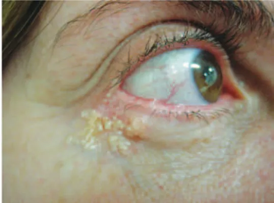
After 2 months, the lesion was still asymptomatic but pro gressing; a new biopsy resulted in a diagnosis of tri -choepithelioma. Afterward, worried about the appearance of the lesion and fearing malignancy, the patient sought advice from another service. Detailed dermatological examination revealed a slightly erythematous plaque of approximately 1.3 cm in diameter in the same place, with multiple cys tic-ap pearing, often translucent, white-yellowish ptic-apules. The papules were not itchy; they were hard, appeared shiny with small, thin vessels on the surface of the grouped cysts, and had a scabby surface (Figure 1). On the same occasion and upon request, the specimen was reanalyzed by an
indepen-dent pathologist who conirmed, as suspected, the diagnosis
of trichoadenoma with extensive squamous metaplasia of the sudoriferous epithelium.
After a conclusive diagnosis was obtained, the treatment strategy consisted of surgical resection of the lesion with narrow margins, followed by cryomicroscopy in the opera-ting room. Before treatment, the patient had also expressed her wish to correct the aging-related changes in her eyelids (Figure 2). Therefore, it was proposed that aesthetic blepha-roplasty would be performed during the same surgical proce-dure as tumor excision and reconstruction.
Surgical Procedure
The surgical procedure was performed under local anes-thesia with bupivacaine hydrochloride 0.5%, using a vaso-constrictor, along with intravenous sedation. The lesion was totally resected with narrow margins, keeping the integrity of the underlying plane of the orbicular muscle. During the operation, the resected piece was forwarded for pathological cryoanalysis, which showed clean, tumor-free margins. The resection created an irregular defect with a horizontal diameter of approximately 15 mm and a vertical diameter of 12 mm (Figure 3).
Next, a myocutaneous graft of the orbicular muscle of the upper eyelid was performed, laterally pedunculated in its preseptal portion (Figure 4). The size of the graft was planned to be not much larger than the defect caused by the resection of the trichoadenoma in the lower eyelid. Canthopexy was subsequently performed, slightly elevating the lateral corner
using “U” stitches with multiilament polyester 4-0 thread (Ethibond – Ethicon, São José dos Campos, SP, Brazil) bet ween the periosteum of the orbital margin/rim and the lateral retinaculum. The donor area was sutured in planes: the muscle portion was sutured using interrupted sutures with
6-0 monoilament nylon thread, and the cutaneous portion,
with the same thread but using a continuous suture. Next, the myocutaneous graft was transposed and sutured to the surgical area in the lower eyelid with 6-0 monoilament nylon thread (Mononylon – Ethicon) using interrupted sutures in
Figure 1 – Preoperative appearance of the trichoadenoma.
its lower portion (Figures 5 and 6). During the same surgical procedure, a small amount of excess skin in the middle two-thir ds of the lower eyelid was resected; after hemostasis was achieved, a continuous suture was placed, using the same thread used in the infraciliary area, in a similar manner as in the blepharoplasty with cutaneous graft. The same suture also attached the ciliary margin of the lower eyelid to the higher part of the myocutaneous graft.
On the contralateral side, blepharoplasty was performed using the same technique, and a segment of the preseptal orbicular muscle was also resected together with the skin in the upper eyelid, associated with the canthopexy (Figure 7). Palpebral bags of all four eyelids were treated with conser-vative resection, followed by hemostasis.
Cefazolin (2 g) was administered 60 minutes before sur -gery for antibiotic prophylaxis. Saline compresses and the
Figure 3 – Defect after tumoral resection.
Figure 4 – Myocutaneous graft in the upper eyelid with lateral peduncle.
Figure 5 – Transposition of the myocutaneous graft.
Figure 6 – Suture in the area of the upper eyelid defect.
usual painkillers were prescribed during the postoperative period, and the sutures were removed after 5 to 7 days.
Histopathology of the specimen revealed multiple cysts,
covered with stratiied, interconnected, corniied epithelium
wi thout atypical features, and dilated sudoriferous glands co -vered with a double layer of epithelial and myoepithelial cells,
replaced by stratiied metaplastic epithelium (Figures 8 and 9).
With the technique used in this case, the results were both functionally and aesthetically satisfactory. The patient was
followed up for 9 months after the surgery and showed no signs
of relapse, retraction, or malocclusion of the eyelid, and was pleased with the appearance of her eyelids (Figures 10 to 12).
DISCUSSION
Given that trichoadenoma is a rare, benign tumor, there
is no consensus or deined algorithm regarding the best treat ment. The few cases described in the literature have been trea -ted through surgery without a detailed protocol for exci sion or reconstruction. There are no described cases of relapse.
With regard to the case described in this article, as soon
as the diagnosis of trichoadenoma was conirmed, the choice
to treat the neoplasia and perform aesthetic blepharoplasty simultaneously was motivated by the benign nature of this type of tumor and by the aesthetic complaints of the patient. The margins of the lesion were narrow because of its benign nature. The reconstruction of the defect and the aesthetic treatment of the eyelids were combined by performing blepharoplasty of the upper right eyelid so it would aid in
Figure 8 – Histopathologic appearance: multiple cysts covered
by stratiied corniied interconnected epithelium, without atypical features (200×, H&E).
Figure 9 – Histopathologic appearance: dilated sudoriferous
glands (in the lower third) covered by double layer of epithelial and myoepithelial cells, replaced by stratiied metaplastic epithelium
(in the two upper thirds) (200×, H&E).
Figure 10 – Appearance 9 months post surgery.
Figure 11 – 9 months post surgery: bipalpebral appearance.
the reconstruction of the lower eyelid. The upper right eyelid showed excessive skin and prominent fat pockets. Making and transposing the graft laterally pedunculated allowed the re construction of the defect resulting from resection of the neoplasia in the lower eyelid, as well as the treatment of the upper eyelid’s excess sagging skin.
In the surgical plan, a graft only slightly larger than the de fect was chosen to compensate for secondary cicatricial re traction, common in these cases. This special consideration aimed at avoiding scleral show or lower eyelid ectropion. The graft donor area also allowed easy access to the fat pockets, the excess of which was removed, besides facilitating can -thopexy aimed at discretely elevating the lower eyelid, in ad dition to improving the lateral tarsal-ligament tension. On the contralateral side, a classical cutaneous blepharoplasty was performed, aiming at achieving symmetry with the side affected by the lesion. A segment of the orbicular muscle was removed, also for the sake of symmetry.
This strategy enabled us to obtain a satisfactory result with regard to both the treatment of the neoplasia and the patient’s aesthetic concerns about her eyelid area. It is known that neighboring grafts offer interesting results because they are similar to the recipient site in texture and coloration, a decisive fact in the planning of our strategy.
CONCLUSIONS
Simultaneously performing aesthetic blepharoplasty and surgical treatment of benign, or even not-very-aggressive malignant cutaneous lesions affecting the eyelid area, may be safe. For this to be achieved, the usual technical safety criteria related to the resection margins and exchange of the surgical material used in tumor resection; the tumor etiology; and mostly, the surgeon’s common sense concerning the indi-cation must be followed. Besides, aesthetic blepharoplasty
may aid in the reconstruction of defects generated by the re section of the lesions, as in the case described here.
Obviously, the decision to simultaneously perform the
blepharoplasty should be based on the irm conviction that it will not harm the patient by making it more dificult to detect
an eventual relapse or by decreasing neighboring skin donor areas that could possibly be needed in future reconstructions.
REFERENCES
1. Gotlib N, Schroh RG, Garcia Fernandez A, Khaski SV. Tricoadenoma. Rev Argent Dermatol. 1987;68(4):214-5.
2. Garcia e Silva L. Tricoadenoma de Nikolowski. Med Cutan Ibero Lat Am. 1982;10(2):85-8.
3. Bañuls J, Silvestre JF, Sevila A, Morell A, Betiloch I, Botella R. Tricoade -noma of Nikolowski. Int J Dermatol. 1995;34(10):711-2.
4. Casas JG. Tumores pilosos [tese de doutorado]. Buenos Aires: Univer-sidad de Buenos Aires, Faculdad de Medicina; 1982. p. 44-6. 5. Duperrat B, Mascaro JM. Essai de classiication dês tumeurs issues du
folliculare pilaire. Ann Derm Syph. 1965;92:241.
6. Mehregan AH, Rahbari H. Benign epithelial tumors of the skin. III: Benign hair follicle tumors. Cutis. 1977;19(5):595-9.
7. Pinkus E, Mehregan AH. A guide to dermatohistopathology. 2nd ed. New
York: Appleton-Century-Crofts; 1976. p. 544.
8. Rahbari H, Mehregan AH. Benign follicular neoplasias. J Dermatol Surg Oncol. 1979;5(4):295-8.
9. Rahbari H, Mehregan A, Pinkus H. Trichoadenoma of Nikolowski. J Cut Pathol. 1977;4(2):90-8.
10. Rueda LA. Histogénesis de los tumores anexiales. Med Cutan Ibero Lat Am. 1973;7:35.
11. Miller CJ, Ioffreda MD, Billingsley EM. Sebaceous carcinoma, basal cell carcinoma, trichoadenoma, trichoblastoma, and syringocystadenoma papilliferum arising within a nevus sebaceus. Dermatol Surg. 2004; 30(12 Pt 2):1546-9.
12. González-Vela MC, Val-Bernal JF, Garcia-Alberdi E, González-López MA, Fernández-Llaca JH. Trichoadenoma associated with an intrader-mal melanocytic nevus: a combined intrader-malformation. Am J Dermatopa-thol. 2007;29(1):92-5.
13. Shields JA, Shields CL, Eagle RC Jr. Trichoadenoma of the eyelid. Am J Ophthalmol. 1998;126(6):846-8.
Correspondence to: Álvaro Júlio de Andrade Sá