A survey, concerning the vitamin supply of people affected by the Chernobyl accident and emergency workers who have received a certain dose, showed a significant reduction in the blood of biologically active form of vitamin B1 (thiamine) – thiamine diphosphate (ThDP) [1]. Without a doubt, this phenomenon is caused by the action of ionizing radiation on the body, as in healthy people TDP content index was at the level of the established norm.
The high sensitivity of thiamine to ionizing radiation and the ability to prevent individual exposure symptoms of radiation sickness in people, preliminarily receiving per os 6 mg of this vitamin, have been noted in earlier studies [2]. More recent studies indicate the development of Wernicke-Korsakoff disease (accompanied by severe thiamine deficiency) in patients, who were subjected to long time radiotherapy [3], as well as the ability of thiamine to prevent genetic damage in human lymphocytes under
the action of X-ray. [4]. Despite these data, the mechanisms of destruction of thiamine by ionizing radiation and its ability to protect the molecule from this exposure were not studied enough. We paid attention to this subject taking into consideration the results of the above studies [1]. They indicate that in the blood of people, who received high radiation doses at the Chernobyl nuclear station and during their stay in the hospital every day took vitamins, most of the ThDP was in oxidized form [5]. It was equivalent to the reduction in the content of the active form of the ThDP and closed the patients to the state of vitamin-B1 deficiency. The reason for this may be the lack of conditions for the reduction reaction of oxidized ThDP because of disturbances of redox balance in tissues.
Taking into consideration above mentioned data, we thought it would be reasonable, using the model of ionizing radiation on the animals, to try to protect the intracellular ThDP from
UDС 577.16:614.876 doi: 10.15407/biotech8.04.063
METOVITAN PREVENTS ACCUMULATION
OF THIAMIN DIPHOSPHATE OXYGENIZED FORM
IN RAT TISSUES UNDER IRRADIATION
Key words: thiamine diphosphate, reactive oxygen species, metovitan.
Yu. M. Parkhomenko1 1Palladin Institute of Biochemistry
G. V. Donchenko1 of the National Academy of Sciences of Ukraine, Kyiv L. I. Chehovskaya1
S. P. Stepanenko1 2Chebotarev Institute of Gerontology
O. A. Mejenskaya1 of Academy of Medical Sciences of Ukraine, Kyiv E. N. Gorban2
E-mail: yupark@ biochem.kiev.ua
Received 29.04.2015
The aim of the research was to test the ability of the drug “Metovitan” to prevent the redox balance disturbance in the tissues and thiamine diphosphate irreversible oxidation upon exposure of ionizing radia-tion on the body. The rats were undergo to a single exposure of the X-ray therapeutic instrument RUM-17 to create a dose of 0.5, 1.0 and 5.0 Gy. Preparation “Metovitan” were administered at a dose of 25 mg per 1 kg body weight for 22–24 h before irradiation. Contents of thiamine diphosphate, reduced SH-groups and reactive oxygen species in blood and brain were determined using previously described methods. It is shown the active form of the thiamine diphosphate content in the blood decreases depending on the doses growth (from 0.5 to 5.0 Gy). In the same time thiamine diphosphate oxidized form content increases. Furthermore the critical changes occur in metabolic processes redox state parameters, namely, the free SH-groups level reduce and the reactive oxygen species level increase. Similar changes were observed in the brain tissue.
the destructive action of X-rays by introducing the animals the drug “Metovitan” for 24 hours before irradiation [6]. The aim was to activate biochemical reactions of antioxidant protection. The preparation consists of several vitamins (B1, B3, E), methionine and zinc salts. It is based on the results of long-term fundamental research of the regulatory impact of vitamins and co-enzymes on key reactions of cellular metabolism. In particular, it was first found the increased synthesis of glutathione from methionine under the influence of vitamin E [7]. The experiments also demonstrated the ability of the drug to reduce the hepatotoxic effect of anti-tuberculosis remedies (ATR), providing a positive effect on the mRNA expression of isoforms of cytochrome P-450 — Cyp3A2, Cyp2S23 and Cyp2E1 [8]. Thanks to the synergistic action of the components of the preparation, it was very active in the intensification of the processes of transsulfurization, transmethylation and as a result, the detoxification of foreign compounds, hydro and endogenous lipid peroxides. It gives the reason to believe in the prospects of using it to correct the disturbance of redox reactions in the tissues under the influence of adverse factors.
The aim of the research was to investigate the ability of the drug “Metovitan” to prevent disturbance of the redox balance and irreversible oxidation of thiamine diphosphate in the tissues under the influence of ionizing radiation. Since it is known that radiation sickness in human is accompanied by nerve disorders, in this study we analyzed the investigated parameters not only in blood, but also in the brain tissue.
Materials and Methods
The experimental model of ionizing radiation. Research was carried out on mongrel white male rats (200–220 g). All manipulations with animals were performed without breaking the generally accepted rules of bioethical treatment of laboratory animals, in accordance with relevant national and international regulations regarding experimental work (European Convention for the Protection of Vertebrate Animals used for experimental and other scientific purposes Strasbourg, 1986); The Law of Ukraine “On protection of animals from cruel behavior” N3447-IV, 2006).
The animals were divided into 4 groups: 1-I — control, 2-, 3- and 4-th group received a dose of 0.5; 1.0 and 5.0 Gray respectively. Only three groups: control, 3rd and 4th were used for
the experiment of prophylactic administration “Metovitan”. Each group of animals was divided in half: one subset got the preparation administered per os in saline in a dose 25 mg per 1 kg for 22–24 hours prior to irradiation, the second — saline only. To achieve the desired dose animals were subjected to a single exposure during the various periods of time. It was used radiotherapeutic equipment RUM-17 under the following conditions: tube voltage — 170 kV, current — 12 mA, dose — 0.833 sGy/s filter — 0.5 mm AL and 1.0 mm Cu, focal length — 45 cm , exposure — 30 (0.5 Gy), 1 m (1.0 Gy) and 5 min (5 Gy). The preparation was administered 22–24 hours prior to exposure to two groups of rats that had received doses of 1 Gy and 5 Gy. Taking into consideration that “the period of semi-recovery” of the body after total exposure in rats is 6–9 days [9], the animals were decapitated on the 6th day after irradiation and it was measured a blood level of SH-groups, and the content of the ROS (reactive oxygen species) and TDP (active and disulfide form).
Determination of the ThDP and the ThDP disulfide. In biological systems the level of formation of ThDP and its disulfide was measured using a previously elaborated express method using yeast pyruvate deсarboxylase apoenzyme (apoPDC) [10]. Enzymatic determination of ThDP based on its recombination as a coenzyme with apoPDK. Then the reaction was carried out with an excess of pyruvate in the presence of alcohol dehydrogenase. The reaction was registered on the oxidation of NAD•H2. To determine ThDP, apoPDK was obtained from brewer’s yeast (Saccharomyces carlsbergensis) in the form of sulfate paste [10], which was stored at –20 C. Immediately prior to the experiment apoenzyme was obtained from the paste. To determine the total amount of ThDP (active form + oxidized form), aliquots of samples were incubated in phosphate buffer for 30 min with dithiothreitol (DTT) as a reducing agent. Active ThDP was found in aliquots that were incubated under the same conditions but without the addition of DTT. The quantity of disulfide ThDP [(ThDP) 2SS] was calculated from the difference between the first and second indicators .
at a wavelength of excitation and emission, respectively, 497 and 525 nm, slit width — 2.5–5.0 nm. Measurements were performed at room temperature (21–23 C).
Determination of SH-groups. The concentration of free SH-groups was determined in tissue homogenates after protein precipitation with ethanol as previously described [12]. Principle of the method: the free SH-group reacts with the 5,5-dithio-bis-(2-nitrobenzoic) acid (Ellman’s reagent), resulting in a yellow colored thionitrophenyl anion in equimolar amounts, with a maximum absorption at 412 nm.
Methods of statistical analysis. The experimental data were processed by standard methods of variation statistics. We calculate the value of arithmetic means (M) and the mean square error (m). To determine significant differences between the averages we used Student’s t-test. The values of P < 0.05 were considered significant. Computer program Microsoft Excel was used for the calculation and graphical representation of the results.
Results and Discussion
Effect of X-ray irradiation on the content of the active (cyclic) and disulfide forms of ThDP.
The key derivative of thiamine, which defines the participation of the vitamin in the metabolic processes, is ThDP – a coenzyme of key enzymes of carbohydrate metabolism. ThDP molecule, as well as of thiamine, is very sensitive to the action of oxidizing factors [2]. Among the oxidized forms of ThDP, its disulfide form dominates. This form is not capable of performing coenzyme function and formed due to disclosure of thiazole ring and releasing of the reactive thiol group [2]. Under physiological conditions thiamine diphosphate disulfide in the presence of reducing agents is
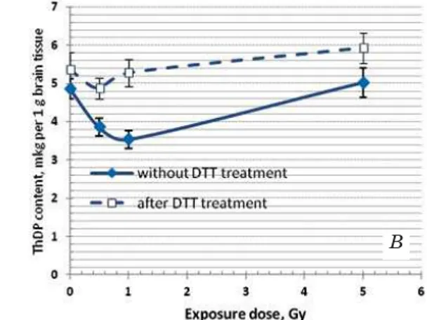
readily converted into a catalytically active, cyclic form. Its accumulation was observed in the tissues of people who have received a certain dose of radiation [5]. It indicates an imbalance of redox processes in tissues. Taken into consideration the crucial role of thiamine-dependent processes in the functioning of neural cells, we found it useful to study the effect of ionizing radiation on the content of the oxidized and active forms of ThDP not only in blood but also in the brain tissue. Fig. 1 shows the average values of these parameters in rats that received high doses of radiation.
These data indicate that the content of the active form of the ThDP in both tissues almost linearly is decreased with increasing of radiation dose to 1.0 Gy, and this decline is largely due to the oxidation of ThDP (ThDP) 2SS. The last compound is not capable of performing the coenzyme function. The accumulation of the oxidized form of the ThDP is especially pronounced in the brain, in blood, obviously, there is the further destruction of (ThDP) 2SS [2].The dose of 5 Gy is out of the overall trends. It is considered to be lethal to humans [13] – the half of the whole amount of irradiated patients die within 1–2 months due to the destruction of the bone marrow. We can assume that ThDP maximum oxidation is achieved at a dose of 1 Gy in blood, and further increase of the dose only accelerates the destruction of already oxidized forms of thiamine derivatives. The different picture is observed in brain tissue. Here all oxidized form of ThDP remains in the tissue, and at a dose of 5 Gy, it is observed even increase in active form of ThDP (Fig. 1, B). The latter can be explained by the fact that over time from exposure to the slaughter of animals, there are adaptation processes to compensate the content of ThDP in the brain tissue. This is in accordance with
Fig. 1. ThDP content determined before and after incubation with dithiothreitol (DTT) in blood (A) and in brain tissue (B) of the irradiated rats
Here and after n = 5–6
the earlier data on animal models of alimentary B1 failure. They show that with insufficiency development in animals, the content of ThDP is gradually reduced in all tissues, except brain. That probably happens due to the continuous redistribution of thiamin from other tissues into the brain [14].
Analyzing the content of the oxidized form of the ThDP in the blood of Chernobyl liquidators (the received doses of radiation were noted in their medical cards), we noticed that the relative content of (ThDP) 2SS in the total content of ThDP (active plus oxidized form) significantly increased with increasing the dose [6]. That was the reason to believe that this indicator can be used to estimate the dose received by a person when the dose was not recorded, but there was a risk of exposure. In order to test this hypothesis, we analyzed the relationship between the two parameters: the dose received and the relative content of (ThDP) 2SS in the total pool of ThDP (%) in the blood samples of the experimental animals.
According to data given in Fig. 2, with increasing doses from 0.5Gy to 5.0 Gy, the relative content of ThDP disulfide in blood, according to preliminary estimation, increases in the logarithmic dependence.
Having at our disposal the rapid enzymatic method for the determination of ThDP in biological fluids, we can talk about the development of a diagnostic test for the determination of the oxidized and reduced forms of ThDP in the blood of people. This indicator can be used for diagnostic purposes and for certain pathologies.
There is also an interesting idea of a group of authors to use the aqueous solutions of thiamine in dosimeters to assess the dose [15].
As it was mentioned above, the formation of disulfide form of ThDP under physiological conditions is a reversible process. The accumulation of oxidized derivatives of thiamine in the irradiated tissue is a marker of breaking of equilibrium of redox reactions. We confirmed it in our study analyzing the indicators in the tissue of irradiated animals — the amount of reduced SH-groups and the AFC.
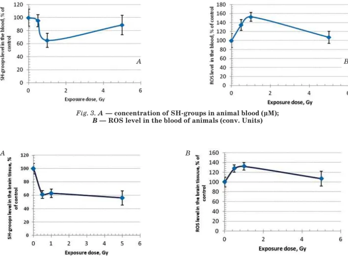
Indicators of redox reactions of tissues In tissue samples that were used for analysis of active and oxidized forms of ThDP, we determined the concentration of reduced SH-groups (parameter actually reflects the level of reduced glutathione) and ROS. Both figures are expressed in % of control in order to avoid a large scatter of data due to small sample size (n = 3). As it is shown at Fig. 3, the concentration of SH-groups is critically reduced at a dose of 1.0 Gy and ROS level is significantly increased at a dose of 0.5 Gy, remaining significantly above control with increasing the dose up to 1 Gy.
Changes of these parameters in the brain of irradiated rats were similar, but the maximal decrease in the concentration of SH-groups was observed at a dose of 0.5 Gy (Fig. 4).
The data shown in Fig. 3 and 4 show an imbalance of redox reactions in the blood and the brain of animals exposed to ionizing radiation at the indicated doses.
Effect of prophylactic introduction of “Metovitan” on the level of the ThDP and (ThDP) 2SS and indicators of redox reactions in the rat tissues. Levels of ThDP and (ThDP) 2SS in the blood and the brain.
The data obtained during the analysis of the content of the ThDP active form in the brain tissue and blood of irradiated rats, which were administered per os drug “Metovitan” or saline, are shown in Fig. 5. They show that for both of the exposure doses “Metovitan” introduction significantly prevents of “Metovitan” it was noted the significant prevention of the decrease of the ThDP active form content in the blood (Fig. 5, A), it is particularly pronounced at a dose of 1.0 Gy.
A different picture was observed in the brain. The effect of drug on ThDP content was significantly more pronounced at both doses. It was observed a significant increase in the active form of the ThDP more than 2-fold (Fig. 5, В). This phenomenon may be explained by three circumstances. Firstly, the preparation of “Metovitan” contains physiological dose of thiamine in its composition and a slight Fig. 2. The percentage of the disulfide form in the
total ThDP pool in blood of irradiated animals, depending on doses
(dotted line — logarithmic trend line)
Fig. 3. A — concentration of SH-groups in animal blood (μM); B — ROS level in the blood of animals (conv. Units)
Fig. 5. ThDP content in blood (A) and brain tissue (B) of the control and experimental animals during prophylactic administration of “Metovitan”
Here in after we designated significant differences from control (without irradiation) at * — P < 0.05 — the figure for the control animals;
"— from the same index without “Metovitan” introduction
Fig. 4. A — concentration of SH-groups in animal brain tissue; B — the level of ROS in the brain tissue
A B
A B
A B
* "
"
increase in its content (and ThDP) in tissues even in the control animals seems to be logical. Secondly, according to the literature data [14] in animals with vitamin B1 deficiency, there is a thiamine redistribution in most tissues, and its arrival in the brain. Thirdly, it can be assumed that the introduction of “Metovitan” not only protects ThDP from the adverse effect of radiation, but also promotes the restoration of its oxidized form in the cells. This is in accordance with the results of SH-group content and ROS level in tissues of animals treated with the drug at the day prior to irradiation.
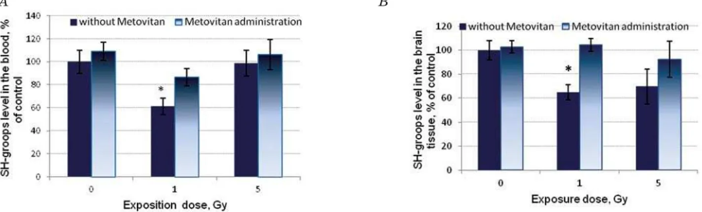
Data concerning the concentration of SH-groups in the tissues of exposed rats having prophylactic administration of “Metovitan” are shown in Fig. 6.
At a dose of 1 Gy, prophylactic admini-stration provided the higher concentration of SH-groups. We can conclude that “Metovitan” effectively prevents the oxidation of low molecular weight thiols under the influence of X-ray irradiation. The same picture was observed at a dose of 1 Gy in brain tissue.
Fig. 7 shows the effect of these preventive administration of “Metovitan” on the another
indicator of the redox balance of the tissues — the level of ROS.
This indicator was significantly increased at a dose of 1 Gy and approached to the control in the tissues of animals, that at the day before irradiation was introduced Metovitan. At the radiation dose of 5 Gy, the positive effect of the drug in the brain tissue was not observed, while its increase was noticed in the blood.
These data suggest that the protective effect of the drug can be shown only at doses up to 1 Gy. At much higher dose of 5 Gy when apparently, the regulation of metabolic processes in cells significantly is disturbed, a single administration of the drug for 24 h before irradiation is not sufficient to protect cells from the effects of radiation, and in some cases even leads to activation of ROS. Taking into account the cumulative effect of “Metovitan”, it is possible to choose the mode of its administration and dosage that will promote a positive effect.
Analysis of the results shows a lack of correlation between radiation dose from 0.5 to 5.0 Gy and indicators of redox reactions, in particular the loss of the general line at the dose of 5.0 Gy. Perhaps this is due to the
Fig. 7. The level of ROS (units) in the blood (A) and brain (B) of control and irradiated animals having prophylactic administration of ”Metovitan”
Fig. 6. The concentration of SH-groups in blood (A) and brain (B) of control and irradiated animals having prophylactic administration of “Metovitan”
A B
A
dynamics of these parameters. They are able to have quick change in both directions. Because the analysis was performed on the 6th day after irradiation, adaptive changes that may occur during this time in the tissues, distort the real picture of the damage occurring during irradiation. The different situation was observed in case of indicators that show the status of the ThDP. As it is followed from the data presented in Fig. 1, the indicator “of an active form of the ThDP content” in the blood is dramatically reduced as compared with the control with the increase of the doses up to 1.0 Gy. The further increase in the radiation dose to 5.0 Gy, have not led to a significant reduction compared to the dose of 1.0 Gy. These results may indicate that, firstly, not all of the ThDP from its general pool in the cell is
capable of undergoing oxidation by the action of ionizing radiation (it was possible that ThDP associated with the enzymatic protein was not oxidized), and, secondly, converting a ThDP in oxidized form under the action of ionizing radiation is irreversible.
In general, given the results of the study, we can conclude that the drug Metovitan can be used for the treatment of personnel involved in the elimination of accidents related to environmental pollution with toxic substances, in particular radioactive, to prevent adverse changes in cellular metabolism under the influence of these factors .
However, more research is needed to optimize the conditions of the drug introduction to personnel and evaluation of the boundary limits of pollution or the received doses.
REFERENCES
1. Spirichev V. B., Donchenko G. V., Blazhee vich N. V., Parkhomenko Iu. M., Aleĭnik S. I., Golub-kina N. A., Vrzhesinskaia O. A., Isaeva V. A., Kodentsova V. M., Pereverzeva O. G., Soko-l’nikov A. E., Iakushina L. M. To the 20th anniversary of the Chornobyl accident study of vitamin status and provision with micro- and macroelements of limited groups of people at different time periods since the accident at Chornobyl nuclear power plant. Ukr. Biokhim. Zh. 2006, 78 (2), 5–26. Review. (In Russian). 2. Parkhomenko Iu. M., Stepuro I. I., Donchenko G. V.,
Stepuro V. I. Oxidized derivatives of thiamine: formation, properties, biological role. Ukr. Biokhim. Zh. 2012, Nov-Dec, 84 (6), 5–24. (In Russian). 3. Rakici S. Y., Erdemli S. D., Yazici Z. A.,
Cengiz E., Acar O. G., Tufan G. Wernicke’s encephalopathy in a patient with unresectable gastric carcinoma and literature review. Int. J. Clin. Exp. Med. 2015, Jan 15, 8 (1),1453– 1459. eCollection 2015.
4. Konopacka M., Rogolinski J. Thiamine prevents X-ray induction of genetic changes in human lymphocytes in vitro. Acta Biochim. Pol. 2004, 51 (3), 839–843.
5. Parkhomenko Yu. M., Chernysh I. Y u., Protasova Z. S., Donchenko G. V. A disturbance in thiamine metabolism in blood of patients with radial pathology and possible connection of this disturbance with the lesion of the nervous system. DAN Ukrainу. 1995, V. 2, P. 112–114. (In Ukrainian).
6. Donchenko G. V., Parkhomenko Yu. M., Pro-ta sova Z. S., Kovalenko V. M., Danevich O. I., Ko zhakina I. P., Chernysh I. Yu., Fedoseeva-Bo ro dina L. O. Preparation for the increase of viability of organism. Patent of Ukraine N 39228 from 15.06. 2001, Biul. N5. 15.06. 2001.
7. Shtutman C. M., Sukharevskaya A. M. Vitamin E, selenium and sulfur-containing amino acids. Chagovec R. V. Vitamins. Kyiv: Naukova dumka. 1975, P. 87–95. (In Russian).
8. Anisimova S. I., Donchenko H. V., Parkho men-ko Iu. M., Kovalenmen-ko V. M. Mechanism of hep-atoprotective action of methionine and com-position “Metovitan” against a background of antituberculosis drug administration to rats. Ukr. Biokhim. Zh. 2013, 85 (2), 59–67.
9. Yarmonenko S. P. Radiobiology humans and animals. Moscow: Higher School. 1988, 424 p. 10. Experimental vitaminology. Red. Yu. M.
Ostrovskiy. Minsk: Science and Technology. 1979, 550 p.
11. Zhang X., Cao J., Jiang L., Zhong L. Sup pres-sive effects of hydroxytyrosol on oxidative stress and nuclear factor-kappa B activation in THP-1 cells. Biol. Pharm. Bull. 2009, V. 32, P. 578–582.
12. Sedlak J., Lindsay R. H. Estimation of total, protein-bound, and nonprotein sulfhydryl groups in tissue with Ellman’s reagent. Anal. Biochem. 1968, V. 25, P. 192–205.
13. nuclphys.sinp.msu.ru/radiation/rad_10.htm 14. Trebukhina R. V., Ostrovsky Y. M.,
Mikhal-tsevich G. N., Velichko M. G., Tumanov V. N. Transketolase, pyruvate and oxoglutarate dehydrogenase activities and [14C]thiamin turnover in tissues of mice fed thiamin-deficient diet. J. Nutr. 1983, 113 (7), 1285–1291.
МЕТОВІТАН ЗАПОБІГАЄ
НАКОПИЧЕННЮ ОКИСНЕНОЇ ФОРМИ ТІАМІНДИФОСФАТУ В ТКАНИНАХ
ЩУРІВ ЗА ДІЇ ІОНІЗУЮЧОГО ОПРОМІНЕННЯ
Ю. М. Пархоменко1, Г. В. Донченко1, Л. І. Чехівська1, С. П. Степаненко1,
О. А. Меженська1, Є. М. Горбань2
1Інститут біохімії ім. О. В. Палладіна
НАН України, Київ
2Інститут геронтології ім. Д. Ф.Чеботарьова
НАМН України
E-mail: yupark@biochem.kiev.ua
Метою роботи було дослідити здатність препарату «Метовітан» запобігати порушен-ню редокс-балансу в тканинах і незворотному окисненню тіаміндифосфату за дії на організм іонізуючого опромінення. Щурів піддавали одноразовому опроміненню упродовж різних проміжків часу за допомогою рентгенотерапе-втичної установки РУМ-17 для створення доз 0,5, 1,0 і 5,0 Гр. Препарат «Метовітан» вводили в дозі 25 мг на 1 кг маси тіла за 22–24 год до опро-мінення. Вміст тіаміндифосфату, вільних SH-груп і активних форм кисню визначали у крові та мозку за допомогою описаних раніше мето-дів. Показано, що вміст активної форми тіамін-дифосфату в крові тварин знижується зі зрос-танням отриманої ними дози (від 0,5 до 5,0 Гр), тимчасом як вміст його окисненої форми зро-стає. При цьому спостерігаються критичні зміни в показниках окиснювально-відновного стану метаболічних процесів, зокрема знижен-ня рівзнижен-ня вільних SH-груп і зростанзнижен-ня рівзнижен-ня ак-тивних форм кисню. Подібні зміни спостеріга-ються і в тканині мозку.
Одноразове введення тваринам препарату «Метовітан» у дозі 25 мг на 1 кг маси тіла за добу до опромінення сприяє збереженню вну-трішньоклітинного тіаміндифосфату і окисню-вально-відновної рівноваги у тканинах тварин (кров, мозок) за негативної дії опромінення в дозах 0,5; 1,0 Гр. Одержані результати да-ють підстави для призначення препарату під час підготовки персоналу, що бере участь в ліквідації аварій, пов’язаних із радіаційним забрудненням. З метою захисту від дії більш високих доз опромінення, ймовірно, потрібен інший режим приймання препарату
Ключові слова: тіаміндифосфат, активні форми кисню, метовітан.
МЕТОВИТАН ПРЕДОТВРАЩАЕТ НАКОПЛЕНИЕ ОКИСЛЕННОЙ ФОРМЫ ТИАМИНДИФОСФАТА В ТКАНЯХ КРЫС
ПРИ ДЕЙСТВИИ ИОНИЗИРУЮЩЕГО ОБЛУЧЕНИЯ
Ю. М. Пархоменко1, Г. В. Донченко1, Л. И. Чеховская1, С. П. Степаненко1,
О. А. Меженская1, Е. Н. Горбань2
1Институт биохимии им. А. В. Палладина
НАН Украины, Киев
2Институт геронтологии им. Д. Ф. Чеботарева
НАМН Украины, Киев
E-mail: yupark@biochem.kiev.ua
Целью работы было исследовать способ-ность препарата «Метовитан» предотвращать нарушение редокс-баланса и необратимое окисление тиаминдифосфата в тканях при дей-ствии на организм ионизирующего облучения. Крыс подвергали однократному облучению в течение разных промежутков времени с по-мощью рентгенотерапевтической установки РУМ-17 для создания доз 0,5, 1,0 и 5,0 Гр. Пре-парат «Метовитан» вводили в дозе 25 мг на 1 кг массы тела за 22–24 ч до облучения. Содержа-ние тиаминдифосфата, свободных SH-групп и активных форм кислорода определяли в крови и мозге животных с помощью описанных ранее методов. Показано, что содержание активной формы тиаминдифосфата в крови снижается по мере возрастания дозы облучения (от 0,5 до 5,0 Гр), в то время как содержание его окис-ленной формы возрастает. При этом наблюда-ются критические изменения в показателях окислительно-восстановительного состояния метаболических процессов, в частности, сни-жение уровня свободных SH-групп и возраста-ние уровня активных форм кислорода. Подоб-ные изменения наблюдаются и в ткани мозга.
Однократное введение животным препа-рата «Метовитан» в дозе 25 мг на 1 кг массы тела за 24 ч до облучения способствует сохра-нению внутриклеточного тиаминдифосфата и окислительно-восстановительного равнове-сия в тканях животных (кровь, мозг) на фоне негативного действия облучения в дозах 0,5; 1,0 Гр. Полученные результаты дают основа-ние рекомендовать использоваоснова-ние препарата при подготовке персонала, участвующего в ликвидации аварий, связанных с радиацион-ным загрязнением. Для защиты от действия более высоких доз облучения, вероятно, нужен иной режим приема препарата.