Nuclear Receptor Ligand and Cofactor Discovery
Jens Tiefenbach1,2, Pamela R. Moll1, Meryl R. Nelson1,2, Chun Hu1, Lilia Baev1, Thomas Kislinger3,4,5, Henry M. Krause1,2*
1Banting and Best Department of Medical Research, The Terrence Donnelly Centre for Cellular and Biomolecular Research (CCBR), University of Toronto, Toronto, Ontario, Canada,2InDanio Bioscience Inc., Toronto, Ontario, Canada,3Department of Medical Biophysics, University of Toronto, Toronto, Ontario, Canada,4Ontario Cancer Institute, University Health Network, Toronto, Ontario, Canada,5Campbell Family Cancer Research Institute, Toronto, Ontario, Canada
Abstract
Nuclear receptors (NRs) belong to a superfamily of transcription factors that regulate numerous homeostatic, metabolic and reproductive processes. Taken together with their modulation by small lipophilic molecules, they also represent an important and successful class of drug targets. Although many NRs have been targeted successfully, the majority have not, and one third are still orphans. Here we report the development of anin vivo GFP-based reporter system suitable for monitoring NR activities in all cells and tissues using live zebrafish (Danio rerio). The human NR fusion proteins used also contain a new affinity tag cassette allowing the purification of receptors with bound molecules from responsive tissues. We show that these constructs 1) respond as expected to endogenous zebrafish hormones and cofactors, 2) facilitate efficient receptor and cofactor purification, 3) respond robustly to NR hormones and drugs and 4) yield readily quantifiable signals. Transgenic lines representing the majority of human NRs have been established and are available for the investigation of tissue- and isoform-specific ligands and cofactors.
Citation:Tiefenbach J, Moll PR, Nelson MR, Hu C, Baev L, et al. (2010) A Live Zebrafish-Based Screening System for Human Nuclear Receptor Ligand and Cofactor Discovery. PLoS ONE 5(3): e9797. doi:10.1371/journal.pone.0009797
Editor:Vincent Laudet, Ecole Normale Supe´rieure de Lyon, France
ReceivedNovember 17, 2009;AcceptedFebruary 19, 2010;PublishedMarch 22, 2010
Copyright:ß2010 Tiefenbach et al. This is an open-access article distributed under the terms of the Creative Commons Attribution License, which permits unrestricted use, distribution, and reproduction in any medium, provided the original author and source are credited.
Funding:The authors would like to thank the Canadian Institutes of Health Research for funding. The funders had no role in study design, data collection and analysis, decision to publish, or preparation of the manuscript.
Competing Interests:The authors (Tiefenbach and Krause) are affiliated with InDanio Bioscience Inc. (www.indanio.com) that has a financial interest and stake in the material discussed in this manuscript. InDanio Bioscience Inc. has been assigned the invention of the authors (Tiefenbach and Krause) that describes ‘‘METHODS AND COMPOSITIONS FOR THE DETECTION AND ISOLATION OF LIGANDS’’ using live zebrafish (PCT/CA2006/002114). The authors have no current consultancies, honoraria, expert testimony, or royalties regarding the material described. Research performed by the authors and described in this manuscript was supported by the Canadian Institutes of Health Research (CIHR). The authors have no affiliations or financial involvement with any other organization or entity with a financial interest in or financial conflict with the subject matter or materials discussed in the manuscript apart from those disclosed. No writing assistance was utilized in the production of this manuscript.
* E-mail: [email protected]
Introduction
Nuclear receptors (NRs) are ligand-activated transcription factors that regulate the expression of specific gene networks by recruiting co-activator or co-repressor complexes. In doing so, they regulate diverse physiological processes such as metabolism, development, growth and reproduction. NRs share a modular structure, which includes highly conserved DNA- and ligand-binding domains (DBDs, LBDs) spaced by a variable hinge region [1,2]. Their activities are modulated by small hydrophobic compounds, such as steroids, fatty acids, retinoids and thyroid hormones. Ligand binding to the LBD alters its conformation, cofactor binding and/or transcriptional activity [3]. About a third of NRs, however, are referred to as orphan receptors, as the identities of their natural ligands are still unknown.
NR ligands, or drugs that mimic them, have been used to deal with many major and debilitating diseases [4,5,6]. However, only a small percentage of NRs have been targeted, and even for these, drugs that act more selectively would provide huge benefits. New drugs capable of modulating orphan NR activities have the potential to control numerous additional disorders such as heart disease, atherosclerosis, metabolic disease, cancer, inflammation, depression and anxiety [7,8].
ligands are highly related, such that in most examined cases, mutual ligand responsiveness has been observed [17,18]. Devel-opmental profiling of zebrafish expression patterns [19] has also demonstrated a high degree of conservation between NR expression patterns in zebrafish and other vertebrate models.
The concepts underlying the screening technology described here were derived from previous studies conducted within the fruitfly, Drosophila melanogaster. In one of these studies [20], we showed it was possible to use fusions between the fly NR ligand binding domains (LBDs) and the DBD of Gal4 to visualize NR ligand and cofactor responsiveness in live animals using a Gal4-dependent GFP reporter. Two other studies, in which the NRs E75 [21] and DHNF4 [22] were affinity purified from insect cells or tissues, suggested that it might be possible to identify bound ligands using newly developed mass spectrometry techniques, provided that sufficient amounts and purities of the bound proteins could be achieved.
Here, we describe a unique combination of NR ligand sensor and affinity chromatography technologies within a vertebrate model. Using our TRb and PPARc lines, we show that they respond within different tissues to endogenous hormones and cofactors, as well as to exogenously added drugs. We also demonstrate the potential of our affinity purification system by isolating a transgenic receptor from zebrafish embryos and validating the function of one of the co-purified cofactors. We
refer to our combination of technologies, within a pharmacolog-ically receptive zebrafish model, as the ‘‘ligand trap’’ system.
Results
Assembly of the ‘ligand trap’ (LT) vector
Construction of the ligand trap (LT) vector (Figure 1a) involved an extensive series of sub-cloning events and configuration comparisons, with the chosen vector described below. The 3X FLAG [23], Strep II [24] and 6X His tags were combined into a single triple-tag cassette, collectively referred to as the ‘FSH’-tag, and inserted in front of a minimal GAL4 DNA-binding domain (DBD: residues 1–132). Following the Gal4 DBD sequence, a series of restriction sites, was used for the in-frame addition of NR cDNAs (hinge+LBD). To reduce potential lethality that might be caused by constitutive expression of the GAL4-LBD fusion proteins, a zebrafish heat shock 70gene promoter [25] was used for inducible expression in any tissue and at any developmental stage. Unique restriction sites flanking thehsp70promoter can be used to swap in tissue-specific promoters, if so desired.
Also within the vector is an enhanced GFP (eGFP; includes a nuclear localization signal) reporter gene that is expressed under the control of a 14x UASGAL4-containing promoter. Expression of
this reporter requires binding and activation of the Gal4-NR-LBD fusion protein. Bracketing the GAL-LBD fusion gene and the
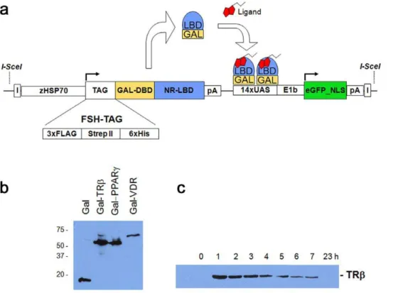
Figure 1. The Ligand Trap (LT) system.(a) Schematic diagram of the multi-component ligand trap (LT) construct. Upon heat pulse, the zebrafish hsp70promoter directs ubiquitous expression of the GAL4 DNA-binding domain (DBD) fused in-frame to ahumannuclear receptor ligand-binding domain (LBD) and an affinity tag cassette (FSH-tag). Upon binding of this fusion protein to the GAL4 UAS (upstream activating sequence) response elements, in the presence of active hormone and cofactors, expression of the reporter gene (nuclear enhanced Green Fluorescent Protein (eGFP)) occurs. Expression of nuclear GFP is used to monitor receptor ligand sensor activity in a cell- and tissue autonomous manner in live zebrafish. The second component makes use of the tags to co-purify bound hormones or cofactors. I = insulator elements; pA = polyadenylation signal; NLS = nuclear localization signal. (b) Western blots of GAL4-NR fusion proteins. Embryos (F2; 72 hpf) were heat pulsed for 30 min at 37uC and recovered for 1 h at room temperature. 10 embryos were pooled and lysed in 50ml of FSH buffer (see material and methods) followed by adding SDS buffer and
boiling. Proteins were detected using the FLAG-M2 antibody. (c) Time course of fusion protein expression. TRbembryos (F2; 72 hpf) were heat induced as in 1b) and recovered for the times indicated. Each sample contained 10 embryos.
eGFP reporter are insulator elements that were introduced to discourage the influence of neighboring gene regulatory elements. Finally, I-SceI meganuclease recognition sites were added at the ends of the vector to facilitate integration into the genomes of transgene and meganuclease co-injected one-cell stage embryos [26].
Induction of LT fusion protein expression
To test the heat inducible expression and activity of LT fusion proteins, and to determine the best parameters for activity screening, we generated stable transgenic lines for each human nuclear receptor by co-injecting the LT-vector together withSceI meganuclease into one-cell stage zebrafish embryos. Injected F0 fish were raised to adulthood and crossed with wild type fish to identify germline transformed animals. Positive progeny (F1) were identified either by target PCR or GFP screening (see Material and Methods). At least two independent lines for each nuclear receptor were obtained, unless otherwise noted. Transgenic LT embryos (F2) were heat pulsed at different developmental stages, temper-atures and durations. Western blot detection using a M2-Flag antibody that recognizes the FSH-tag of each LT fusion protein revealed uniquely sized proteins with expected molecular weights (Figure 1b). Figure 1c shows the robust expression observed in homozygous transgenic (F2) Thyroid Receptor-b (TRb; NR1A2) embryos at successive time points following a temperature shift to 37uC. As with most LT fusion proteins, peak expression was observed ,1 hr following the heat pulse (Figure 1c). A thirty
minute heat pulse at 37uC also proved adequate for the production of robust GFP fluorescence in the presence of endogenous or exogenously added ligands (see below).
LT fusion proteins interact with fish ligands and cofactors
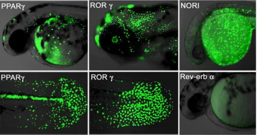
Different NR-derived lines should exhibit unique GFP responses during development in response to their respective ligand and cofactor distributions. To verify this, we induced expression of our Peroxisome Proliferator-Activated Receptor-gamma (PPARc; NR1C3), Retinoic acid-related Orphan Recep-tor-gamma (RORc: NR1F3), Neuron-derived Orphan
Receptor-1 (NORReceptor-1: NR4A3) and Rev-erb alpha (Rev-erba: NR1D1) transgenic lines and documented their patterns of GFP expression. The PPARc embryos show strong GFP expression in the tail bud epidermis, as well as the brain, posterior spinal cord and heart (Figure 2; left panel). RORc embryos show ubiquitous GFP expression over the entire embryonic epidermis during the first three days of development, along with brain and retina later on (Figure 2 middle panel). GFP expression in NOR1 embryos occurs in the epidermis and CNS (Figure 2; upper right picture). In contrast, Rev-erba LT embryos show no GFP expression (Figure 2; lower right), consistent with its role as a transcriptional repressor [27].
To verify that GFP expression effectively reflects endogenous signaling activity, we injected a morpholino (MO) oligo-nucleo-tide, complementary to the translational start site of the transgene, into one-cell stage PPARc, RORcand TRbembryos (Figure S1). GFP expression was lost or dramatically reduced in these MO injected embryos, even in the presence of control agonist.
Further documentation of these sites of hormone activity, and those of the other LT lines, should provide many new insights into novel NR functions and relationships during development. They will also serve as useful tools to genetically and chemically probe corresponding cellular and developmental processes, and to mark cells and tissues for co-expression and lineage analyses.
Purification of anin vivoTRbprotein complex from early stage embryos
While a large number of nuclear receptor cofactors have been identified, their means of identification have generally been limited to immunoprecipitation from cultured cell extracts or yeast two-hybrid screens. The ability to identify cofactors throughout development or in different tissues where NRs are responding to unique cofactors, and/or ligands, would lead to the identification of many new cofactors, cofactor complexes and associated functions. To validate the usefulness of our tags, we induced expression of the TRb fusion protein in 5–7 hpf embryos (Blastula/Gastrula stage) and purified the receptor protein complex from a whole embryo extract (see Methods for details).
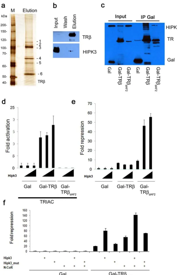
Figure 3a shows the silver-stained SDS PAGE gel of the final elution of the TRb purification. Several proteins (bands 1–5) in addition to the bait protein (band 6; Gal-hTRb) can readily be seen. These and others were identified by MALDI-ToF and Orbitrap mass spectrometry (Dataset S1). Identified proteins included several known nuclear receptor interacting proteins, such as specific heat shock proteins and lung resistance protein [28,29]. Nuclear proteins were in less abundance, due most likely to the use of whole cell extracts for this purification.
To test the validity and relevance of one of the novel TRb
cofactors identified, we focused on the protein Homeodomain-interacting Protein Kinase 3 (HIPK3). HIPK3 is a Ser/Thr kinase that affects transcriptional regulation, cell differentiation, growth and apoptosis [30,31,32,33]. Notably, it has already been shown to bind and modulate two other NRs; Steroidogenic Factor-1 (SF1) and Androgen Receptor (AR) [30,31].
Western blot analysis of the zebrafish extract and purified fractions (Figure 3b) confirms the presence of HIPK3 in the elution. Clearly though, the majority of HIPK3 present in the whole animal extract is not bound to the TRb bait protein, consistent with its enzymatic participation in numerous other protein complexes.
The specificity and nature of this interaction was also tested using transiently expressed GAL, GAL-TRb or GAL-TRbDAF2
fusion proteins. The latter protein lacks the C-terminal AF2 helix, which is required for co-activator binding. The fusion proteins were expressed together with HIPK3 in human HEK 293 cells. Both full-length and AF2-deleted GAL-TRbfusion proteins were able to pull down the HIPK3 protein (Figure 3c). Although the intensity of the HIPK3 band co-purified by the TRbDAF2deleted
protein appears less intense, this is largely due to the presence of additional co-migrating bands in the full-length TRb pull down, which may be due to subsequent modifications of the non-deleted LBD protein. We conclude that the AF-2 helix is not required for HIPK3 complex formation.
HipK3 negatively modulates TRbtranscriptional activity
Ligand activation of TR is associated with the displacement of co-repressors and recruitment of co-activators. To investigate whether HIPK3 affects the transcriptional activity of TRb, a cell culture co-transfection assay was employed using Gal4-TRband Gal-TRbDAF-2fusion protein. Deletion of the AF2 domain of TRb
results in continued co-repressor binding even in the presence of hormone, a phenomenon referred to as ‘Resistance to Thyroid Hormone’ (RTH) [34]. This type of deletion represents the most severe genetic form of RTH.
Addition of the thyroid hormone analog, TRIAC (3,5,39 -triiodothyroacetic acid) strongly stimulates the activity of the full LBD fusion protein, but has no effect on the non-activating AF2-deleted form (Figure 3d). Addition of HIPK3 led to a modest (,1.3X) increase in the transcriptional activation activity of the
WT fusion protein (Figure 3d). No effect was seen upon addition of HIPK3 on the activation activity of the AF2 deleted protein, consistent with the need for the AF2 motif for agonist-based transcriptional activation.
As TRbrepresses the expression of target genes in the absence of hormone, we also looked for effects of HIPK3 on Gal4-LBD fusion protein activity using a reporter plasmid with significant basal transcription activity, making it suitable for the observation of repression (Figure 3e and Figure S2a). As with the transcrip-tional activation assay with ligand present, HIPK3 had only a modest affect on GAL4-TRb activity (,1.3X lower). However,
with the AF2 removed, HIPK3 increased transcriptional repres-sion by an impressive 5–6 fold. This suggests that HIPK3 acts
primarily as an enhancer of TRb- mediated transcriptional repression.
To further probe the mechanistic nature of this HIPK3 affect, we examined its ability to augment repression mediated by the known TRb co-repressor, N-CoR [35,36,37]. Co-expression of HIPK3 or N-CoR with the WT TRb fusion protein increased repression of the UAS-containing reporter approximately 4- and 3- fold respectively (Figure 3f and Figure S2b). When added at the same time, a further,2-fold increase in repression mediated by
the TRbDAF2mutant was observed. These interactions and effects
were not observed with a kinase-defective HIPK3 protein (Figure 3f). These results are consistent with the activities of other HIPK family members on target gene expression and co-repressor function [38,39].
Zebrafish ligand trap activities are hormone-inducible
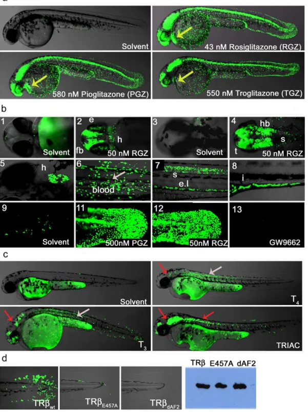
To test whether our LT lines are responsive to externally provided hormones or drugs, we treated the PPARc line with readily available agonists and antagonists (Figure 4a–b). PPARc
fish treated with the receptor specific agonists Rosiglitazone (RGZ), Pioglitazone (PGZ) or Troglitazone (TGZ)) increased the GFP reporter response from the relatively restricted signal in tail epidermis and posterior spine to include strong expression in the CNS, heart, blood, renal tube and eye (Figure 4a). Higher magnification of the embryo head shows specific and detailed activation in the presence of drug in the brain, eye and heart (Figure 4b panels 2 and 4). Older embryos (5–6 dpf) also show strong GFP expression in other tissues including heart, blood and intestine (Figure 4b, panels 5–8). Treatment of PPARcfish with PPARa- or PPARd-specific agonists showed no increase in reporter activation when used at their receptor-specific EC50
concentrations (Figure S3a). However, as seen in Gal4-PPAR cell based assays [40], higher concentrations of the beta or alpha agonist do result in partial activation of the PPARcLT embryos (Figure S3a). Treatment with the selective PPARc antagonist GW9662 either decreases or completely blocks the GFP responses to endogenous ligand(s) (Figure 4b-13 and Figure S3b). When added together with Rosiglitazone, in a classical agonist/ antagonist competition experiment, GW9662 was clearly able to displace the potent PPARcagonist (Figure S3b).
As further validation of drug responsiveness, we also treated TRb fish with known agonists (Figure 4c). Vehicle treated TRb
embryos show the same response seen in untreated embryos. TRb
fish treated with either Thyroxine (T4) or Triiodothyronine (T3)
induced reporter activation in the eye, heart, epidermis, blood, muscle and brain (Figure 4c). As expected, T3 activates the
reporter more strongly than T4. Treatments with TRIAC, also
yielded strong responses at dosages similar to those used in mammalian tissues. The eye activity of TRb ligand trap fish correlates well with previously reported expression of endogenous TRb in retina at 48 hpf [19]. However, our finding of receptor activity in other tissues such as the brain and anterior spinal cord are novel. Interestingly, in a recent profiling of zebrafish NR expression patterns, Bertrand and colleagues found that nuclear receptors expressed in the retina, except of TRb, are also expressed in the brain and/or anterior spinal cord. We expect that many of the novel activity patterns observed in our LT lines will represent novel roles in early development, opening the door for new avenues of study.
LT drug treatments reveal tissue selective nuclear receptor activities
regulated tissues. SuchSelectiveNR Modulators (SNRMs) would yield fewer side effects and have additional uses. Interestingly, each of the TR agonists tested above exhibited significant tissue-selective activities, as indicated by the colored arrows in Figure 4c. For example, T3 and T4elicited strong GFP responses in dorsal
muscle while TRIAC did not. Conversely, TRIAC elicited responses in the spinal cord while T3and T4did not. Differential
responses were also seen in the eye, heart, epidermis brain and blood. Many of these sites of action have not been previously documented.
To further verify that GFP expression in the TRb lines are ligand- and cofactor-dependent, we made two additional trans-genic lines carrying mutations in the TRbLBD (Figure 4d). These include the previously described LT-TRbDAF2 and a construct
carrying a single AF2 point mutation, LT-TRbE457A. Both
mutations result in impaired co-activator recruitment upon ligand binding [34]. As already shown (Figure 4c), TRb WT embryos respond strongly to the addition of TRIAC. However, TRIAC-treated TRbE457Afish show dramatically reduced GFP expression,
with signal restricted mainly to epidermal cells of the tail bud, and TRbDAF2 embryos show no detectable GFP whatsoever
(Figure 4d). Western blot analysis using anti-FLAG antibody shows that both fusion proteins are well expressed. We conclude that the signals observed with these lines are ligand and co-activator dependent.
GFP signals are readily quantified and suitable for drug screening
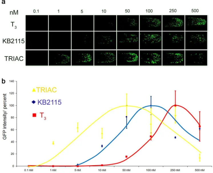
To test whether drug effects can be quantified with our ligand trap fish, we subjected TRb LT embryos to increasing concentrations of the agonists T3, TRIAC or KB2115 [41], and
the antagonist Amiodarone. Figure 5a shows a series of tail regions of the treated TRbembryos. Note that the number and intensity of GFP responding nuclei increase in proportion to hormone concentration but decrease again as levels become teratogenic.
Figure 5b shows averaged GFP response curves for increasing concentrations of all three ligands. TRIAC proved to be the most effective, then KB2115, and lastly T3. Amiodarone treatment
yielded no detectable GFP response as expected. Importantly our dose-response curves for the various TRbagonists tested correlate well with previously determined animal and human data [42,43].
Discussion
We have taken advantage of the unique properties of zebrafish as a model organism to engineer a powerful in vivo screening system for new Nuclear Receptor ligands and cofactors (uses summarized in Figure 6). Embryos and larvae produced by the transgenic fish are readily arrayed, drugged and assayed within microtiter plates, making them suitable for high throughput screens. As discussed below, this whole-animal approach will facilitate the identification of ligands and cofactors that could not
otherwise be identified by previously usedin vitro or cell based approaches. Many of these ligands and cofactors will act tissue- or stage-specifically, providing new options for NR study and manipulation. In turn, the tissue-specific GFP responses produced by these novel endogenous and exogenous ligands will also provide the bases for subsequent genetic, molecular and visual screens that provide new insights into how these tissues develop and function.
A major problem with current drugs that target NRs is the side-effects that arise due to unwanted modulation of the targeted NR in tissues that are not the source of the problem, or to the targeting of other NR members of the same class. The identification of more selective compounds that act more NR- and tissue-specifically has the potential to circumvent these problems. Although such selective NR modulators (SNRMs) have been discovered, they are extremely difficult to screen for and new techniques and models are needed to succeed in the discovery of beneficial new drugs. With the LT system, known or potential ligands can be tested in all tissues and during any stage of development. As shown here, this readily reveals the SNRM properties of all active compounds.
The LT lines developed here should also be immensely useful for the identification of ‘endocrine disruptors’ within industrial, agricultural and municipal wastes [44]. The majority of these molecules emulate NR ligands, thereby interfering with normal human and animal development and function. Fish are one of the major targets and concentrators of endocrine disrupting com-pounds, and as such, are a highly appropriate test bed for their presence [45].
Several methods for isolating native protein complexes have been described in yeast, bacteria, cell culture, plants and insects [46]. Here, we show that the affinity tag cassette used in the LT system is capable of purifying protein complexes from whole vertebrate tissues with sufficient yield and purity for subsequent cofactor purification and identification. With appropriate modifi-cations, the identities of bound ligands should also be readily determined. Indeed, the use of multiple affinity chromatography steps followed by mass spectrometry has already yielded ligands for the Drosophila NR E75 [21], and the mouse NR PPARa [47]. Given the number of NRs that remain as orphans, this has great potential for the timely identification of their recalcitrant ligands. The ability to identify directly bound ligands also provides a means to determine whether active compounds identified in compound screens are directly bound and unmodified. In some cases, the active molecule may be a metabolite of the original compound added. Thus, the LT system has the potential to identify ‘pro-drugs’, something that is difficult or impossible to achieve by other means.
Our results with TRb suggest that its major role during early development is primarily as a repressor. This was evidenced by the lack of GFP activity in the absence of supplemented agonists, and the isolation of repressive cofactors. Among the novel cofactors
bait protein. Presence of the TRbprotein in the same fractions, detected using Flag-M2 antibody, is shown above. (c) HEK293 cells were transfected with FLAG-HIPK3 and one of three different Gal expression constructs containing the FSH-tag: Gal, Gal-TRb(aa 189–461) and TRbDAF2(aa 189–451).
After 48 h of transfection, the cells were harvested in IP buffer and Gal and Gal fusion proteins immunoprecipitated using Streptactin Sepharose. Western blotting using a FLAG M2 antibody shows HIPK3 in the Gal-TRband Gal-TRbDAF2pull downs. (d) Transcriptional activity ofTRbproteins. 293
cells were transfected with a 2xUAS-Luciferase reporter construct (0.2mg/well) and Gal or Gal-TRbconstructs (0.05mg/well): Gal-TRb(aa 189–461) and
TRbDAF2(aa 189–451) in the presence of 100 nM TRIAC. Fold activation was measured in the presence (0.05 or 0.1mg/well) or absence of HIPK3.
Luciferase values were normalized againstb-Gal. (e) Effects of HIPK3 on Gal-TRbinduced repression. 293 cells were transfected with a 2xUAS-Luciferase reporter construct and either Gal, Gal-TRbor GAL-TRbDAF2. Fold repression was measured in the presence or absence of HIPK3. Luciferase
values were normalized againstb-Gal. Transfections as under 3d. (f) The effect of HIPK3 was investigated in the absence or presence of the co-repressor N-CoR. Transfections were performed as in 3d using the Gal-TRbDAF2(aa 189–451) construct. Fold repression of the TRbmutant in the
presence of HIPK3 and/or N-CoR (0.05mg/well) are shown.
Figure 4. Ligand trap activities are drug responsive and reveal SNRM activities.(a) LT-PPARcembryos show strong reporter activation in the presence of receptor specific agonist (uses as indicated). After heat pulse followed by overnight incubation with solvent or drug GFP expression in cells of the epidermis, CNS, eye, blood and heart (48 hpf; lateral view) is induced. Yellow arrow indicates GFP expression in heart. (b) Upper panel shows magnifications of embryo heads in ventral (b-1-2) and dorsal (b-3-4) orientation of 2 dpf fish treated with solvent or 50 nM Rosiglitazone (RGZ). B-2 shows reporter expression in eye (e), forebrain (fb), heart (h) and b-4 indicates GFP expression in hindbrain (hb), tectum (t) and anterior spinal cord (s). Middle panel shows drug responses in tissues of older embryos: Expression in heart (b-5; 5 dpf ventral view), in blood (b-6; 8 dpf), in the spinal cord (s) and endothelium layer ((el), b-7; 4 dpf, lateral view) and in the intestine ((I, b-8; 6 dpf). Embryos were subjected to a 30 minute heat pulse at 37uC, followed by incubation with 50 nM Rosiglitazone for 24 h. The lower panels show responses to: endogenous hormone in the tail epidermis of 3 dpf embryos treated with solvent (b-9); 500 nM Pioglitazone (b-11), 500 nM RGZ (b-12) and treatment with the antagonist GW9662 (500 nM). (c) TRbLT embryos show tissue-specific responses to hormones and drugs. After 40 min heat induction, embryos (24 hpf) were incubated with compounds for 28 h in a 28uC incubator in the dark. Solvent (Ethanol) treated embryos show no GFP expression. T4(2.5mM) treated embryos
show strong reporter activity in muscle and to a lesser amount in epidermis, brain and eye retina. T3(2.5mM) treatments result in similar responses
identified was the kinase HIPK3, which we found to act as a potentiator of target gene repression. Given the ligand-indepen-dent nature of this interaction, it may provide additional means to treat or circumvent ‘resistance to thyroid hormone’ (RTH) conditions. Previous studies on HIPK proteins have also shown important functions during development [48], and in some cases via interactions with well characterized co-repressors [38]. As the dissociation and disposal of these co-repressors appears to require phosphorylation and/or ubiquitination [49,50,51], HIPK3 may be functioning in a related capacity. It should be noted, however, that while HIPK3 enhances N-CoR-mediated repression of TRb, it
has also been shown to mediate ligand dependent transcriptional activation by the Androgen and SF1 Nuclear Receptors, while having no apparent effect on Glucocoritcoid Receptor mediated transcription [30,31]. As such, NR modulation by HIP kinases may serve as an alternative means of differentially controlling multiple NR activities.
Taken together, this study shows that the LT system provides enormous potential for the acceleration of NR functional analyses. Its amenability to systems biology scale analyses that access all tissues and developmental processes will reveal many new unpredictable and unexpected results.
Figure 5. LT drug responses are quantifiable.(a) Dose dependent GFP expression Shown are the dose responses for T3, KB2115 and TRIAC in
TRbtail epidermis (48 hpf) treated embryos. Treatments were done from 0.1–500 nM. GFP images show anterior-posterior tail view (imaged at 1506 magnification) (b) The graph indicates average intensity of GFP signals for each treatment in percent. GFP expression was quantified using nuclei count software from MetaX files. Values for drug treated fish are shown in yellow (TRIAC), in blue (KB2115) and in red (T3).
doi:10.1371/journal.pone.0009797.g005
in brain, heart, eye, epidermis and muscle, and in addition, anterior spinal cord. Overlay pictures of bright field and GFP (75% transparent) are shown. Arrows indicate tissue-specific GFP responses: grey = muscle and red = brain. (d) TRbresponses in transgenic fish are dependent on a functional LBD. TRbwt, TRbE457Aand TRbdAF2embryos (24 hfp, F1) were heat pulsed and soaked in TRIAC (100 nM) as described in Figure 4c. The upper row
shows strong (wt), weak (E457A) and no (dAF2) epidermal GFP expression in the tail. Western blot detected proteins show similar levels of TRb transgene expression (right side).
Materials and Methods
pLT plasmid construction
To generate the ligand trap vector, p-LT, we first combined the SV40 late polyadenlyation site from pCS2+ (cloned XbaI/Hind III) and the Gal4 DBD (aa 1–132; cloned into XbaI) into pBluescript (Stratagene). PmeI and NheI restriction sites were introduced in the primer as cloning sites for NR ligand binding domains. An EcoRV restriction site was designed N-terminal to the Gal4DBD for introducing the FSH-triple tag. Oligos for the triple tag were designed with EcoRI restriction sites on both ends and annealed together followed cloning into EcoRI digested pBSII. The tag was then PCR amplified with primers containing EcoRV restriction sites and a Kozak consensus sequence followed by an ATG start codon. This fragment was then introduced into the EcoRV restriction site of pBSGal1-132-pA, resulting in pBS-tripletag-Gal1-132-pA. Next, twoI-SceImega nuclease recognition sites were introduced by site directed mutagenesis flanking the fragment.
The eGFP-NLS gene was PCR amplified from the pUAS Stinger vector [52] and cloned into pBluescript II containing a SV40 polyA to generate pB-eGFP-pA. A fragment containing multi-UAS Gal DBD binding sites followed by the basal adenovirus promoter, E1b, was PCR amplified from pBUASEIB [13] and inserted into pB-eGFP-pA. The UASE1b-eGFP-pA fragment was then cut out by KpnI digestion and cloned into KpnI digested pB SceI-Tag-Gal1-132-pA. The resulting plasmid was SacII/NotI cut to insert a SacII/NotI fragment of the zebrafish Hsp70 promoter amplified from pzHSP70/4prom [25]. Finally, the gypsy insulator elements from the UAS-Stinger GFP transformation vector were PCR amplified and inserted upstream of the Hsp70 promoter into SacII restriction sites or downstream of the eGFP reporter into HindIII/ApaI.
Fish microinjections and maintenance
Zebrafish were maintained at 28.5uC on a 14/10 hour light/ dark cycle and staged according to hours (h) or days (d) postfertilization [53]. To generate pLT lines, plasmids were pre-digested with I-SceI (0.8mg DNA, 1ml I-SceI (New England
Biolabs) 2ml 10x I-SceI buffer, 1ml BSA) for 20 min at 37uC. Digested DNA was adjusted with 0.1% phenol red. 4.6 nl of DNA solution was injected into the blastomeres of early one-cell stage embryos [26]. Morpholino (MO) injection: 4.6 nl of MO (0.5 mM) in 0.1% phenol red was injected into the one-cell stage. F0 fish were crossed with WT fish to identify germ-line transformed animals, as determined by fin cut PCR with GFP primers or control agonist treatments. F1 progeny showing strong and consistent GFP responses were selected for F2 homozygote production. To avoid reporter GFP silencing of stable transgenic LT lines [54,55], F2 lines showing strong and consistent GFP responses were selected for continued propagation of the line. These lines have been maintained for several generations with no loss of responsiveness. At least two independent stable lines of each LT construct were generated, however for LT-PPARconly one germline fish was identified.
All experiments were approved by the institutional review board at University of Toronto.
Plasmids, cell culture transfection
FSH-Gal1-132 from pLT plasmid (NotI/PmeI) was cloned in frame into pcDNA3.1 (Invitrogen) to generate pcDNA3 FSH-GalDBD. The N-CoR plasmid has been described previously [36]. pFLAG-HIPK3 (2–1191) and pFLAG-HIPK3 (K226R) were provided by J.J. Palvimo (University of Helsinki). The 14xUA-SE1B plasmid was a gift of Dr. Sue (Banting and Best Department of Medical Research/originally provided by Dr. R.W. Ko¨ster; Helmholtz Zentrum, Germany). The plasmids for reporter gene assays 2X-GAL4 binding-site luciferase, pSV40b-Gal) have been described previously [37,56]. 293 cell line transfections were done with LipofectaminTM2000 (Invitrogen).
Compound screening
Embryos were heat induced (28R37uC) for 30 minutes and then arrayed in 24 well plates (10 per well). The water was removed and 500ml of embryo water/well including dissolved small molecules or solvent was added shortly after heat pulse. Embryos were incubated at 28uC for 14–20 h and then monitored using a fluorescent dissection scope (SteREO Lumar.V12 Carl
Figure 6. Schematic representation of LT platform functions.
Zeiss). For analyzing GFP fluorescent pattern, embryos, larvae and adult fish were anesthetized with Tricaine (Sigma, Cat.#A-5040). Embryos were mounted in 1% AgarPlaque PlusTM Agarose (# 21403A) purchased from PharminGen.
Drugs used
Sigma-Aldrich: Amiodarone hydrochloride (A8423); T3
(T2877); T4 (T2376) and TRIAC (T7650); Alexis Biochemicals:
Pioglitazone (ALX-270-367); Cayman Chemicals: Rosiglitazone (#71740); Troglitazone (#71750); GW 9662 (#70785); KB2115 (#10011054), GW 0742 (#10006798) and GW 590735 (#10009880).
Affinity purification of proteins
Purifications were performed at 4uC. 5-6 hpf LT-TRbembryos (128 g) were heat induced (37uC) for 20 min and recovered for 2 h. Homogenization was carried out in 100 ml FSH buffer (100 mM Tris-Cl pH 8, 150 mM NaCl, 0.1% Triton X-100 and Roche complete Mine protease inhibitor cocktail) including 2.5 mM DTT, 10 nM Avidin and 1 mM EDTA. After 20 min incubation the lysate was centrifuged for 10 min at 9000 rpm. 15 ml of prewashed (FSH buffer) Agarose-bead slurry (ABT; 2% plain Agarose beads) was incubated with the centrifugation supernatant (220 ml) for 30 minutes. After this incubation and again centrifugation the supernatant (200 ml) was incubated with 5 ml prewashed Strep TactinH(IBA; Cat: 2-1201-0251) slurry for 2 h. After incubation the beads were transferred to 2 gravity disposable columns (Bio-Spin columns, Cat: 732–6008) and washed 6 times with 2 ml of FSH-buffer including 1 mM EDTA. Bound proteins were eluted using volume 0.5 ml of 2.5 mM d-desthiobiotin containing FSH-buffer and 1 mM EDTA. For triple step purifications the Strep elutions were incubated for 2 hrs with FLAG M2 monoclonal antibody matrix (Sigma), and then washed 3times with FSH buffer followed by elution with FSH buffer containing 300mg/ml 3x FLAG peptide (Sigma). Flag elutions were incubated for 1 hrs with 60ml of Talon beads in an
Eppendorf tube, and then washed two times with FSH buffer followed elution with 2xSDS buffer. For immunoprecipitations, HEK 293 cells were grown in 10 cm dishes and DNA (9mg of
FLAG-HIPK3 with either 4mg of pcDNA3-FSH-GAL, -GAL-TRb or -GAL-TRbDAF2/empty vector was used to adjust the
DNA concentration) was transfected with Lipofectamin following the manufacturer’s instructions. Cells were harvested in 1 ml IP buffer (50 mM Tris pH 8, 150 mM NaCl, 1 mM EDTA, 0,75% NP-40, 1 mM DTT and Roche complete Mine protease inhibitor cocktail). After 10 min centrifugation at 13,000 rpm, 600ml of
cleared extract was incubated with 100ml Strep TactinHslurry for 2 h; followed by 3 washes with 1 ml of IP buffer. Immunopre-cipitated proteins were separated by SDS PAGE and detected by FLAG-M2 Western blotting.
Western blotting
Expression of ligand trap constructs was verified using a monoclonal mouse aM2 antibody (dilution 1:10 000; Sigma). Heat pulsed and recovered transgenic embryos were homogenized in 5ml FSH buffer/embryo and denatured with SDS PAGE loading buffer followed separation on 10% SDS-polyacrylamide gels. Detection of HIPK3 was done with aHIPK3 from Abgent (AP 7500b).
Mass spectrometry
Eluted proteins were separated by SDS PAGE, silver stained and individual bands excised, digested with Trypsin and analyzed
by MALD-TOF or ESI-MS tandem mass spectrometry. All samples for ESI-MS were analyzed by a 2-hour LC gradient using a split-free nano-LC (EasyLC, Proxeon, Odense, Denmark) coupled to a LTQ-Orbitrap XL, as recently described [57]. Raw data was converted to m/z XML using ReAdW and searched on a Swquest SorcererTM, using both Sequest and X!Tandem, against a zebrafish IPI protein sequence database (version 3.38) also containing the protein sequences of known contaminants (i.e. human keratins) and trypsin and the bait protein (human thyroid receptor). Searches were performed with a fragment ion mass tolerance of 0.8 Da and a parent ion tolerance of 50 ppm. Complete tryptic digestion was assumed. The iodoacetamide derivative of cysteine was specified in Sequest and X!Tandem as a fixed modification. The oxidation of methionine was specified as a variable modification. Scaffold (version Scaffold 2.1.1, Proteome Software Inc., Portland, OR) was used to validate MS/MS based peptide and protein identifications. Peptide identifications were accepted if they could be established at greater than 95.0% probability as specified by the Peptide Prophet algorithm and contained at least 1 identified peptide. Proteins that contained similar peptides and could not be differentiated based on MS/MS analysis alone were grouped to satisfy the principles of parsimony.
Oligos used to clone pLT:
pB Gal1-132-pA:
5’ATTCATCTAGAGATATCAAGCTACTGTCTTCTATCGAACA-AGC 3’ATTATCTAGAGTTTAAAC5’ATTCATCTAGAGATATCAAGCTACTGTCTTCTATCGAACA-AGCT5’ATTCATCTAGAGATATCAAGCTACTGTCTTCTATCGAACA-AGCTGATGATGTCGCA- 3’ATTATCTAGAGTTTAAACAGCTAGCTGATGATGTCGCA-CTTATTCTATGC
pB II triple tag:
sense –AATTCGACTACAAAGACCATGACGGTGATTATAAAGATCATGACATCG - ACTACAAGGATGACGATGACAAGGAGAACCTGTACTTCCAGTCCAACTGGAGCC -ACCCGCAGTTCGAAAAGCATCACCATCACCATCACG -antisense -ATGATCTTTATAATCACCGTCATGGTCTTT-GTAGTCG -AGTTGGACTGGAAGTACAGGTTCTCCTTGTCATCGTCATCC-TTGTAGTCGATGTC -AATTCGTGATGGTGATGGTGATGCTTTTCGAACTGCGGGTG-GCTCC
pB triple tag-Gal1-132-pA
5’ ATTATGATATCgccaccatgGACTACAAAGACCATGACGG 3’ ATTATGATATCGTGATGGTGATGGTGATGC
SceI pB triple tag-Gal1-132-pA
T7 5’ GACTCACTATAGGGCTAGGGATAACAGGGTAATGAA-
TTGGGTACCGGG-T7 3’ CCCGGTACCCAATTCATTACCCTGTTATCCCTAGCCC-
TATAGTGAGTC-T3 5’ CGGTGGAGCTCCAGTAGGGATAACAGGGTAATCTTTT-
GTTCCCTTTAGTG-T3 3’ CACTAAAGGGAACAAAAGATTACCCTGTTATCCCTAC- TGGAGCTCCACCG-pB eGFP-pA 5’ ATTATCTAGAACCATGGTGAGCAAGGGC 3’ ATTATCTAGATTACTTGTACAAGTAGCG pB UASE1b-eGFP-pA 5’ ATTATCCGCGGGGTACCCTCCAAGGCGGAGTACTGTCC 3’ ATAATCGGCCGGTGTGGAGGAGCTCAAAGTGAGGC pLT ; SceI pB zHsp70 triple tag Gal-1-132-pA-UASE1b-eGFP-pA)
pLT gypsy; SceI gypsy pB zHsp70 triple tag Gal-1-132-pA-UASE1b-eGFP-pA)
5’ Hsp ATAACCGCGGTCACGTAATAAGTGTGCG 3’ Hsp ATAACCGCGGAGATCTATACTAGAATTGATCGGC 5’ Gfp ATAAAAGCTT TCACGTAATAAGTGTGCG 3’ Gfp ATAAGGGCCCATACTAGAATTGATCGGC Morpholino oligo sequence:
5’ to 3’ and complementary to the translational start of the FSH Gal-NR:
ACCGTCATGGTCTTTGTAGTCCATG Primes used to clone NR cDNAs: NR1A2wt/E457A/dAF2
5’ ATTAGCTAGCATGACTCCCAACAGTATGAC NR1A2wt 3’ ATTAGTTTAAACCTAATCCTCGAACACTTCC NR1A2E457A 3’ ATTAGTTTAAACCTAATCCTCGAACACTGCCAAGAACAAA-GG NR1A2dAF2 3’ ATTAGTTTAAACCTACTATTCTGTGGGGCATTCCACC NR1C3 5’ ATTAGCTAGCGAGAAGGAGAAGCTGTTGG 3’ ATTAGTTTAAACCTAGTACAAGTCCTTGTAGATCTCC NR1F3 5’ ATTAGCTAGCATGTCCAAGAAGCAGAGGG 3’ ATTAGTTTAAACTCACTTGGACAGCCCCACAGG NR1D1 5’ ATTAGCTAGCATGCTTGCTGAGATGCAGAGTGCC 3’ ATTAGTTTAAACTCACTGGGCGTCCACCCGGAAGG NR4A3
5’ GATCGCTAGCCCATTACAACAGGAACCTTCTCAG 3’ CACCGTTTAAACTTAGAAAGGTAGGGTGTCCAGG
Supporting Information
Figure S1 Ligand trap signals are NRs specific A Morpholino (MO) against the FSH-tag of the GAL-NR transgene was injected into one-cell stage F3 embryos of LT-PPARc, RORc and LT-TRb. Endogenous and agonistic drug responses were compared between control and MO injected embryos. The upper row shows 48 hpf PPARcembryos in their chorions, and tail close ups of the same conditions are shown in the middle row. The lower row shows RORc (F2) or homozygous TRb F3 embryos in the presence of 250 nM T3 with control or MO injections.
Found at: doi:10.1371/journal.pone.0009797.s001 (0.70 MB TIF)
Figure S2 (a/b) Expression of HIPK3 and Gal- and Gal-TR-fusion proteins in reporter assays (Figure 3e and 3f) verified by Western Blot using Flag M2 antibody.
Found at: doi:10.1371/journal.pone.0009797.s002 (1.62 MB TIF)
Figure S3 Selective PPARcdrug responses (a) At 24 hpf PPARc
embryos were subjected to a 30 minute heat shock at 37uC and then incubated for 24 hr in the presence of agonists specific for one of the three PPAR isoforms (Rosiglitazone forc, GW0742 for
d/b and GW9578 or GW590735 for a). The concentrations chosen represent known EC50 values for the appropriate targets, along with significantly higher levels to test for cross-reactivity. Lateral views of 48 hpf embryos, anterior to the left, are shown. (b) PPARc agonist/antagonist replacement. At 24 hpf PPARc
embryos were subjected to a 30 minute heat induction at 37uC and either incubated for 24 hr in the presence of solvent or 1000 nM GW9662 (upper row) or 50 nM Rosiglitazone (RGZ) alone or 50 nM RGZ and increasing concentrations of GW9662 (20 nM and 500 nM). Two embryos for each treatment showing lateral views at 48 hpf, anterior to the left, are shown.
Found at: doi:10.1371/journal.pone.0009797.s003 (2.76 MB TIF)
Dataset S1 Table of mass spectrometry-identified bait protein (Gal-hTRb) and interacting proteins are shown. Eluted proteins were separated by SDS PAGE, silver stained and individual bands (band 1–6) excised, digested with Trypsin and analyzed by MALD-TOF or ESI-MS tandem mass spectrometry. Accession number, molecular weight (MW) and description of identified proteins are listed. The purified bait (Gal-hTRb) and the investigated cofactor are underlined in yellow.
Found at: doi:10.1371/journal.pone.0009797.s004 (0.04 MB XLS)
Acknowledgments
We would like to thank Structural Genomics Canada, T. Heinzel (University of Jena), D. Mangelsdorf (Howard Hughes Medical Institute) and C. L. Cummins (University of Toronto) for providing NR cDNAs, F. Vizeacoumar for helping with MetaX software.
Author Contributions
Conceived and designed the experiments: JT HMK. Performed the experiments: JT PRM MN TK. Analyzed the data: JT PRM MN TK HMK. Contributed reagents/materials/analysis tools: JT PRM MN TK HMK. Wrote the paper: JT HMK. Technical work: LB.
References
1. Aranda A, Pascual A (2001) Nuclear hormone receptors and gene expression. Physiol Rev 81: 1269–1304.
2. Gronemeyer H, Gustafsson JA, Laudet V (2004) Principles for modulation of the nuclear receptor superfamily. Nat Rev Drug Discov 3: 950–964.
3. Nagy L, Schwabe JW (2004) Mechanism of the nuclear receptor molecular switch. Trends Biochem Sci 29: 317–324.
4. Dias JM, Go NF, Hart CP, Mattheakis LC (1998) Genetic recombination as a reporter for screening steroid receptor agonists and antagonists. Anal Biochem 258: 96–102.
5. Glickman JF, Wu X, Mercuri R, Illy C, Bowen BR, et al. (2002) A comparison of ALPHAScreen, TR-FRET, and TRF as assay methods for FXR nuclear receptors. J Biomol Screen 7: 3–10.
6. Weatherman RV, Chang CY, Clegg NJ, Carroll DC, Day RN, et al. (2002) Ligand-selective interactions of ER detected in living cells by fluorescence resonance energy transfer. Mol Endocrinol 16: 487–496.
7. Blumberg B, Evans RM (1998) Orphan nuclear receptors–new ligands and new possibilities. Genes Dev 12: 3149–3155.
8. Kliewer SA, Lehmann JM, Willson TM (1999) Orphan nuclear receptors: shifting endocrinology into reverse. Science 284: 757–760.
9. Ciana P, Di Luccio G, Belcredito S, Pollio G, Vegeto E, et al. (2001) Engineering of a mouse for the in vivo profiling of estrogen receptor activity. Mol Endocrinol 15: 1104–1113.
10. Maggi A, Ciana P (2005) Reporter mice and drug discovery and development. Nat Rev Drug Discov 4: 249–255.
11. Nagel SC, Hagelbarger JL, McDonnell DP (2001) Development of an ER action indicator mouse for the study of estrogens, selective ER modulators (SERMs), and Xenobiotics. Endocrinology 142: 4721–4728.
12. Solomin L, Johansson CB, Zetterstrom RH, Bissonnette RP, Heyman RA, et al. (1998) Retinoid-X receptor signalling in the developing spinal cord. Nature 395: 398–402.
13. Luria A, Furlow JD (2004) Spatiotemporal retinoid-X receptor activation detected in live vertebrate embryos. Proc Natl Acad Sci U S A 101: 8987–8992. 14. Yamaguchi M, Saito H, Suzuki M, Mori K (2000) Visualization of neurogenesis in the central nervous system using nestin promoter-GFP transgenic mice. Neuroreport 11: 1991–1996.
15. Dooley K, Zon LI (2000) Zebrafish: a model system for the study of human disease. Curr Opin Genet Dev 10: 252–256.
16. Fishman MC (2001) Genomics. Zebrafish–the canonical vertebrate. Science 294: 1290–1291.
17. Machuca I, Esslemont G, Fairclough L, Tata JR (1995) Analysis of structure and expression of the Xenopus thyroid hormone receptor-beta gene to explain its autoinduction. Mol Endocrinol 9: 96–107.
from Xenopus thyroid hormone receptor beta gene promoter in response to thyroid hormone in Xenopus tadpoles in vivo. Proc Natl Acad Sci U S A 93: 1205–1209.
19. Bertrand S, Thisse B, Tavares R, Sachs L, Chaumot A, et al. (2007) Unexpected novel relational links uncovered by extensive developmental profiling of nuclear receptor expression. PLoS Genet 3: e188.
20. Palanker L, Necakov AS, Sampson HM, Ni R, Hu C, et al. (2006) Dynamic regulation of Drosophila nuclear receptor activity in vivo. Development 133: 3549–3562.
21. Reinking J, Lam MM, Pardee K, Sampson HM, Liu S, et al. (2005) The Drosophila nuclear receptor e75 contains heme and is gas responsive. Cell 122: 195–207.
22. Yang P, Sampson HM, Krause HM (2006) A modified tandem affinity purification strategy identifies cofactors of the Drosophila nuclear receptor dHNF4. Proteomics 6: 927–935.
23. Wegner GJ, Lee HJ, Corn RM (2002) Characterization and optimization of peptide arrays for the study of epitope-antibody interactions using surface plasmon resonance imaging. Anal Chem 74: 5161–5168.
24. Voss S, Skerra A (1997) Mutagenesis of a flexible loop in streptavidin leads to higher affinity for the Strep-tag II peptide and improved performance in recombinant protein purification. Protein Eng 10: 975–982.
25. Shoji W, Yee CS, Kuwada JY (1998) Zebrafish semaphorin Z1a collapses specific growth cones and alters their pathway in vivo. Development 125: 1275–1283.
26. Thermes V, Grabher C, Ristoratore F, Bourrat F, Choulika A, et al. (2002) I-SceI meganuclease mediates highly efficient transgenesis in fish. Mech Dev 118: 91–98.
27. Adelmant G, Begue A, Stehelin D, Laudet V (1996) A functional Rev-erb alpha responsive element located in the human Rev-erb alpha promoter mediates a repressing activity. Proc Natl Acad Sci U S A 93: 3553–3558.
28. Abbondanza C, Rossi V, Roscigno A, Gallo L, Belsito A, et al. (1998) Interaction of vault particles with estrogen receptor in the MCF-7 breast cancer cell. J Cell Biol 141: 1301–1310.
29. Kimmins S, MacRae TH (2000) Maturation of steroid receptors: an example of functional cooperation among molecular chaperones and their associated proteins. Cell Stress Chaperones 5: 76–86.
30. Lan HC, Li HJ, Lin G, Lai PY, Chung BC (2007) Cyclic AMP stimulates SF-1-dependent CYP11A1 expression through homeodomain-interacting protein kinase 3-mediated Jun N-terminal kinase and c-Jun phosphorylation. Mol Cell Biol 27: 2027–2036.
31. Moilanen AM, Karvonen U, Poukka H, Janne OA, Palvimo JJ (1998) Activation of androgen receptor function by a novel nuclear protein kinase. Mol Biol Cell 9: 2527–2543.
32. Rochat-Steiner V, Becker K, Micheau O, Schneider P, Burns K, et al. (2000) FIST/HIPK3: a Fas/FADD-interacting serine/threonine kinase that induces FADD phosphorylation and inhibits fas-mediated Jun NH(2)-terminal kinase activation. J Exp Med 192: 1165–1174.
33. Zhang Q, Yoshimatsu Y, Hildebrand J, Frisch SM, Goodman RH (2003) Homeodomain interacting protein kinase 2 promotes apoptosis by downregu-lating the transcriptional corepressor CtBP. Cell 115: 177–186.
34. Ortiga-Carvalho TM, Shibusawa N, Nikrodhanond A, Oliveira KJ, Machado DS, et al. (2005) Negative regulation by thyroid hormone receptor requires an intact coactivator-binding surface. J Clin Invest 115: 2517–2523. 35. Cohen RN, Putney A, Wondisford FE, Hollenberg AN (2000) The nuclear
corepressors recognize distinct nuclear receptor complexes. Mol Endocrinol 14: 900–914.
36. Horlein AJ, Naar AM, Heinzel T, Torchia J, Gloss B, et al. (1995) Ligand-independent repression by the thyroid hormone receptor mediated by a nuclear receptor co-repressor. Nature 377: 397–404.
37. Tiefenbach J, Novac N, Ducasse M, Eck M, Melchior F, et al. (2006) SUMOylation of the corepressor N-CoR modulates its capacity to repress transcription. Mol Biol Cell 17: 1643–1651.
38. Choi CY, Kim YH, Kwon HJ, Kim Y (1999) The homeodomain protein NK-3 recruits Groucho and a histone deacetylase complex to repress transcription. J Biol Chem 274: 33194–33197.
39. Lee W, Andrews BC, Faust M, Walldorf U, Verheyen EM (2009) Hipk is an essential protein that promotes Notch signal transduction in the Drosophila eye by inhibition of the global co-repressor Groucho. Dev Biol 325: 263–272. 40. Willson TM, Brown PJ, Sternbach DD, Henke BR (2000) The PPARs: from
orphan receptors to drug discovery. J Med Chem 43: 527–550.
41. Berkenstam A, Kristensen J, Mellstrom K, Carlsson B, Malm J, et al. (2008) The thyroid hormone mimetic compound KB2115 lowers plasma LDL cholesterol and stimulates bile acid synthesis without cardiac effects in humans. Proc Natl Acad Sci U S A 105: 663–667.
42. Messier N, Langlois MF (2000) Triac regulation of transcription is T(3) receptor isoform- and response element-specific. Mol Cell Endocrinol 165: 57–66. 43. Schueler PA, Schwartz HL, Strait KA, Mariash CN, Oppenheimer JH (1990)
Binding of 3,5,39-triiodothyronine (T3) and its analogs to the in vitro translational products of c-erbA protooncogenes: differences in the affinity of the alpha- and beta-forms for the acetic acid analog and failure of the human testis and kidney alpha-2 products to bind T3. Mol Endocrinol 4: 227–234. 44. Zoeller RT, Tan SW (2007) Implications of research on assays to characterize
thyroid toxicants. Crit Rev Toxicol 37: 195–210.
45. Segner H (2009) Zebrafish (Danio rerio) as a model organism for investigating endocrine disruption. Comp Biochem Physiol C Toxicol Pharmacol 149: 187–195.
46. Chang IF (2006) Mass spectrometry-based proteomic analysis of the epitope-tag affinity purified protein complexes in eukaryotes. Proteomics 6: 6158–6166. 47. Chakravarthy MV, Lodhi IJ, Yin L, Malapaka RR, Xu HE, et al. (2009)
Identification of a physiologically relevant endogenous ligand for PPARalpha in liver. Cell 138: 476–488.
48. Harvey RP (1996) NK-2 homeobox genes and heart development. Dev Biol 178: 203–216.
49. Hermanson O, Jepsen K, Rosenfeld MG (2002) N-CoR controls differentiation of neural stem cells into astrocytes. Nature 419: 934–939.
50. Hong SH, Privalsky ML (2000) The SMRT corepressor is regulated by a MEK-1 kinase pathway: inhibition of corepressor function is associated with SMRT phosphorylation and nuclear export. Mol Cell Biol 20: 6612–6625.
51. Perissi V, Rosenfeld MG (2005) Controlling nuclear receptors: the circular logic of cofactor cycles. Nat Rev Mol Cell Biol 6: 542–554.
52. Barolo S, Carver LA, Posakony JW (2000) GFP and beta-galactosidase transformation vectors for promoter/enhancer analysis in Drosophila. Biotech-niques 29: 726, 728, 730, 732.
53. Westerfield M (1993) The zebrafish book a guide for the laboratory use of zebrafish Danio (Brachydanio) rerio. Eugene, OR: Institute of Neuroscience University of Oregon.
54. Goll MG, Anderson R, Stainier DY, Spradling AC, Halpern ME (2009) Transcriptional silencing and reactivation in transgenic zebrafish. Genetics 182: 747–755.
55. Halpern ME, Rhee J, Goll MG, Akitake CM, Parsons M, et al. (2008) Gal4/ UAS transgenic tools and their application to zebrafish. Zebrafish 5: 97–110. 56. Heinzel T, Lavinsky RM, Mullen TM, Soderstrom M, Laherty CD, et al. (1997)
A complex containing N-CoR, mSin3 and histone deacetylase mediates transcriptional repression. Nature 387: 43–48.