Submitted18 August 2015 Accepted 14 December 2015 Published25 January 2016
Corresponding author Shigeru Tadano,
Academic editor Arti Ahluwalia
Additional Information and Declarations can be found on page 11
DOI10.7717/peerj.1562
Copyright 2016 Endo et al.
Distributed under
Creative Commons CC-BY 4.0
OPEN ACCESS
Structural strength of cancellous
specimens from bovine femur under
cyclic compression
Kaori Endo1, Satoshi Yamada2, Masahiro Todoh2, Masahiko Takahata1,
Norimasa Iwasaki1and Shigeru Tadano2
1Department of Orthopaedic Surgery, Hokkaido University Graduate School of Medicine, Sapporo, Japan 2Division of Human Mechanical Systems and Design, Faculty of Engineering, Hokkaido University, Sapporo,
Japan
ABSTRACT
The incidence of osteoporotic fractures was estimated as nine million worldwide in 2000, with particular occurrence at the proximity of joints rich in cancellous bone. Although most of these fractures spontaneously heal, some fractures progressively collapse during the early post-fracture period. Prediction of bone fragility during progressive collapse following initial fracture is clinically important. However, the mechanism of collapse, especially the gradual loss of the height in the cancellous bone region, is not clearly proved. The strength of cancellous bone after yield stress is difficult to predict since structural and mechanical strength cannot be determineda priori. The purpose of this study was to identify whether the baseline structure and volume of cancellous bone contributed to the change in cancellous bone strength under cyclic loading. A total of fifteen cubic cancellous bone specimens were obtained from two 2-year-old bovines and divided into three groups by collection regions: femoral head, neck, and proximal metaphysis. Structural indices of each 5-mm cubic specimen were determined using micro-computed tomography. Specimens were then subjected to five cycles of uniaxial compressive loading at 0.05 mm/min with initial 20 N loading, 0.3 mm displacement, and then unloading to 0.2 mm with 0.1 mm displacement for five successive cycles. Elastic modulus and yield stress of cancellous bone decreased exponentially during five loading cycles. The decrease ratio of yield stress from baseline to fifth cycle was strongly correlated with bone volume fraction (BV/TV,r=0.96,p<0.01) and structural model index (SMI, r= −0.81,p<0.01). The decrease ratio of elastic modulus from baseline to fifth cycle was also correlated with BV/TV (r=0.80,p<0.01) and SMI (r= −0.78,p<0.01). These data indicate that structural deterioration of cancellous bone is associated with bone strength after yield stress. This study suggests that baseline cancellous bone structure estimated from adjacent non-fractured bone contributes to the cancellous bone strength during collapse.
SubjectsBioengineering, Orthopedics
INTRODUCTION
Osteoporosis is a common disease which is characterized by low bone mass and deteriora-tion of bone tissue, resulting in an increased risk of fracture, particularly at the proximity of the joint rich in cancellous bone (Riggs & Melton 3rd, 1995). Among the estimated nine million osteoporotic fractures worldwide in 2000, 1.6 million were in the hip, 1.7 million in the forearm, and 1.4 million were vertebral fractures (Johnell & Kanis, 2006). The hip and spine fractures in osteoporosis patients often include those occurring because of falls from a standing height and less due to progressive collapse. Progressive collapse is characteristically found in deformities after compress fractures with continuous loss of the height in the whole fracture bone area. For example, progressive vertebral collapse can be defined in this way: the vertebral collapse fracture is found in deformities after initial fracture, followed by continuous loss of vertebral body height. Within a femoral fracture, the occult fracture of the femur and the subchondral insufficiency fracture (SIF) of the femoral head are also manifested in the cancellous bone region with rapid progression of the collapse, such as seen in rapidly progressive arthritis of SIF (Bangil et al., 1996;
Yamamoto, 2012). Severe collapse of these areas can lead to chronic pain, depression, an inability to perform daily life activities, and in extreme cases can be life-threatening (Riggs & Melton 3rd, 1995;Poole & Compston, 2006;Ioannidis et al., 2009;Adachi et al., 2010). Thus, prevention of the collapse in rich area of cancellous bone, especially proximal femur and spine fracture, is clinically critical.
Cancellous bone plays an important role as the primary load-carrying component that absorbs energy (Fyhrie & Schaffler, 1994). Characteristic stress–strain (S–S) curves indicate that the mechanical competence of cancellous bone depends mainly on the trabeculae network and material properties of the tissue. The post-yield behavior of cancellous bone has been well-documented in previous studies and linked to strain and density (Keaveny et al., 1994a;Keaveny et al., 1994b;Keaveny, Wachtel & Kopperdahl, 1999). These studies also investigated failure patterns in specimens of cancellous bone and showed similar S–S curves to those obtained.Fyhrie & Schaffler (1994)conducted on compressive tests of cancellous cubic bone and concluded that S–S curves after linear region were considered to have three phases which correspond with: (1) the initial
trabeculae yield; (2) the secondary trabeculae yield and buckle; and (3) the plateau region. Mechanical properties in three phases decreased and finally plateaued, while the strain increased and the height loss progressively collapsed in areas rich with cancellous bone without specific trauma.
According to macroscopic experiments using compression tests, the stiffness of cancellous bone is correlated with its apparent density (Keller, 1994;Morgan & Keaveny, 2001;Morgan, Bayraktar & Keaveny, 2003;Follet et al., 2004). In addition, mechanical properties that contribute to bone strength are determined by bone geometry (size and shape of trabecular bone) and microarchitecture using noninvasive three-dimensional micro-computed tomography (micro-CT) evaluation (Kopperdahl & Keaveny, 1998;
Ulrich et al., 1999;Ahlborg et al., 2003;Todoh et al., 2004). However, some studies suggest that detection of bone geometries and microarchitecture, as well as structural indices, is
associated with fracture in the distal radius and tibia (Genant et al., 1996;Mackey et al., 2007;Link & Lang, 2014). Therefore, the geometry and microarchitecture of cancellous bone contributes to the bone structural strength and would likely enable estimation of osteoporotic fracture risk.
However, the means by which the mechanical properties of cancellous bone were decreasing after the elastic region, and which structural factors affected the decreased mechanical properties, has not been well documented. This is because the non-linear response of cancellous bone post-yielding is more difficult to interpret than the linear response of the cortical diaphysis. In addition, the thin cortical shell covers 45% of the mid-transverse section to as low as 15% at the endplates (Eswaran et al., 2006). To advance our knowledge of the mechanism of progressive collapse, it is necessary to understand functional adaptation of cancellous bone to post-yield stress tendency. Moreover, identifying the relationship between the structural indices of cancellous bone and decreased mechanical properties post-yield may contribute to understanding the gradual decrease of the height in progressive collapse. Hence, the aim of this study was to investigate how cancellous bovine bone specimens taken from different areas of the femur change in elastic modulus and yield stress during cyclic compressions, and to examine the relationships between structural indices and decreased strength after the elastic region.
METHODS
Specimen preparation
A total 15 specimens were extracted from two bovines (A and B); 8 specimens were obtained from the right femur of bovine A and 7 specimens were obtained from the left femur of bovine B as shown inTable 1. Specimens were grouped (n=5/groups) in accordance with their extraction regions: the proximal metaphysis, neck, and head (Fig. 1). The femurs were first sliced along the coronal plane and then into 5-mm cubic shapes using a diamond wheel saw (model 650; South Bay Technology, San Clemente, CA, USA). All specimens containing cancellous bone were retrieved where one axis of the specimen corresponded to the longitudinal axis and 2 mm away from the cortical bone and epiphysis. Bone marrow was removed from the specimens using a pressure water jet, brushes, and automatic ultrasound device (US-1; Samsung, Tokyo, Japan). Specimens were stored in a container at−35◦C until experimentation. Preliminary experiments demonstrated that the bone volume fraction (BV/TV) increased in direct proportion with the proximity of the specimen from the femur metaphysis to the head. In accordance with this finding, specimens were grouped into three parts according to BV/TV, differing by approximately 10% between each part, as shown inTable 1.
Microstructure analysis using micro-CT
The micro-CT technique and image processing approach were based on current guide-lines and a previous study (Bouxsein et al., 2010). All specimens were scanned using a micro focus CT instrument (InspeXio SMX-90CT; Shimadzu, Kyoto, Japan) at a 22µm
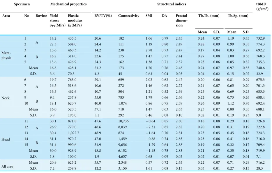
Table 1 Mechanical properties and structural indices are shown.These data are shown as mean and standard deviation.
Specimen Mechanical properties Structural indices tBMD
(g/cm3)
Area No Bovine Yield
stress σY1(MPa)
Elastic modulus E1(MPa)
BV/TV(%) Connectivity SMI DA Fractal dimen-sion
Tb.Th. (mm) Tb.Sp. (mm)
Mean S.D. Mean S.D.
1 14.2 435.5 20.6 182 1.66 0.79 2.45 0.24 0.07 1.19 0.45 732.9 2 A 22.3 504.0 24.4 111 1.19 0.80 2.49 0.28 0.09 0.99 0.35 774.3 3 15.6 460.3 14.2 238 2.78 0.73 2.47 0.17 0.04 0.83 0.27 692.2 4 18.2 314.0 22.6 175 1.47 0.77 2.44 0.27 0.08 1.00 0.38 768.3 5
B
13.6 426.9 24.3 162 1.38 0.71 2.57 0.23 0.06 0.85 0.32 735.3 Mean 16.8 428.1 21.2 173 1.70 0.76 2.48 0.24 0.07 0.97 0.35 740.6
Meta-physis
S.D. 3.6 70.5 4.2 45 0.63 0.04 0.05 0.04 0.02 0.15 0.07 32.9 6 19.7 763.0 29.1 659 2.02 0.62 2.47 0.20 0.06 0.81 0.29 675.3 7 16.5 518.6 40.6 272 1.46 0.62 2.72 0.24 0.07 0.65 0.20 701.3 8
A
16.3 462.6 40.7 804 1.21 0.52 2.69 0.25 0.06 0.69 0.25 683.3 9 9.4 237.8 35.0 783 1.79 0.66 2.66 0.22 0.06 0.73 0.26 688.4 10 B 18.1 620.7 40.0 1,074 0.86 0.75 2.59 0.26 0.09 1.12 0.76 692.4 Mean 16.0 520.5 37.1 718 1.47 0.63 2.63 0.23 0.07 0.80 0.35 688.1 Neck
S.D. 3.9 195.0 5.1 292 0.46 0.08 0.10 0.02 0.01 0.19 0.23 9,8 11 30.1 871.8 47.6 10,736 −0.64 0.85 2.80 0.18 0.08 0.29 0.18 726.8 12 26.9 779.0 48.6 8,039 −2.31 0.85 2.82 0.20 0.08 0.31 0.19 722.8 13
A
30.4 1,022.7 48.9 874 −1.64 0.70 2.81 0.23 0.05 0.45 0.18 724.3 14 31.1 970.2 47.0 1,459 −0.88 0.74 2.85 0.23 0.06 0.41 0.16 716.0 15 B 31.4 990.6 51.9 9,656 −1.79 0.64 2.88 0.19 0.08 0.32 0.17 709.4 Mean 30.0 926.9 48.8 6,152 −1.45 0.75 2.83 0.21 0.07 0.35 0.18 719.9 Head
S.D. 1.8 100.0 1.9 4,657 0.68 0.09 0.03 0.02 0.01 0.07 0.01 7.1 Mean 20.9 625.2 35.7 2,348 0.57 0.72 2.65 0.22 0.07 0.71 0.29 716.2 All area S.D. 7.2 258.9 12.2 3,150 1.61 0.08 0.15 0.03 0.01 0.27 0.15 28.3
Figure 1 Cubic specimens were obtained along the bovine femur bone axes from the (A) head, (B) neck, and (C) metaphysis.
imaging reference micro-CT phantom. A review of the literature indicated that filtration does not necessarily require software-based beam hardening corrections for imaging of cancellous bone (Waarsing, Day & Weinans, 2004;Meganck et al., 2009). In preliminary assessments, a cover of only 0.1 mm Cu as an X-ray filter could reduce the induced beam hardening enough to calculate tBMD and structural indices. To assess the volumetric density and microarchitecture of cancellous bone, Bone J software can be used to calculate a number of structural indices (Doube et al., 2010). The structural indices determined in this study included BV/TV, degree of anisotropy (DA), trabecular thickness (Tb.Th.), trabecular space (Tb.Sp.), structure model index (SMI), and number of connections (connectivity).
Cyclic compression test
Uniaxial compression tests were conducted on the specimens in the longitudinal direction to measure the apparent elastic modulus and yield stress using a mechanical testing machine (model 3365; Instron, Grove City, PA, USA) at room temperature. Loading parameters were established during preliminary experiments using bone samples.Figure 2 shows that the S–S curves obtained in this experiment also had the three phases described previously in the introduction (Fyhrie & Schaffler, 1994). A strain of 0.06 (0.3 mm displacement) was chosen to ensure failure of the three specimens in the head groups, as this exceeds the failure strain by an average 1.5 times.
Figure 2 Stress–strain curve of successful uniaxial compression.
Figure 3 Method and stress–strain curves of cyclic compression.(A) Total displacement of each cycle increased with 0.1-mm displacement. (B) Stress–strain curves under cyclic compression; yield stress was set to the maximum point, and elastic modulus were calculated from 50% to 70% of the maximum load in each cycle.
for five successive cycles (Fig. 3A). The five compression cycles were set to auto control regulation with preloading of 20 N and a displacement rate of 0.05 mm/min. The apparent elastic modulus was calculated from 50% to 70% of maximum loading in each cycle. Yield stress was defined as the inflection point that existed between the linear region to the plateau or the stress decreasing region (Fig. 3B).
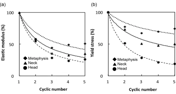
Figure 4 (A) Elastic modulus and (B) yield stress of each cycle are expressed relative to those of the first cycle.
Statistical analysis
Pearson’s correlation coefficient between the mechanical properties and structural indices were calculated and considered to be significant atp<0.01. JMP Pro software (Version 11.0.0; SAS Institute Inc., Cary, NC, USA) was used to perform statistical analyses.
RESULTS
Mechanical properties, structural indices and tBMD of the specimens are shown in Table 1. BV/TV correlated negatively with Tb.Sp.Mean and positively with connectivity. SMI identified plate, rod and concave shapes in the trabeculae of the metaphysis, neck and head groups, respectively. There were no significant differences with respect to the mean DA and Tb.Th between the groups. However, elastic modulus was significantly different between the groups after the first cycle, with values of 428.1±70.5 MPa in the metaphysis group, 520.5±195.0 MPa in the neck group, and 926.9±100.0 MPa in the head group. Yield stress at the first cycle was 16.8±3.6 MPa in the metaphysis group, 16.0±3.9 MPa in the neck group, and 30.0±1.8 MPa in the head group. Bone strength was considerably reduced after the first yield stress, and then decreased more moderately with subsequent cycles (Fig. 4).
The elastic modulus ratio between the fifth and first cycle (E5/E1) was 26.0±13.4%
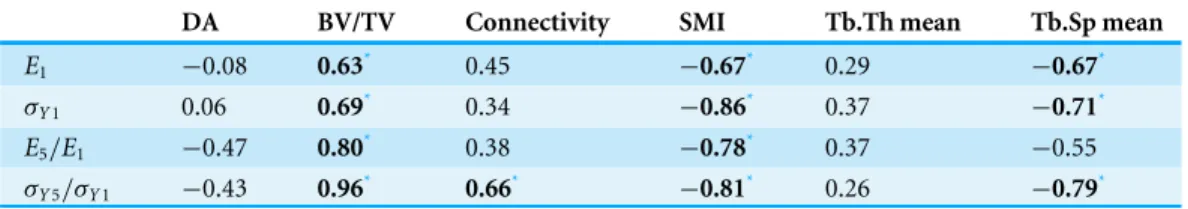
Table 2 Correlation coefficients between structural indices and mechanical properties.
DA BV/TV Connectivity SMI Tb.Th mean Tb.Sp mean
E1 −0.08 0.63* 0.45 −0.67* 0.29 −0.67*
σY1 0.06 0.69* 0.34 −0.86* 0.37 −0.71*
E5/E1 −0.47 0.80* 0.38 −0.78* 0.37 −0.55 σY5/σY1 −0.43 0.96* 0.66* −0.81* 0.26 −0.79*
Notes.
*p<0.01.
Figure 5 showed that BV/TV was strongly correlated withE5/E1(r=0.80) andσY5/σY1 (r=0.96). SMI was negatively correlated withE5/E1(r= −0.78) andσY5/σY1 (r= −0.81).
σY5/σY1 was also correlated with connectivity (r=0.66) and Tb.Sp.Mean (r= −0.79).
DISCUSSION
The present study elucidated two important post yielding mechanical phenomena of cancellous bone in order to understand the progressive collapse. Firstly, both elastic modulus and yield stress initially decreased rapidly and then decreased more gradually in line with the exponential approximation. In addition, both of the decrease ratios of elastic modulus and yield stress were inversely proportional to BV/TV. Following the exposure of cancellous bone to compressive loads high enough to initiate buckling of the trabeculae, mechanical strength decreases and progressive collapse occurs much easier in groups with low BV/TV. Thus, breakage of the trabeculae following load stress may impair the strength of the cancellous bone. From this viewpoint, the collapse in osteoporosis patients might progress rapidly as a consequence of the significant decrease of cancellous bone strength. Secondly, the mechanical properties at the first cycle and the decrease ratio correlated strongly with BV/TV and SMI, and weakly with connectivity and Tb.Sp.Mean. The novel relationships between the decrease ratio of mechanical properties and structural indices allowed estimations of the decrease in mechanical strength. Hence, these results indicated that baseline cancellous bone structure could help to understand cancellous bone strength during collapse. It has recently been shown that clinical CT can determine structural indices with a resolution of 0.1 mm (Burghardt et al., 2007). This study suggests that baseline bone structure estimated from the adjacent non-fractured bone side can contribute to the cancellous bone strength during clinical collapse. This bone fragility of cancellous bone during progressive collapse is only one part of the whole bone strength; however, medical institutions could more correctly evaluate the permissible pressure of the fracture area and more confidentially allow each osteoporosis patient to undertake the appropriate levels of rehabilitation without progressive collapse.
This first experiment in terms of using cyclic compression testing helps to understand the post-yield tendency of progressive collapse in areas rich with cancellous bone. Additionally, our study used successive compression testing which increased strain in every cycle.Keaveny et al. (1994b)aimed at determining how the modulus and strength decreased after being subjected to yield stress with various initial loading strains. Figure 4 shows that the
Figure 5 Correlations between mechanical properties and structural indices.These graphs showed (A)
E5/E1and BV/TV (r=0.80), (B)σY5/σY1and BV/TV (r=0.96), (C)E5/E1and SMI (r= −0.78), and
(D)σY5/σY1and SMI (r= −0.81).
approximate amount of decrease ratio in our study is similar in elastic modulus but different in yield stress compared to their study. The reason for this may be because of differences in the specimens preparations, experimental settings or in cycle numbers.
(Goulet et al., 1994;Dux et al., 2010;Slyfield et al., 2012;Hernandez et al., 2014). Other studies have also included a broader range of BV/TV, but under different preparations; for example, after nephrectomy, ovariectomy or loading (Cory et al., 2010;Morgan, Bostrom & Van der Meulen, 2015). Destructive mechanical tests would be more likely to result in complicated S–S curves. Limiting the focus of structural indices would simplify interpretations superior in accuracy to prevent errors occurring from other factors. Therefore, the novelty of our study compared to other studies is broad range of BV/TV with the same preparations.
For further improvements towards more accurate estimation of cancellous bone strength in our in vitro model, other additional points were considered. First, the mechanical properties of single trabeculae consisting of cancellous bone had to be considered.Yamada, Tadano & Fukuda (2014) previously reported on single trabecular strength in bovine femurs and the relationship between mechanical properties and nanostructure. To more accurately predict bone strength, the structural strength of the whole cancellous bone was applied in our work, with fine distinctions in material strength also applied using their technique. Second, many studies (Tang & Vashishth, 2010;Lambers et al., 2013;Hernandez et al., 2014;Lambers et al., 2014) reported that the damaged bone fraction affected elastic modulus and yield stress. Use of these microscopic techniques to detect the damaged bone fraction might anticipate the three phases in post-yielding. More specifically the second and third phases, being the secondary trabecular yield and buckle and the plateau region, respectively. In fact, our experiments attempted to understand the process of trabecular buckling using micro-CT imaging, but obvious trabecular buckling was not observed from the first yield to the start of plateau region. In future experiments, measuring the damaged bone fractions might help to interpret the mechanism of decreased bone strength after the first trabecular yielding in our experiments.
There were two limitations; the first limitation was that this study selected the direction of compression only in the longitudinal direction. Previous studies have also used cubic specimens from various parts and animals but have compared the elastic modulus compressed in three directions (Vahey, Lewis & Vanderby Jr, 1987; Goulet et al., 1994;
Frich et al., 1997;Morgan, Bostrom & Van der Meulen, 2015). These studies showed that the elastic modulus in the gravity direction was between 2 and 4 times greater than in the other directions. Conversely, the strongest elastic modulus in our two bovine specimens was not necessarily along the gravity direction as the specimens were relatively isotropic. The uniaxial compression tests of the cubic specimens were conducted in each of three directions and the maximum elastic modulus divided by minimum elastic modulus was 1.57 ±0.47. This study also showed that the degree of anisotropy was not correlated with the mechanical properties in cases where the specimens were limited to bovine femurs. Therefore, this study ignored the effect of compression direction. For subsequent investigations, we will consider how the mechanical properties of the cancellous bone are influenced by the main trabecular direction of the human anatomical bone microstructure, using the mean intercept length method (Odgaard et al., 1997;Perilli et al., 2008).
The second limitation was that the low number of specimens (n=15) obtained from only two young bovines with epiphysis. However, the latter limitation was also the scheme
of our study to minimize the complicated errors in establishing the relationship between structural indices and the decrease ratio of mechanical properties at first. The reason was that healthy and young bovines tend to have little risk of secondary osteoporosis. In future studies we will need to consider other complex factors such as BMD, collagen quality and bone turnover. The experimental conditions must be achieved by a carefully designedin vivostudy and partly referring to our data of bovine specimens in this study. Other studies have shown that the elastic modulus and yield stress in some patients are not compromised by reduced BMD; for example, in severe osteoporosis, metastasis (including after irradiation), necrosis, and secondary osteoporosis (Lavernia, Sierra & Grieco, 1999;Fazzalari et al., 1998;Wu et al., 2008;Dux et al., 2010;Keaveny et al., 2014). Hence, structural indices have a great potential to become disease-specific predictors. In the future, structural indices might become the first predictors for the risk of progressive collapse in rich area of cancellous bone after an initial fracture to enable better treatments, external fixation, and more appropriate levels of activity and rehabilitation.
CONCLUSIONS
Elastic modulus and yield stress were significantly decreased at the first trabeculae yield, and then decreased more gradually as the number of compression cycles was increased. Subsequent decrease ratio in elastic modulus and yield stress was significantly correlated with bone volume fractions and connectivity of cancellous bone. These results contribute to one of the predictions of cancellous bone strength during progressive collapse, from baseline cancellous bone structure estimated from the adjacent non-fractured side bone.
ADDITIONAL INFORMATION AND DECLARATIONS
Funding
This work was supported by a Grant-in-Aid for Scientific Research (A), MEXT (No. 15H02207). The funders had no role in study design, data collection and analysis, decision to publish, or preparation of the manuscript.
Grant Disclosures
The following grant information was disclosed by the authors: Grant-in-Aid for Scientific Research (A), MEXT: 15H02207.
Competing Interests
The authors declare there are no competing interests.
Author Contributions
• Kaori Endo conceived and designed the experiments, performed the experiments, analyzed the data, contributed reagents/materials/analysis tools, wrote the paper, prepared figures and/or tables.
• Masahiro Todoh and Shigeru Tadano contributed reagents/materials/analysis tools, reviewed drafts of the paper.
• Masahiko Takahata contributed reagents/materials/analysis tools, wrote the paper, reviewed drafts of the paper.
• Norimasa Iwasaki reviewed drafts of the paper.
Animal Ethics
The following information was supplied relating to ethical approvals (i.e., approving body and any reference numbers):
IRB approval was not required because the specimens were obtained from only bovine femurs. We obtained these femurs from the meat industry after butchery (immediately frozen).
Data Availability
The following information was supplied regarding data availability: The raw data can be found inTable 1.
REFERENCES
Adachi JD, Adami S, Gehlbach S, Anderson Jr FA, Boonen S, Chapurlat RD, Compston JE, Cooper C, Delmas P, Diez-Perez A, Greenspan SL, Hooven FH, LaCroix AZ, Lindsay R, Netelenbos JC, Wu O, Pfeilschifter J, Roux C, Saag KG, Sambrook PN, Silverman S, Siris ES, Nika G, Watts NB. 2010.Impact of prevalent fractures on quality of life: baseline results from the global longitudinal study of osteoporosis in women.Mayo Clinic Proceedings85:806–813DOI 10.4065/mcp.2010.0082.
Ahlborg HG, Johnell O, Turner CH, Rannevik G, Karlsson MK. 2003.Bone loss and bone size after menopause.New England Journal of Medicine349:327–334 DOI 10.1056/NEJMoa022464.
Bangil M, Soubrier M, Dubost JJ, Rami S, Carcanagues Y, Ristori JM, Bussiere JL. 1996.Subchondral insufficiency fracture of the femoral head.Revue du Rhumatisme 63:859–861.
Bouxsein ML, Boyd SK, Christiansen BA, Guldberg RE, Jepsen KJ, Muller R. 2010. Guidelines for assessment of bone microstructure in rodents using micro-computed tomography.Journal of Bone and Mineral Research25:1468–1486 DOI 10.1002/jbmr.141.
Burghardt AJ, Wang Y, Elalieh H, Thibault X, Bikle D, Peyrin F, Majumdar S. 2007. Evaluation of fetal bone structure and mineralization in IGF-I deficient mice using synchrotron radiation microtomography and Fourier transform infrared spectroscopy.Bone40:160–168DOI 10.1016/j.bone.2006.06.017.
Cory E, Nazarian A, Entezari V, Vartanians V, Muller R, Snyder BD. 2010.Compressive axial mechanical properties of rat bone as functions of bone volume fraction,
apparent density and micro-ct based mineral density.Journal of Biomechanics 43:953–960DOI 10.1016/j.jbiomech.2009.10.047.
Doube M, Klosowski MM, Arganda-Carreras I, Cordelieres FP, Dougherty RP, Jackson JS, Schmid B, Hutchinson JR, Shefelbine SJ. 2010.BoneJ: free and extensible bone image analysis in ImageJ.Bone47:1076–1079DOI 10.1016/j.bone.2010.08.023. Dux SJ, Ramsey D, Chu EH, Rimnac CM, Hernandez CJ. 2010.Alterations in damage
processes in dense cancellous bone following gamma-radiation sterilization.Journal of Biomechanics43:1509–1513DOI 10.1016/j.jbiomech.2010.01.042.
Eswaran SK, Gupta A, Adams MF, Keaveny TM. 2006.Cortical and trabecular load shar-ing in the human vertebral body.Journal of Bone and Mineral Research21:307–314. Fazzalari NL, Forwood MR, Smith K, Manthey BA, Herreen P. 1998.Assessment of
cancellous bone quality in severe osteoarthrosis: bone mineral density, mechanics, and microdamage.Bone22:381–388DOI 10.1016/S8756-3282(97)00298-6. Follet H, Boivin G, Rumelhart C, Meunier PJ. 2004.The degree of mineralization is
a determinant of bone strength: a study on human calcanei.Bone34:783–789 DOI 10.1016/j.bone.2003.12.012.
Frich LH, Jensen NC, Odgaard A, Pedersen CM, Sojbjerg JO, Dalstra M. 1997.Bone strength and material properties of the glenoid.Journal of Shoulder and Elbow Surgery6:97–104 DOI 10.1016/S1058-2746(97)90029-X.
Fyhrie DP, Schaffler MB. 1994.Failure mechanisms in human vertebral cancellous bone. Bone15:105–109DOI 10.1016/8756-3282(94)90900-8.
Genant HK, Engelke K, Fuerst T, Gluer CC, Grampp S, Harris ST, Jergas M, Lang T, Lu Y, Majumdar S, Mathur A, Takada M. 1996.Noninvasive assessment of bone mineral and structure: state of the art.Journal of Bone and Mineral Research 11:707–730.
Goulet RW, Goldstein SA, Ciarelli MJ, Kuhn JL, Brown MB, Feldkamp LA. 1994.The relationship between the structural and orthogonal compressive properties of trabec-ular bone.Journal of Biomechanics27:375–389DOI 10.1016/0021-9290(94)90014-0. Hernandez CJ, Lambers FM, Widjaja J, Chapa C, Rimnac CM. 2014.Quantitative
relationships between microdamage and cancellous bone strength and stiffness.Bone 66:205–213DOI 10.1016/j.bone.2014.05.023.
Ioannidis G, Papaioannou A, Hopman WM, Akhtar-Danesh N, Anastassiades T, Pickard L, Kennedy CC, Prior JC, Olszynski WP, Davison KS, Goltzman D, Thabane L, Gafni A, Papadimitropoulos EA, Brown JP, Josse RG, Hanley DA, Adachi JD. 2009.Relation between fractures and mortality: results from the Canadian Multicentre Osteoporosis Study.Canadian Medical Association Journal 181:265–271DOI 10.1503/cmaj.081720.
Johnell O, Kanis JA. 2006.An estimate of the worldwide prevalence and disability associated with osteoporotic fractures.Osteoporosis International17:1726–1733 DOI 10.1007/s00198-006-0172-4.
Keaveny TM, Guo XE, Wachtel EF, McMahon TA, Hayes WC. 1994a.Trabecular bone exhibits fully linear elastic behavior and yields at low strains.Journal of Biomechanics 27:1127–1136DOI 10.1016/0021-9290(94)90053-1.
Radcliffe HS, Libanati C. 2014.Femoral and vertebral strength improvements in postmenopausal women with osteoporosis treated with denosumab.Journal of Bone and Mineral Research29:158–165DOI 10.1002/jbmr.2024.
Keaveny TM, Wachtel EF, Guo XE, Hayes WC. 1994b.Mechanical behavior of damaged trabecular bone.Journal of Biomechanics27:1309–1318
DOI 10.1016/0021-9290(94)90040-X.
Keaveny TM, Wachtel EF, Kopperdahl DL. 1999.Mechanical behavior of human trabecular bone after overloading.Journal of Orthopaedic Research17:346–353 DOI 10.1002/jor.1100170308.
Keller TS. 1994.Predicting the compressive mechanical behavior of bone.Journal of Biomechanics27:1159–1168DOI 10.1016/0021-9290(94)90056-6.
Kopperdahl DL, Keaveny TM. 1998.Yield strain behavior of trabecular bone.Journal of Biomechanics31:601–608DOI 10.1016/S0021-9290(98)00057-8.
Lambers FM, Bouman AR, Rimnac CM, Hernandez CJ. 2013.Microdamage caused by fatigue loading in human cancellous bone: relationship to reductions in bone biome-chanical performance.PLoS ONE8:e83662DOI 10.1371/journal.pone.0083662. Lambers FM, Bouman AR, Tkachenko EV, Keaveny TM, Hernandez CJ. 2014.The
effects of tensile-compressive loading mode and microarchitecture on microdamage in human vertebral cancellous bone.Journal of Biomechanics47:3605–3612
DOI 10.1016/j.jbiomech.2014.10.011.
Lavernia CJ, Sierra RJ, Grieco FR. 1999.Osteonecrosis of the femoral head.Journal of the American Academy of Orthopaedic Surgeons7:250–261.
Link TM, Lang TF. 2014.Axial QCT: clinical applications and new developments.Journal of Clinical Densitometry17:438–448DOI 10.1016/j.jocd.2014.04.119.
Mackey DC, Eby JG, Harris F, Taaffe DR, Cauley JA, Tylavsky FA, Harris TB, Lang TF, Cummings SR, Health A, Body Composition Study G. 2007.Prediction of clinical non-spine fractures in older black and white men and women with volumetric BMD of the spine and areal BMD of the hip: the Health, Aging, and Body Composition Study.Journal of Bone and Mineral Research22:1862–1868 DOI 10.1359/jbmr.070807.
Meganck JA, Kozloff KM, Thornton MM, Broski SM, Goldstein SA. 2009.Beam hardening artifacts in micro-computed tomography scanning can be reduced by X-ray beam filtration and the resulting images can be used to accurately measure BMD. Bone45:1104–1116DOI 10.1016/j.bone.2009.07.078.
Morgan EF, Bayraktar HH, Keaveny TM. 2003.Trabecular bone modulus-density relationships depend on anatomic site.Journal of Biomechanics36:897–904 DOI 10.1016/S0021-9290(03)00071-X.
Morgan TG, Bostrom MP, Van der Meulen MC. 2015.Tissue-level remodeling simula-tions of cancellous bone capture effects ofin vivoloading in a rabbit model.Journal of Biomechanics48:875–882DOI 10.1016/j.jbiomech.2014.12.011.
Morgan EF, Keaveny TM. 2001.Dependence of yield strain of human trabecular bone on anatomic site.Journal of Biomechanics34:569–577
DOI 10.1016/S0021-9290(01)00011-2.
Odgaard A, Kabel J, Van Rietbergen B, Dalstra M, Huiskes R. 1997.Fabric and elastic principal directions of cancellous bone are closely related.Journal of Biomechanics 30:487–495DOI 10.1016/S0021-9290(96)00177-7.
Perilli E, Baleani M, Ohman C, Fognani R, Baruffaldi F, Viceconti M. 2008. Depen-dence of mechanical compressive strength on local variations in microarchitecture in cancellous bone of proximal human femur.Journal of Biomechanics41:438–446 DOI 10.1016/j.jbiomech.2007.08.003.
Poole KE, Compston JE. 2006.Osteoporosis and its management.BMJ 333:1251–1256 DOI 10.1136/bmj.39050.597350.47.
Riggs BL, Melton 3rd LJ. 1995.The worldwide problem of osteoporosis: insights afforded by epidemiology.Bone17:505S–511SDOI 10.1016/8756-3282(95)00258-4.
Slyfield CR, Tkachenko EV, Fischer SE, Ehlert KM, Yi IH, Jekir MG, O’Brien RG, Keaveny TM, Hernandez CJ. 2012.Mechanical failure begins preferentially near resorption cavities in human vertebral cancellous bone under compression.Bone 50:1281–1287DOI 10.1016/j.bone.2012.02.636.
Tang SY, Vashishth D. 2010.Non-enzymatic glycation alters microdamage formation in human cancellous bone.Bone46:148–154DOI 10.1016/j.bone.2009.09.003.
Todoh M, Ihara M, Matsumoto T, Tanaka M. 2004.Relationship between mechanical property of cancellous bone and hardness of trabeculae.JSME International Journal Series C-Mechanical Systems Machine Elements and Manufacturing 47:1075–1078 DOI 10.1299/jsmec.47.1075.
Ulrich D, Van Rietbergen B, Laib A, Ruegsegger P. 1999.The ability of
three-dimensional structural indices to reflect mechanical aspects of trabecular bone.Bone 25:55–60DOI 10.1016/S8756-3282(99)00098-8.
Vahey JW, Lewis JL, Vanderby Jr R. 1987.Elastic modulus, yield stress, and ultimate stress of cancellous bone in the canine proximal femur.Journal of Biomechanics 20:29–33DOI 10.1016/0021-9290(87)90264-8.
Waarsing JH, Day JS, Weinans H. 2004.An improved segmentation method for in vivomicroCT imaging.Journal of Bone and Mineral Research19:1640–1650 DOI 10.1359/JBMR.040705.
Wu ZX, Lei W, Hu YY, Wang HQ, Wan SY, Ma ZS, Sang HX, Fu SC, Han YS. 2008. Effect of ovariectomy on BMD, micro-architecture and biomechanics of cortical and cancellous bones in a sheep model.Medical Engineering & Physics30:1112–1118 DOI 10.1016/j.medengphy.2008.01.007.
Yamada S, Tadano S, Fukuda S. 2014.Nanostructure and elastic modulus of single trabecula in bovine cancellous bone.Journal of Biomechanics47:3482–3487 DOI 10.1016/j.jbiomech.2014.09.009.