Morphological and Chemical Analysis of Bone
Substitutes by Scanning Electron Microscopy and
Microanalysis by Spectroscopy of Dispersion Energy
Gabriela Alessandra da CRUZ Sérgio de TOLEDO Enilson Antonio SALLUM Antonio Fernando Martorelli de LIMA
Department of Prosthodontics and Periodontics,
School of Dentistry of Piracicaba, State University of Campinas, Piracicaba, SP, Brazil
This study evaluated the morphological and chemical composition of the following bone substitutes: cancellous and cortical organic bovine bone with macro and microparticle size ranging from 1.0 to 2.0 mm and 0.25 to 1.0 mm, respectively; inorganic bovine bone with particle size ranging from 0.25 to 1.0 mm; hydroxyapatite with particle size ranging from 0.75 to 1.0 mm; and demineralized freeze-dried bone allograft with particle size ranging from 0.25 to 0.5 mm. The samples were sputter-coated with gold in an ion coater, the morphology was observed and particle size was measured under vacuum by scanning electron microscopy (SEM). The chemical composition was evaluated by spectroscopy of dispersion energy (EDS) microanalysis using samples without coating. SEM analysis provided visual evidence that all examined materials have irregular shape and particle sizes larger than those informed by the manufacturer. EDS microanalysis detected the presence of sodium, calcium and phosphorus that are usual elements of the bone tissue. However, mineral elements were detected in all analyzed particles of organic bovine bone except for macro cancellous organic bovine bone. These results suggest that the examined organic bovine bone cannot be considered as a pure organic material.
Key Words: scanning electron microscopy, x-ray microanalysis, bone substitute, bovine bone, human bone, hydroxyapatite.
Correspondence: Dra. Gabriela Alessandra da Cruz, Faculdade de Odontologia de Piracicaba, UNICAMP, Avenida Limeira, 901, Areião, 13414-018 Piracicaba, SP, Brasil. Tel: +55-19-430-5299. e-mail: [email protected]
INTRODUCTION
Bone substitutes have been used to fill periodon-tal osseous defects around teeth affected by periodonperiodon-tal disease (1). Four categories of grafts are commonly used in the these days: autografts (bone from the same patient that is transferred from an intra or extra-oral donor site); allografts (bone obtained from a different donor of the same specie); xenograft (bone obtained from a different donor of another specie); and aloplastic materials or inert implants (2,3).
Bone is a metabolically active organ composed of both mineral and organic phases. The mineral phase of the skeleton contributes about two thirds of its weight; the remaining one third is organic matrix, primarily consisting of collagen and small amounts of proteoglycan, lipid and several noncollagenous proteins (4).
Bone substitutes are commercially available in Brazil with different forms: organic and inorganic, cortical or cancellous, or macro and microparticle
Scanning electron microscopy (SEM) provides rich visual details and accuracy for measurements of particle size. Microanalyses by spectroscopy of disper-sion energy (EDS) allows quantitative and qualitative evaluations of the mineral components when the device is coupled with a correcting system (Proza) that assesses the atomic number (Z), absorption (A) and fluorescence (F) of the elements in the samples. EDS microanalyses allow verifying a small particle region without physically separating this part from the material (13).
In view of the lack of published information regarding the actual particle size and chemical compo-sition of commercially available bone substitutes, the purpose of this study was to analyze, by SEM and EDS microanalysis, the shape, size and chemical composi-tion of the particles of the following bone substitutes: cancellous and cortical organic bovine bone with macro and microparticle size; inorganic bovine bone; hy-droxyapatite; and freeze-dried bone allograft.
MATERIAL AND METHODS
Particle batches of the following materials were obtained: 1. organic cortical bovine bone with macro and microparticle sizes ranging from 1.0 to 2.0 mm and 0.25 to 1.0 mm, respectively (Gen-ox®; Baumer S/A,
Division of Biomaterials, Mogi Mirim, SP, Brazil); 2. organic cancellous bovine bone with macro and microparticle sizes ranging from 1.0 to 2.0 mm and 0.25 to 1.0 mm, respectively (Gen-ox®); 3. inorganic bovine
bone with particle size ranging from 0.25 to 1.0 mm (Bio-Oss, Geistlich-Pharma, Wolhusen, Switzerland); 4. demineralized freeze-dried bone allograft with par-ticle size ranging from 0.25 to 0.50 mm. (Dembone, Pacific Coast Tissue Bank, Los Angeles, CA, USA). 5. natural mineral hydroxyapatite bovine bone with rough granulation particle size ranging from 0.75 to 1.0 mm (Pro-Ha, Pró-line, São Paulo, SP, Brazil). Samples were purchased from materials’ commercial representatives.
SEM Analysis
The materials’ particles were fixed on stubs with carbon tape or adhesive containing powdered graphite (Ceil, São Paulo, SP, Brazil) and sputter-coated with gold in an ion coater (Denton Desk II, Denton Vacuum LLC, Moorestown, NJ, USA). Particle size and mor-phology were examined under vacuum with a scanning
electron microscope (JSM 5600LV, Jeol, Tokyo, Ja-pan). Visual morphological analysis was done using a specific software (SEM Control User Interface, version 1.27, Jeol). Particle size measurements were under-taken in 20 particles of each bone substitute, according to the highest longitudinal dimension and were ex-pressed using descriptive statistics.
EDS Microanalysis
The chemical composition of the particles was evaluated by EDS microanalysis (Noran Vantage EDS, version 1.4, Noran Instruments, Inc., Tokyo, Japan) under vacuum on uncoated samples fixed on stubs with carbon tape or adhesive containing powdered graphite (Ceil). The analysis was performed after selecting an area on standard SEM photomicrographs acquired with 15 kVp, 20 mm of distance and spot sized 33. The quantitative analysis of bone substitutes was performed with a correcting system (PROZA) (Noran Vantage EDS, version 1.4, Noran Instruments, Inc.) that assesses the atomic number (Z), absorption (A) and fluores-cence (F) of the elements present in the samples areas.
Statistical Analysis
Particle size data were expressed by descriptive statistics using specific softwares (Statistix for Win-dows, v. 7.0, 2000, StatSoft South America, São Caetano do Sul, SP, Brazil and Minitab for Windows, v. 13.1, 2001, Minitab, Inc., State College, PA, USA). EDS microanalysis data were expressed as the percent-age of elements in the samples and by the spectrographics showing counter time and electron volt energy.
RESULTS AND DISCUSSION
organic bovine bone. The hydroxyapatite showed no porosity (Fig. 2), which is in accordance with previous studies (15,16). However, it has been suggested that it is possible to modify the hydroxyapatite structure to obtain porosity, allowing bone cell infiltration as it seems that pore colonization is favored by diameters larger than 50-100 mm or even 250-300 mm (17). However, a previous study (7) did not report bone growth in pores smaller than 50 mm. Figure 2 shows the presence of pores with different sizes in the tested materials.
Particle size analysis showed some discrepancy between the obtained values and the values informed on the product label. The microparticles of cancellous organic bovine bone showed a mean size of 1.19 ± 0.29 mm (label information: microparticles ranging from 0.25 to 1.0 mm and macroparticles ranging from 1.0 to 2.0 mm), whereas the smallest particle size was pre-sented by the demineralized freeze-dried bone (0.97 ± 0.31 mm) (label information: 0.25 to 0.50 mm range). Such a discrepancy has been previously reported by
Schwartz et al. (4) while evaluating the size of deminer-alized freeze-dried bone particles. The largest particle sizes were presented by the macroparticle cancellous organic bovine bone (2.54 ± 0.31 mm) and macroparticle cortical organic bovine bone (2.01 ± 0.35mm). The microparticle size of cortical organic bovine bone (1.33 ± 0.28), microparticle size of inorganic bovine bone (1.31 ± 0.41 mm) and hydroxyapatite (1.43 ± 0.41 mm) showed intermediate length values (label information: particles ranging from 0.75 to 1.0 mm). These data suggest that all evaluated materials showed larger par-ticle sizes than those informed by the manufacturers.
It is well accepted that the particle size may interfere with the success of the regenerative therapy. When demineralized freeze-dried bone is used, particle sizes ranging from 0.12 mm to 1.00 mm possess a higher osteoinductive effect than do particles below 0.12 mm (5). Apparently, the ideal particle size should range from 0.10 mm to 0.30 mm. Very small particle sizes of demineralized freeze-dried bone may elicit a
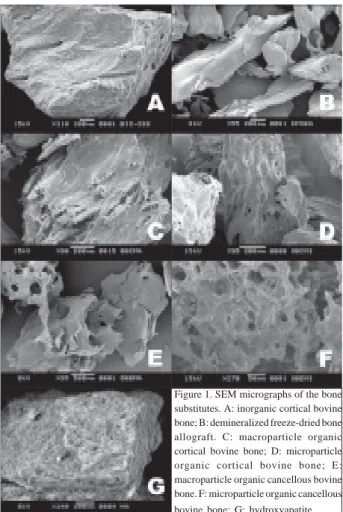
Figure 1. SEM micrographs of the bone substitutes. A: inorganic cortical bovine bone; B: demineralized freeze-dried bone allograft. C: macroparticle organic cortical bovine bone; D: microparticle organic cortical bovine bone; E: macroparticle organic cancellous bovine bone. F: microparticle organic cancellous
bovine bone; G: hydroxyapatite
Figure 2. Pores of the bone substitutes. A: inorganic cortical bovine bone; B: demineralized freeze-dried bone allograft. C: macroparticle organic cortical bovine bone; D: microparticle organic cortical bovine bone; E: macroparticle organic cancellous bovine bone. F: microparticle organic cancellous
macrophage response and are rapidly resorbed with little or no new bone formation (5). However, Fucini et al. (14) found no differences after clinical application of two different sizes of demineralized freeze-dried bone (0.25 and 0.50 mm; and 0.85 and 1.00 mm).
EDS microanalysis of bovine bone matrix, hu-man bone and hydroxyapatite detected elements, like calcium, phosphorus, aluminum and sulfur (Fig. 3). These are expected findings (18) as these chemical elements could be incorporated during bone develop-ment and Ca/P ratio may vary according to nutritional conditions. Normal bone composition should include
magnesium, sodium, potassium and carbonate salts (18). Table 1 displays the quantitative analysis with PROZA correcting system. Mineral elements were detected in micro and macro cortical organic bovine bone and micro cancellous organic bovine bone.
Thus, it may be assumed that the evaluated organic bovine bone could not be considered as a pure organic material (i.e., collagen and proteins) except for the macro cancellous bovine bone that did not show any mineral elements. The analysis of inorganic bovine bone and hydroxyapatite detected calcium and phosphor that are usual chemical elements of their compositions.
Merkx et al. (19) found similar morphostructural and chemical elements in the inorganic bovine bone and concluded that bovine bone composition seems to be the same as that of human bone. The presence of aluminium could be attributed to the type of stub used. Carbon and oxygen could not be quantified because their anatomic numbers are lower than that of sodium and the equip-ment was not calibrated for this type of analyses (19). SEM analysis showed that the particles of the bone substitutes have irregular shape and size. Particle sizes tended to be lager than those mentioned on product label. EDS microanalyses detected the presence of chemical elements that are typical of bone tissue. Further research is required to thoroughly describe the
Table 1. Percentage of the chemical elements detected in the particles of inorganic cortical bovine bone (BIO-OSS), demineralized freeze-dried bone allograft (DFDBA), macroparticle organic cortical bovine bone (MaOCoBB), microparticle organic cortical bovine bone (MiOCoBB), macroparticle organic cancellous bovine bone (MaOCaBB), microparticle organic cancellous bovine bone (MiOCaBB), hydroxyapatite particles (HA), stub, carbon tape and adhesive.
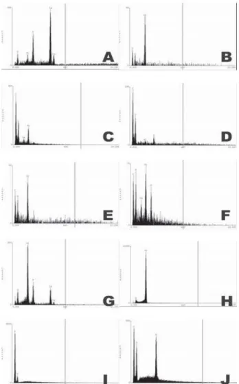
Figure 3. Spectrographic showing the chemical elements detected in the samples by EDS microanalysis. A: inorganic cortical bovine bone; B: demineralized freeze-dried bone allograft; C: macroparticle organic cortical bovine bone; D: microparticle organic cortical bovine bone; E: macroparticle organic cancellous bovine bone; F: microparticle organic cancellous bovine bone; G: hydroxyapatite; H: stub; I: carbon tape; J: adhesive.
Chemical elements (%)
Materials Time (s) Na P Ca S Al
BIO-OSS 150 - 17.59 79.45 - 2.96
DFDBA 50 - - - - 100.0
MaOCoBB 300 21.19 - 78.81
MiOCoBB 190 - - - 81.79 18.21
MaOCaBB 40 - - - - 100.0
MiOCaBB 70 16.50 37.88 - - 45.62
HA 200 - 22.08 26.39 - 44.40
Stub 11000 - - - - 100.0
Carbon tape 3000 - - - - 100.0
-characteristics of these materials and to determine their role in the treatment of periodontal osseous defects.
Under the tested conditions, it may be concluded that: 1. The evaluated materials presented particles of irregular shape and size. 2. The mean measured particle sizes were larger than the sizes informed by the manu-facturers. 3. The analysis of chemical composition by EDS detected the presence of sodium, calcium and phosphorus that are common chemical elements on bone tissue composition; 4. Mineral elements were detected on all particles of organic bovine bone, except for macro cancellous organic bovine bone, which suggest that the evaluated organic bovine bone could not be considered as a pure organic material.
RESUMO
Neste estudo foram avaliados a morfologia, o tamanho e a composição química dos seguintes substitutos ósseos: osso bovino orgânico cortical e esponjoso com micropartículas medindo entre 0,25 e 1,0 mm e macropartículas medindo entre 1,0 e 2,0 mm; osso bovino cortical inorgânico com partículas medindo entre 0,25 e 1,0 mm; hidroxiapatita com partículas medindo entre 0,75 e 1,0 mm; e osso humano descalcificado, congelado e seco medindo entre 0,25 a 0,5 mm. Para a analise da morfologia e tamanho das partículas, as amostras foram preparadas em porta-espécime, metalizadas em ouro e analisadas a vácuo em microscopia eletrônico de varredura (MEV). Para a análise da composição química, as partículas não foram metalizadas e foram analisadas por microanálise por espectroscopia por dispersão de energia (EDS). A análise em MEV, demonstrou que as partículas substitutos ossos apresentaram formato irregular e tamanho variável, maior do que o mencionado pelo fabricante. A microanálise por EDS detectou a presença de elementos como sódio, cálcio e fósforo, que são comuns à composição do tecido ósseo, porém revelaram a presença de elementos químicos nas partículas de osso bovino orgânico, exceto para a macropartícula de osso bovino orgânico esponjoso. Esses resultados sugerem que o osso bovino orgânico não pode ser considerado um material orgânico puro.
ACKNOWLEDGEMENTS
To Professor Ivan Balducci, University School of Dentistry of São José dos Campos/UNESP, for assistance with statistical analysis.
REFERENCES
1 . Persson GR, Falk H, Laurell L. A retrospective radiographic outcome assessment study of intra-bony defects treated by osseous surgery or by bone graft procedures. J Periodontol 2000;27:104-108.
2 . Betz, R.R. Limitations of autograft and allograft: new syn-thetic solutions. Orthopedics 2002;25:561-570.
3 . Cordioli G, Mazzocco C, Schepers E, Brugnolo E, Majzoub Z.
Maxillary sinus floor augmentation using bioactive glass gran-ules and autogenous bone with simultaneous implant place-ment. Clinical and histological findings. Clin Oral Impl Res 2001;12:270-278.
4 . Schwartz Z, Goultschin J, Dean DD, Boyan BD. Mechanisms of alveolar bone destruction in periodontitis. Periodontology 2000 1997;14:158-172.
5 . American Academy Periodontology. Position paper. Tissue banking of bone allografts used in periodontal regeneration. J Periodontol 2001;72:834-838.
6 . Melloning JT. Freeze-dried bone allografts in periodontal reconstructive surgery. Den Clin North Amer 1991;35:505-522.
7 . Ikeda N, Kawanabe K, Nakamura T. Quantitative comparison of osteoconduction of porous, dense A-W glass ceramic and hydroxyapatite granules (effects of granules and pore size). Biomaterials 1999;20:1087-1089.
8 . Suh H, Han D, Park J, Lee DH, Lee WS, Han CD. A bone replaceable artificial bone substitute: osteoinduction by com-bining with bone inducing agent. Artificial Organs 2001;25:459-466.
9 . Artzi Z, Tal H, Dayan D. Porous bovine bone mineral in healing of human extraction sockets: 2. Histochemical obser-vations at 9 months. J Periodontol 2001;72:152-159. 10. Artzi Z, Nemcovsky CE, Dayan D. Bovine-HA spongiosa
blocks and immediate implant placement in sinus augmenta-tion procedures. Histopathological and histomorphometric observations on different histological staining in 10 consecu-tive patients. Clin Oral Impl Res 2002;13:420-427. 11. Stephan EB, Jiang D, Lynch S, Bush P, Dziak R. Anorganic
bovine bone supports osteoblastic cell attachment and prolif-eration. J Periodontol 1999;70:364-369.
12. Tadjoedin ES, De Lange GL, Bronkers ALJJ, Layaruu DM, Burger EH. Deproteinized cancelous bovine bone (Bio-OSS)
as bone substitute for sinus floor elevation. A retrospective, histomorphometrical study of five cases. J Clin Periodontol 2003;30:261-270.
13. Goldstein JI, Newburry DE, Joy DC, Lyman CE, Echlin P, Lifshin E et al. Scanning electron microscopy and x-ray microanalysis. 3rd ed. New York: Plenum Press; 2003. 14. Fuccini ES, Quintero G, Gher ME, Black BS, Richardson AC.
Small versus large particles of demineralized freeze-dried bone allografts in human intrabony periodontal defects. J Periodontol 1993;64:844-847.
15. Frank RM, Klewansky P, Hemmerle J, Tenenbaum H. Ultra structural demonstration of the importance of crystal size of bioceramic powers implanted into human periodontal lesions. J Clin Periodontol 1991;18:669-680.
16. Valdre G, Mongiorgi R, Ferrieri P, Corvo G, Cattaneo V, Tartaro GP. Scanning electron microscopy (SEM) and mi-croanalysis (EDS) applied to the study of biomaterial for dental use. Minerva Stomatol 1995;44:55-68.
17. Engin NO, Tas AC. Manufacture of macroporous calcium hydroxyapatite bioceramics. J Eur Ceram Soc 1999;19:2569-2572.
18. Guyton AC. Textbook of Medical Physiology. 8th ed. Phila-delphia: WB Saunders, 1993.
19. Merkx AWM, Maltha JC, Freihofer MH, Kuijpers-Jagtman AM. Incorporation of particulated bone implants in the facial skeleton. Biomaterials 1999;20:2029-2035.