T-Cells in Healthy Adults
Aamir Aslam1, Helen Chapel2, Graham Ogg1*
1MRC Human Immunology Unit, Oxford NIHR Biomedical Research Centre, Weatherall Institute of Molecular Medicine, University of Oxford, Oxford, United Kingdom,
2Clinical Immunology Unit, Nuffield Department of Medicine, University of Oxford, Oxford, United Kingdom
Abstract
Streptococcus pneumoniae is an encapsulated bacterium that causes significant global morbidity and mortality. The nasopharynxes of children are believed to be the natural reservoir of pneumococcus and by adulthood nasopharyngeal carriage is infrequent; such infrequency may be due to demonstrable pneumococcal specific T and B-cell responses. HLA Class 2 tetrameric complexes have been used to characterise antigen specific T-cell responses in a variety of models of infection. We therefore sought to determine the frequency and phenotype of pneumococcal specific T-cells, using a novel HLA-DRB1*1501 tetramer complex incorporating a recently defined T-cell epitope derived from the conserved pneumococcal serine/threonine kinase (StkP). We were able to detect directex-vivoStkP446–60-tetramer binding in
HLA-DRB1*1501 adults. These StkP446–60-tetramer binding T-cells had increased CD38 expression and were enriched in
CCR7-CD45RA+expression indicating recent and on-going activation and differentiation. Furthermore, these StkP446–60-tetramer
binding T-cells demonstrated rapid effector function by secreting interferon-gamma on stimulation with recombinant StkP. This is the first study to directly enumerate and characterise pneumococcal specific T-cells using HLA class 2 tetrameric complexes. We found thatex-vivopneumococcal-specific T cells were detectable in healthy adults and that they were enriched with cell surface markers associated with recent antigen exposure and later stages of antigen-driven differentiation. It is likely that these activated pneumococcal specific T-cells reflect recent immunostimulatory pneumococcal exposure in the nasopharynx and it is possible that they may be preventing subsequent colonisation and disease.
Citation:Aslam A, Chapel H, Ogg G (2011) Direct Ex-Vivo Evaluation of Pneumococcal Specific T-Cells in Healthy Adults. PLoS ONE 6(10): e25367. doi:10.1371/ journal.pone.0025367
Editor:Ray Borrow, Health Protection Agency, United Kingdom
ReceivedMarch 28, 2011;AcceptedSeptember 2, 2011;PublishedOctober 24, 2011
Copyright:ß2011 Aslam et al. This is an open-access article distributed under the terms of the Creative Commons Attribution License, which permits unrestricted use, distribution, and reproduction in any medium, provided the original author and source are credited.
Funding:The study was funded by the Medical Research Council grant code: HBRRYDT, http://www.mrc.ac.uk/index.htm; and supported by the NIHR Biomedical Research Centre Programme. The funders had no role in study design, data collection and analysis, decision to publish, or preparation of the manuscript.
Competing Interests:The authors have declared that no competing interests exist. * E-mail: [email protected]
Introduction
Streptococcus _neumonia(pneumococcus) is an extracellular bacte-rium that causes significant mortality and morbidity globally [1]. Young children are often nasally colonised and also have the highest incidence of pneumococcal infections. However with time, the rate of colonisation and infection falls and by late childhood the prevalence of nasal colonisation reaches a low-point – a state that persists into adulthood, although the incidence of pneumo-coccal infection increases in the elderly despite their maintaining relatively low rates of colonisation [2,3,4]. Pneumococcal exposure can lead to the generation of both B-cell and T-cell immune responses to polysaccharide and protein antigens [5,6,7], and although anti-capsular antibody responses generated by vaccina-tion in children can prevent subsequent colonisavaccina-tion, the natural acquisition of immunity to pneumococcus precedes detectable rises in anticapsular antibody responses [8]. Furthermore, in adults the possession of high titre anti-capsular antibody responses does not necessarily protect against pneumococcal disease in selected patients [9]. T-cells can play an important role in the development and maintenance of class switched antibody responses, although T-cell independent B cell class switching can also occur. Indeed, anti-pneumococcal protein antibody responses are T-cell depen-dant [10] and T-cell responses, as expected, are detectable in adults and children to both whole pneumococcus and pneumococcal
proteins and peptides; these have been demonstrated by measuring T-cell proliferation and cytokine secretion [6,7,8]. In addition to influencing antibody production by B-cells, T-cells can activate cell mediated immunity via the secretion of IL-17, IL-22 and IFN-gamma. It is likely that these responses are important in clearing mucosal colonisation in children and maintaining protective immunity in adults [11,12]. Unlike children, young adults are rarely colonised with pneumococcus and have a relatively low incidence of pneumococcal infection. It is possible that pneumococcal specific T-cell immunity is contributing to this and we therefore sought to evaluate direct ex-vivo pneumococcal T-cells in healthy adults. Having previously defined an HLA-DRB1*1501 restricted MHC Class 2 epitope within StkP, we used StkP-HLA-DRB1*1501 tetrameric complexes to enumerate pneumococcal specific T-cells directly ex-vivo from healthy adults and to characterise these cells further in terms of maturity and activation status [12]. We found that pneumococcal specific T-cells were detectable in most healthy adults. Furthermore, these T-cells have increased expression of CD38, suggesting that they have been recently activated.
Results
Identifying pneumococcal specific T-cells
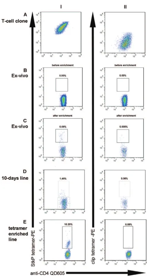
to a pneumococcal specific IFN-gamma secreting T cell clone from HV1 (figure 1A); this clone had been generated by its ability to secrete IFN-gamma in response to the StkP HLA-DRB1*1501 restricted epitope QSFQISNYVGRKSSD (StkP446–60).
Back-ground non-specific tetramer staining was determined using the HLA-DRB1*1501-CLIP tetramer which contains the CLIP peptide (PVSKMRMATPLLMQA), that associates with HLA-Class 2 molecules during antigen processing. We next determined whether we could detect pneumococcal specific T-cells in healthy HLA-DRB1*1501 expressing adults and, as shown in figure 1b, theex-vivofrequency was undetectable using PBMC from HV2. As ex-vivo epitope specific T-cell responses are often found at low frequencies, we enriched StkP446–60tetramer binding cells in HV2
using anti-PE magnetic beads as has been done previously [13]. By determining the absolute CD4 T-cell count, we were able to calculate the percentage StkP446–60-tetramer binding and were
able to show detectable ex-vivo StkP CD4+ T-cell responses following enrichment (figure 1C).
Proliferative and effector capacity of tetramer binding cells
After expanding StkP-specific T-cells further, by incubating PBMC from HV2 with StkP446–60 peptide for 10-days, the
proportions of StkP446–60-tetramer binding T-cells were increased
(figure 1D), which suggests that tetramer binding cells are able to proliferatein vitro. Approximately one thousand of these tetramer binding cells were sorted by flow cytometry and, after further rounds of expansion, there was a marked increase in the frequency and absolute numbers (.107 CD4 T-cells) of these StkP446–60
-tetramer-binding CD4+T-cells (figure 1E). This confirms that the tetramer binding CD4+ T-cells are capable of substantial proliferation in response to stimulation. In addition to their proliferative capacity, StkP446–60-tetramer-binding CD4+ T-cell
lines and clones were also able to secrete interferon-gamma. A StkP446–60-tetramer-binding CD4+ T-cell line and clone were
stimulated with both StkP446–60 peptide and recombinant StkP
and interferon gamma secretion was demonstrated using the ELISPot assay (figure 2).
Frequency and phenotype of pneumococcal specific T-cells
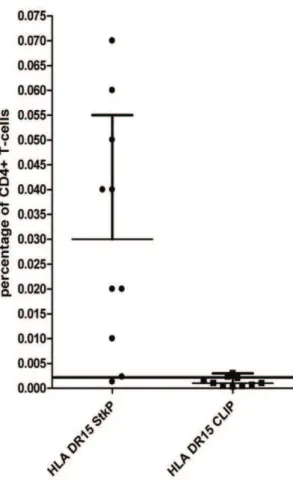
Using the StkP446–60-tetramer to enumerate the direct ex-vivo
frequency of StkP-specific CD4+T-cells, we found that 8 out of 10 healthy DRB1*1501 expressing adults had detectable responses (figure 3), with staining similar to that seen in HV2 after enrichment (figure 1C). Concomitant staining of cell surface proteins was used to determine the differentiation and activation status of these pneumococcal specific cells in these 8 healthy adults. The expression of CCR7 and CD45RA allows for the categorisa-tion of T-cells into central memory (CM), effector memory (EM), naive and a mature CCR7 negative, CD45RA positive subset (figure 4) [14,15]. The proportions of StkP446–60-tetramer binding
CD4+ T-cells were enriched within CD45RA+
CCR72 CD4+
T-cells and consequently reduced in CD45RA-CCR7+ CM CD4+T-cells (figure 4, p = 0.04 and p = 0.01, respectively). There was no significant difference when comparing the StkP446–60
-tetramer binding frequency of the other T-cell subsets to the tetramer negative population of T-cells, nor was there any difference when using the T-cell markers of differentiation, CD27 and CD28 (figure 4). Interestingly, using the expression of CD38 as a marker of T-cell activation, there was a higher frequency of CD38 expression in the StkP446–60-tetramer binding
cells than in the tetramer negative population (figure 4).
Discussion
This is the first report to use HLA-class 2 tetramers for the direct ex-vivo enumeration and characterisation of pneumococcal specific T-cells in healthy individuals. We have shown that 8 out of 10 HLA DRB1*1501 healthy adults have detectable StkP446–60
-tetramer binding CD4+T-cells with a range in frequency (0.01– 0.07%) similar to frequencies observed in other models of exposure to viral and intra-cellular bacterial infections as well as following desensitisation therapy in allergic disease [16,17,18,19]. HLA-class 2 tetramers identify CD4+cells irrespective of the T-cells ability to proliferate or secrete cytokine. The increased frequency of StkP446–60-tetramer binding in StkP stimulated lines
after 10 days expansion, as well as in StkP446–60-tetramer sorted
and subsequently expanded T-cell lines, indicates that these StkP446–60-tetramer binding CD4 T-cells were able to proliferate in vitro. Furthermore both StkP446–60-tetramer binding CD4 T-cell
lines and clones secreted IFN-gamma in response to the StkP446–60
peptide and recombinant StkP. Indeed, pneumococcal specific interferon-gamma responses have been detected in adenoidal tissue from children [20]. Thus, StkP446–60-tetramer binding cells
CD4+ T-cells have rapid effector function in terms of interferon gamma secretion and proliferation.
The identification of antigen specific T-cells by HLA-class 2-tetramers allows the direct characterisation of ex-vivo T-cells without altering their phenotype as occurs when using antigen-based activation assays. We determined the expression of cell surface markers, CD45RA, CCR7, CD27 and CD28 that have been used to categorise CD4+T-cells into distinct subsets. There is evidence that emergence of these distinct subsets of antigen experienced cells follows a progressive differentiation model. Accordingly, antigen experienced cells that express CCR7, CD27 and CD28 traffic to lymphoid tissue and are enriched with IL-2 secreting cells and represent early-differentiated cells. In contrast, as antigen experienced cells progress through sequential rounds of cell division, a substantial proportion will progressively lose CCR7, CD28 and CD27, with the eventual re-expression of CD45RA marking terminally differentiated cells [14,15]. The loss of CCR7 allows for these late-differentiated cells to home to inflamed peripheral tissue where they can directly participate in the effector immune response to infectious agents. Our demon-stration that HLA-class 2 tetramer binding cells are enriched in CCR7- CD45RA+ expression implies that they are terminally differentiated CD4+T-cells. We also found that CD38 expression was increased on StkP446–60-tetramer binding T-cells. CD38
expression is an activation marker, suggesting that the StkP446–
60-tetramer binding cells have recently encountered antigen. These
CD38 positive cells readily produce cytokine and are more likely to apoptose, a feature that is typical of effector T-cells [21].
We know that the prevalence of pneumococcal nasopharyngeal colonisation is low in adults, ranging between 4–5.9%, although we did not directly determine if any our healthy volunteers were colonised [2,22,23]. It is therefore likely that the activation of the StkP446–60-tetramer binding T-cells reflects recent
immunostimula-tory exposure to pneumococci. It is possible that these activated T-cells may be more than markers of pneumococcal exposure, being directly involved in preventing such exposure from progression to sustained nasopharyngeal invasion. The loss of CCR7 expression -which favours trafficking of T-cells to peripheral tissue – is also consistent with a direct role for these StkP446–60-tetramer binding
T-cells in pneumococcal immunity in the nasopharynx. Indeed, others have demonstrated that pneumococcal specific T-cell cytokine and proliferative responses are associated with nasopharyngeal pneu-mococcal sterility in children [24]. These subclinical, non (or very
Figure 1. Identifying pneumococcal specific T-cells with a Class 2 tetrameric complex.Dot plots showing tetramer binding of CD4+T-cells from various cellular sources. Column I shows binding of CD4+T-cells to the StkP446–60-HLA-DRB1*1501 tetrameric complexes and column II indicates
staining with a control CLIP DRB1*1501 tetramer. StkP446–60-tetramer and control tetramer binding of a known StkP446–60-specific IFN –gamma
secreting T cell clone from HV1 is shown in A. As StkP446–60-tetramer staining ofex-vivosamples (B) was not detectable in HV2, anti-PE magnetic
beads were used to enrich with consequent detection of StkP446–60-tetramer binding (C). StkP446–60-tetramer binding CD4+ T-cells were also
identified after 10 days expansion of HV2 PBMC with StkP446–60 peptide (D). This StkP446–60-tetramer binding population was sorted by flow
cytometry and further expandedin vitrofor 2 weeks with a subsequent further enrichment of StkP446–60-tetramer binding cells (E).
brief) colonising exposures may not only activate T-cells but may also drive their differentiation into the CD45RA re-expressing compartment.
We have used HLA-class 2 tetramers to show that healthy adults have detectable direct StkP446–60responses to pneumococci. Our
observation that these cells have been recently activated indicates that in healthy adults there is a dynamic T-cell response to presumed frequent exposures to pneumococci that do not progress to detectable nasopharyngeal colonisation or invasion. It would be of great interest to use Class 2 tetramers to enumerate and characterise the pneumococcal specific T-cell response during invasive pneumococcal infection. Given the increased risk of pneumococcal infection in the elderly, it would also be interesting to determine if there are any age-related changes in the frequency and phenotype of StkP446–60-tetramer binding cells, as occurs in
other situations of persistent/ongoing exposure [16,17].
Materials and Methods
Ethics Statement
The study was approved by Oxfordshire Research Ethics Committee (REC); all subjects gave informed consent.
Subjects
Ten healthy adult volunteers (HV1-10) who expressed HLA-DRB1*1501 were recruited. Peripheral blood mononuclear cells (PBMCs) were separated from heparinized peripheral blood by density gradient using Lymphoprep (Nycomed, Roskilde, Den-mark). PBMCs were then washed in RPMI supplemented with
penicillin, streptomycin and l-glutamine (R0) and resuspended in RPMI with penicillin, streptomycin, l-glutamine and 10% fetal calf serum (FCS; R10).
HLA Typing
All laboratory volunteers were HLA typed. Genomic DNA Puregene DNA isolation kit (Gentra Systems, USA) was used to isolate DNA from whole blood. HLA-A, -B, -C, DRB1, DRB3, DRB4, DRB5 and DQB1 specificities were determined using sequence specific primers by our in-house HLA typing service [25].
Antigens
The StkP446–60 peptide QSFQISNYVGRKSSD was
synthe-sized in-house in an automated synthesizer using 9-fluorenyl-methoxycarbonyl chemistry and the purity of the peptides was determined to be greater than 90% by high-performance liquid chromatography (Gilson, Middleton, WI, USA) analysis [26,27]. The purity of the peptide were confirmed by matrix-assisted laser desorption mass spectrometer on a Bruker Daltonics Ultraflex TOF/TOF mass spectrometer (Bruker, Billerica, MA, USA) [28].
T-cell lines and clones
An interferon-gamma secreting StkP446–60specific T-cell clone
that had been previously generated was used to confirm specificity Figure 2. StkP446–60-HLA-DRB1*1501-tetramer binding T-cells
secrete IFN-gamma.A tetramer binding T-cell line (unfilled colums) and a separate T-cell clone from HV2 were stimulated with StkP446–60
peptide (10 micromolar final concentration-vertical lines), recombinant StkP protein (10 ug/ml final concentration-chequered) or media (control-no pattern) for 16 hours. Interferon gamma secreting cells were enumerated by the ELISPot assay.
doi:10.1371/journal.pone.0025367.g002
Figure 3. Frequency of Pneumococcal specific T-cells. The percentage of StkP446–60-tetramer and the control CLIP-tetramer
binding was calculated in 10 healthy HLA-DRB1*1501 adults. The limit of detection (mean of the CLIP-tetramer binding +2SD) of specific
StkP446–60-tetramer binding after magnetic-bead enrichment is 0.002%
HV1-8 had detectable tetramer binding-i.e.frequencies greater than the limit of detection.
doi:10.1371/journal.pone.0025367.g003
of the StkP446–60 tetramer [12]. StkP446–60 specific T-cells were
expandedin vitroby incubating 46106PBMC in 2mls R10* in
24-well plates (Corning) for 10 days with StkP446–60 peptide (final
concentration 2mM). Interleukin-2 was added on days 3 and 7 at a concentration of 100 units/ml. All cell lines were maintained at 37uC, in 5% CO2.
A StkP446–60tetramer enriched T-cell line was generated by first
sorting StkP446–60-tetramer positive CD4+T-cells from a 10-days
expanded T-cell line. Approximately 1000 of these cells were sorted into a single well of a round-bottomed 96-well plate using a MoFlo cell sorter. 100ml of irradiated feeder cells – (1:1:1 of
PBMC from 3 different individuals) at 16106 cells/ml in R10*
containing IL-2 (100 IU/ml) and PHA (10mg/ml) were added to the well.
IFN-gamma ELISpot
ELISpot plates (Millipore Corp., Bedford, MA, USA) were coated with anti-human interferon (IFN)-c overnight (Mabtech
AB, Nacka, Sweden). The plates were washed six times with RPMI-1640 and blocked for 1 h with RPMI-1640 supplemented with 2 mM L-glutamine, 100 IU/ml penicillin and 100mg/ml plus 10% human serum (R10*). 40,000 T-cell blasts fromin vitro
Figure 4. Phenotype of Pneumococcal specific T-cells.The phenotype of tetramer binding T-cells HV1-8, who had detectable tetramer binding was evaluated using concomitant staining of T-cell surface markers. T-cell memory subsets were identified on the basis of CCR7, CD45RA, CD27 and CD28 surface expression, (an example of which is shown in A). The frequencies of central memory (CM), effector memory, naive and mature CD45RA+
CCR7- subsets were determined in the StkP446–60-tetramer binding and StkP446–60-tetramer-negative populations in 8 healthy adults (B). CD38
expression was also determined in the StkP446–60-tetramer binding and the StkP446–60-tetramer -negative CD4+T-cells (shown in HV3 in C, grey
outline is tetramer positive cells and black outline is tetramer negative CD4+T-cells). D shows CD38 expression in StkP446–60-tetramer positive (grey
filled column) and negative (unfilled) cells from 8 healthy adults with a significant difference in the mean expression (Wilcoxon signed rank test, p = 0.02).
expanded T-cell lines or 1000 T-cell clones were added to each well to which StkP446–60peptide (10 micromolar final
concentra-tion), recombinant StkP protein (10 ug/ml final concentration) or media (control) was added. Wells were set-up in duplicate. After overnight incubation at 37uC and 5% CO2, plates were washed
66 in PBS-Tween 0.05% and incubated with 1mg/ml of biotin-linked anti-IFN-c (Mabtech AB) for 2 hours. After washing66 in PBS-Tween 0.05%, the plates were incubated for a further 1 hour with streptavidin-alkaline phosphatase (Mabtech AB). Spots were visualized using an alkaline phosphatase conjugate substrate kit (Biorad, Hercules, CA, USA) and enumerated using an automated ELISpot reader. Results were expressed as spot-forming cells per total number of cells after subtracting the background (cells alone).
Tetramer staining
DRB1*1501 MHCII tetramer and hCLIP peptide HLA DRB1*1501 negative control tetramer were provided by the NIH Tetramer Core Facility at Emory University in Atlanta, GA, USA. DRB1*1501-PE tetramer was complexed to the StkP446–60peptide
QSFQISNYVGRKSSD, a previously defined HLA-DRB1*1501 restricted T-cell epitope [12] . Cell lines, T-cell clones and PBMC were incubated with 0.2mg/ml HLA class II tetramer for 120 min at 37uC in R10* before staining with cell surface marker antibodies at room temperature for 20 minutes, including: anti-CD3 pacific orange, anti-CD4-quantum dot 605, anti-CD45 quantum dot 655, anti-CD27-FITC, LIVE/DEADHFixable Violet Dead Cell Stain (Invitrogen, Carlsbad, CA, USA), CD14 pacific blue, anti-CD19 pacific blue (Biolegend, San Diego, CA, USA), anti-CCR7 Alexa647, anti-CD38 PercP Cy 5.5 and anti-CD28 PE Cy5 (Becton,
Dickinson and Company, Franklin Lakes, NJ,USA). Stained cells
were washed with phosphate-buffered saline (PBS) and fixed in 0N5% PBS/formaldehyde. Cells were acquired on a BDTMLSR II (BD) and analysed using FlowJo software (Tree Star, Inc. OR, USA). Gating strategy, singlet cells were first gated using FSC vs. FSC (area). Dead, CD14 and CD19 positive cells were then excluded using violet1 versus FSC.
Tetramer enrichment:
CD4+ T-cells were first enriched by negative selection using RosetteSepH (STEMCELL Technologies, France) from whole blood. The absolute CD4+ T-cell count was then determined using TrucountTM(BD) beads before staining with tetramer. The CD4+ T-cells were then incubated with anti-PE beads and positively selected using a magnet-based separation (Miltenyi Biotec, Germany). The tetramer enriched cells were then stained with antibodies directed to cell surface markers as mentioned above, before the entire sample was run through a BDTMLSR II
(BD). The percentage of CD4 T-cells that were tetramer binding was calculated by dividing the total number of tetramer positive events by the total number of CD4+ T-cells that had been enumerated prior to enrichment.
Statistics
Statistical tests were used to determine if the null hypothesis could be rejected at a probability of ,0.05. Non-parametric statistical tests were used; Wilcoxon signed rank test using the statistical software package GraphPad Prism 4.
Author Contributions
Conceived and designed the experiments: AA GO HC. Performed the experiments: AA. Analyzed the data: AA GO. Wrote the paper: AA GO.
References
1. O’Brien KL, Wolfson LJ, Watt JP, Henkle E, Deloria-Knoll M, et al. (2009) Burden of disease caused by Streptococcus pneumoniae in children younger than 5 years: global estimates. Lancet 374: 893–902.
2. Flamaing J, Peetermans WE, Vandeven J, Verhaegen J (2010) Pneumococcal colonization in older persons in a nonoutbreak setting. J Am Geriatr Soc 58: 396–398.
3. Bogaert D, De Groot R, Hermans PW (2004) Streptococcus pneumoniae colonisation: the key to pneumococcal disease. Lancet Infect Dis 4: 144–154. 4. (2010) Active Bacterial Core Surveillance Report, Emerging Infections
Pro-gram Network, Streptococcus pneumoniae, 2009. http://www.cdc.gov/abcs/ reports-findings/survreports/spneu09.pdf. Centers for Disease Control and Prevention.
5. McCool TL, Schreiber JR, Greenspan NS (2003) Genetic variation influences the B-cell response to immunization with a pneumococcal polysaccharide conjugate vaccine. Infect Immun 71: 5402–5406.
6. Soininen A, Pursiainen H, Kilpi T, Kayhty H (2001) Natural development of antibodies to pneumococcal capsular polysaccharides depends on the serotype: association with pneumococcal carriage and acute otitis media in young children. J Infect Dis 184: 569–576.
7. Goldblatt D, Hussain M, Andrews N, Ashton L, Virta C, et al. (2005) Antibody responses to nasopharyngeal carriage of Streptococcus pneumoniae in adults: a longitudinal household study. J Infect Dis 192: 387–393.
8. Lipsitch M, Whitney CG, Zell E, Kaijalainen T, Dagan R, et al. (2005) Are anticapsular antibodies the primary mechanism of protection against invasive pneumococcal disease? PLoS Med 2: e15.
9. Malley R, Lipsitch M, Bogaert D, Thompson CM, Hermans P, et al. (2007) Serum antipneumococcal antibodies and pneumococcal colonization in adults with chronic obstructive pulmonary disease. J Infect Dis 196: 928–935. 10. Khan AQ, Lees A, Snapper CM (2004) Differential regulation of IgG
anti-capsular polysaccharide and antiprotein responses to intact Streptococcus pneumoniae in the presence of cognate CD4+T cell help. J Immunol 172: 532–539.
11. Lu YJ, Gross J, Bogaert D, Finn A, Bagrade L, et al. (2008) Interleukin-17A mediates acquired immunity to pneumococcal colonization. PLoS Pathog 4: e1000159.
12. Aslam A, Mason A, Zemenides S, Chan H, Novakova L, et al. (2010) Rapid effector function of circulating CD4+T cells specific for immunodominant regions of the conserved serine/threonine kinase found in Streptococcus pneumoniae (StkP) in healthy adults. FEMS Immunol Med Microbiol 60: 113–122.
13. Scriba TJ, Purbhoo M, Day CL, Robinson N, Fidler S, et al. (2005) Ultrasensitive detection and phenotyping of CD4+T cells with optimized HLA class II tetramer staining. J Immunol 175: 6334–6343.
14. Appay V, van Lier RA, Sallusto F, Roederer M (2008) Phenotype and function of human T lymphocyte subsets: consensus and issues. Cytometry A 73: 975–983.
15. Amyes E, Hatton C, Montamat-Sicotte D, Gudgeon N, Rickinson AB, et al. (2003) Characterization of the CD4+T cell response to Epstein-Barr virus during primary and persistent infection. J Exp Med 198: 903–911.
16. Malavige GN, Jones L, Black AP, Ogg GS (2008) Varicella zoster virus glycoprotein E-specific CD4+T cells show evidence of recent activation and effector differentiation, consistent with frequent exposure to replicative cycle antigens in healthy immune donors. Clin Exp Immunol 152: 522–531. 17. Aslam A, Chan H, Warrell DA, Misbah S, Ogg GS (2010) Tracking
antigen-specific T-cells during clinical tolerance induction in humans. PLoS ONE 5: e11028.
18. Lucas M, Ulsenheimer A, Pfafferot K, Heeg MH, Gaudieri S, et al. (2007) Tracking virus-specific CD4+T cells during and after acute hepatitis C virus infection. PLoS ONE 2: e649.
19. Wei H, Wang R, Yuan Z, Chen CY, Huang D, et al. (2009) DR*W201/P65 tetramer visualization of epitope-specific CD4 T-cell during M. tuberculosis infection and its resting memory pool after BCG vaccination. PLoS ONE 4: e6905.
20. Zhang Q, Bernatoniene J, Bagrade L, Paton JC, Mitchell TJ, et al. (2006) Regulation of production of mucosal antibody to pneumococcal protein antigens by T-cell-derived gamma interferon and interleukin-10 in children. Infect Immun 74: 4735–4743.
21. Sandoval-Montes C, Santos-Argumedo L (2005) CD38 is expressed selectively during the activation of a subset of mature T cells with reduced proliferation but improved potential to produce cytokines. J Leukoc Biol 77: 513–521. 22. Gill CJ, Mwanakasale V, Fox MP, Chilengi R, Tembo M, et al. (2008) Impact of
human immunodeficiency virus infection on Streptococcus pneumoniae colonization and seroepidemiology among Zambian women. J Infect Dis 197: 1000–1005.
23. Regev-Yochay G, Raz M, Dagan R, Porat N, Shainberg B, et al. (2004) Nasopharyngeal carriage of Streptococcus pneumoniae by adults and children in community and family settings. Clin Infect Dis 38: 632–639.
24. Zhang Q, Bagrade L, Bernatoniene J, Clarke E, Paton JC, et al. (2007) Low CD4 T cell immunity to pneumolysin is associated with nasopharyngeal carriage of pneumococci in children. J Infect Dis 195: 1194–1202.
25. Bunce M, O’Neill CM, Barnardo MC, Krausa P, Browning MJ, et al. (1995) Phototyping : comprehensive DNA typing for HLA-A, B, C, DRB1, DRB3, DRB4, DRB5 & DQB1 by PCR with 144 primer mixes utilizing sequence-specific primers. Tissue Antigens 46: 355–367.
26. Chan WW, P (1999) Fmoc Solid Phase Peptide Synthesis: A Practical Approach. Oxford: Oxford University Press.
27. Coin I, Beyermann M, Bienert M (2007) Solid-phase peptide synthesis: from standard procedures to the synthesis of difficult sequences. Nat Protoc 2: 3247–3256.