Malagasy
Conostigmus
(Hymenoptera:
Ceraphronoidea) and the secret
of scutes
Istva´n Miko´1, Carolyn Trietsch1,*, Emily L. Sandall1,*,
Matthew Jon Yoder2, Heather Hines1,3and Andrew Robert Deans1 1Frost Entomological Museum, Department of Entomology, Pennsylvania State University,
University Park, PA, USA
2Illinois Natural History Survey, University of Illinois at Urbana-Champaign, Champaign, IL, USA
3Department of Biology, Pennsylvania State University, University Park, PA, USA
*These authors contributed equally to this work.
ABSTRACT
We revise the genusConostigmus Dahlbom1858 occurring in Madagascar, based on data from more specimens than were examined for the latest world revision of the genus. Our results yield new information about intraspecific variability and the nature of the atypical latitudinal diversity gradient (LDG) observed in Ceraphronoidea. We also investigate cellular processes that underlie body size polyphenism, by utilizing the correspondence between epidermal cells and scutes, polygonal units of leather-like microsculpture. Our results reveal that body size polyphenism in Megaspilidae is most likely related to cell number and not cell size variation, and that cell size differs between epithelial fields of the head and that of the mesosoma. Three species,Conostigmus ballescoracasDessart, 1997,C. babaiax Dessart, 1996 andC. longulusDessart, 1997, are redescribed. Females ofC. longulusare described for the first time, as are nine new species:C. bucephalusMiko´ and Trietsch sp. nov.,C. clavatusMiko´ and Trietsch sp. nov.,C. fianarantsoaensisMiko´ and Trietsch sp. nov.,C. lucidusMiko´ and Trietsch sp. nov.,C. macrocupula, Miko´ and Trietsch sp. nov.,C. madagascariensisMiko´ and Trietsch sp. nov.,C. missyhazenae Miko´ and Trietsch sp. nov.,C. pseudobabaiaxMiko´ and Trietsch sp. nov., and C. toliaraensisMiko´ and Trietsch sp. nov. A fully illustrated identification key for Malagasy Conostigmusspecies and a Web Ontology Language (OWL) representation of the taxonomic treatment, including specimen data, nomenclature, and phenotype descriptions, in both natural and formal languages, are provided.
Subjects Biodiversity, Bioinformatics, Entomology, Evolutionary Studies, Taxonomy
Keywords Microscopy, Morphology, Taxonomy, CLSM, LDG, Imaginal disks, Phenotypic plasticity, Male genitalia
INTRODUCTION
With 162 extant species,ConostigmusDahlbom, 1858 is the second most species-rich genus of Megaspilidae (Ceraphronoidea), a hymenopteran family showing a reverse latitudinal diversity gradient (LDG) in species richness (Johnson & Musetti, 2004;
Noyes, 1989). Since ceraphronoid faunistic and taxonomic studies mostly focus on the
Submitted25 July 2016 Accepted13 October 2016 Published13 December 2016 Corresponding author
Istva´n Miko´, izm2@psu.edu
Academic editor
Ilaria Negri
Additional Information and Declarations can be found on page 80
DOI10.7717/peerj.2682
Copyright
2016 Mikó et al.
Distributed under
Holarctic fauna, it is possible that sample bias is the reason for this atypical
distribution. This might be especially true forConostigmus, given that the only taxonomic review of the genus focused exclusively on non-Nearctic, non-European species (n = 36) and was based on only 145 specimens (Dessart, 1997). The large number of Malagasy specimens examined in the present study will not only double the number of specimens of non-Nearctic, non-European Conostigmusbut will also provide a reasonable data set for comparing the Malagasy fauna with that of a similarly sized area in Europe, the Atlantic Archipelago (British Isles;Broad & Livermore, 2014). Madagascar is considered a hotspot of biodiversity (Myers et al., 2000), and if reverse LDG is false and based on sample bias, we would be able to document more species in Madagascar than in the AA.
Ceraphronoids likely belong to the basal apocritan Evaniomorpha, and exhibit mostly ancestral sets of phenotypes (Heraty et al., 2011;Vilhelmsen, Miko´ & Krogmann, 2010). The complexity of the ceraphronoid ovipositor system and male genitalia is unparalleled among Apocritans (Miko´ et al., 2013;Ernst, Miko´ & Deans, 2013), and the leather-like microsculpture covering their head and mesosoma (Miko´, Yoder & Deans, 2011;Burks, Miko´ & Deans, 2016) is hypothesized to be an ancestral trait in Insecta (Hinton, 1970).
Besides the ten-fold interspecific body length variability from 0.37 mm
(Microceraphron subterraneusSzele´nyi, 1935) to 4.5 mm, (Megaspilus armatusSay, 1836), up to four-fold intraspecific variability has been reported in Ceraphronoidea (Mackauer & Chow, 2015;Dessart & Ga¨rdenfors, 1985;Liebscher, 1972). Similar intraspecific
variability is common among microhymenoptera and can be stimulated by alternative host species with different nutritional quality (Nalepa & Grisell, 1993;Medal & Smith, 2015) gregariousness with variable brood sizes (Harvey et al., 1998), and climatic differences, such as temperature (Wu et al., 2011). Ceraphronoids are parasitoids on insect parasitoid and predator larvae (Haviland, 1920;Withycombe, 1924;Kamal, 1939) and have a broad host range (Gilkeson, McLean & Dessart, 1993;Sullivan & Vo¨lkl, 1999). Dendrocerus carpenteri, for example, has been reared from >70 aphidiine (Braconidae) species (Fergusson, 1980). Based on the few studies with appropriate rearing conditions, gregariousness might also be not uncommon among Ceraphronoidea (Kamal, 1939;
Bennett & Sullivan, 1978;Mackauer & Chow, 2015;Cooper & Dessart, 1975;Dessart, 1997;
Liebscher, 1972).
Environmental factors impact development and determine final adult body size (Nijhout & Callier, 2015) by altering different cellular processes. Temperature and oxygen level usually impact cell size (Azevedo, French & Partridge, 2002;Harrison & Haddad, 2011;Heinrich et al., 2011), while nutrition mostly regulates cell division (Emlen, Lavine & Ewen-Campen, 2007).Conostigmusspecies are relatively large ceraphronoids (0.8–2.2 mm) making it feasible to observe and examine scutes (Meyer, 1842;Cals, 1974;Krell, 1994), elements of the aforementioned leather-like sculpture. Scutes likely have a one-to-one correspondence to epidermal cells in arthropods (Moretto, Minelli & Fusco, 2015;
Research related to geographic distribution and polyphenism requires a stable taxonomic framework. We revise the MalagasyConostigmus, Dahlbom, 1858 and use this system to explore the apparently anomalous ceraphronoid diversity patterns and possible reasons for body size polyphenism.
MATERIALS AND METHODS
Specimens for the present study (Table S1) were obtained from Malaise trap samples and were loaned to the authors from the California Academy of Sciences (CAS). Morphological characters were scored with an Olympus SZX16 stereo-microscope equipped with an Olympus SDF PLAPO 2XPFC objective, resulting in 230
magnification. Specimens are deposited in the CAS, in the Frost Entomological Museum (FEM) and in the Royal Museum of Central Africa (MRAC) (Table S1).
Brightfield images of dried specimens were taken with an Olympus BX43 compound microscope equipped with an Olympus DP73 digital camera. Image stacking was performed with Zerene Stacker (Version 1.04 Build T201404082055; Zerene Systems LLC, Richland, WA, USA) and extended focus images were annotated and modified with Adobe Photoshop 6TM(Adobe Systems, San Jose, CA, USA) using Adjust/Filter/Unsharp
mask and Image/Adjustments/Exposure (Gamma correction) tools.
Metasomata were removed from the specimens and placed in 35% H2O2for 20 min,
rinsed in distilled water for 30 min and dehydrated with 25 and 50% ethanol for 15–15 min, then transferred to a glycerol droplet on a concavity slide (Sail Brand Ltd., West Yorkshire, UK) and dissected. This protocol preserves muscle tissue while
bleaching melanized structures, making them transparent for confocal laser scanning microscopy (CLSM).
Sample preparation for CLSM followed Miko´ & Deans (2013): male genitalia were temporarily mounted between two coverslips (1.5mm, 2260) in a glycerin droplet, which did not reach the edge of the coverslip. We used Blu-tack (Bostik, Wauwatosa, WI, USA) as spacer as this material does not interact with glycerol and provides an adjustable, appropriate distance between the coverslips. Specimens were examined with an
Olympus FV10i desktop CLSM using a 60X objective.
Soft and sclerotized anatomical structures in arthropods tend to fluoresce with different intensities at different wavelength intervals (Miko´ & Deans, 2013). CLSM tissue-specific contrast is gained by exciting specimens using multiple excitation
wavelengths and/or recording the fluorescence on multiple channels assigned to different laser wavelength intervals. In previous research (Miko´ et al., 2013;Popovici et al., 2014;
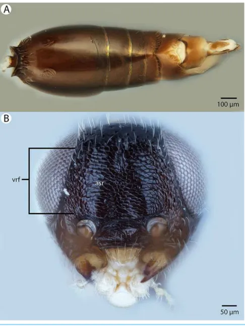
For the morphometric analysis on scute patterns, extended focal images of the frons and the mesocutellar-axillar complex of 14Conostigmus longulusDessart, 1997specimens were taken using an Olympus BX43 compound microscope equipped with an Olympus DP73 digital camera on 200magnification. Extended focal images were generated using the online “extended focal imaging” (efi) tool of an Olympus CellsensTMsoftware.
Measurements (Table S2) were taken using the same software. First, a 9,636mm2;
rectangular area was assigned on the extended focal images for recording scute pattern. The lateral vertices of the medially-positioned rectangle were adjacent with the
scutoscutellar sulcus on the mesonotum, while the rectangle was positioned medially on the frons with equal distance from the anterior ocellus and the intertorular carina. Scutes overlapping this area (including scutes adjacent to the margin of the rectangle) were counted and the longest diameter of each scute was measured. Measurements were taken on the images while constantly checking their accuracy on live view at 200–500 magnification (Figs. 2Aand2B). Body length largely depends on the relative orientations of the tagmata. The head of most species is flattened dorsoventrally and attached at its posterior end to the thorax (compare the position of the head onFigs. 31Aand31B). We used the interorbital distance (IOS,http://purl.obolibrary.org/obo/HAO_0000432) to infer body size in our statistical analysis.
Volume rendered CLSM micrographs and media files and scaleable vector (.msi) annotated extended focal images of the frons and mesoscutum, complete with scute measurements, IOS and rectangles can be accessed from Figshare (http://dx.doi.org/10. 6084/m9.figshare.3848544;http://dx.doi.org/10.6084/m9.figshare.3848547.v1).
Abbreviations of anatomical structures used in Figures are listed in Table S3. Intraspecific variation in scute diameter and relative size was scrutinized by linear regression analyses. Intra-individual variation was scrutinized via Mann-Whitney sum-rank test (Mann & Whitney, 1947). The relationship between morphometric variables was inspected through linear regression analyses. Statistical analyses were carried out and Boxplots were generated in R version 3.2.2 (R Development Core Team, 2015) (Fig. S1).
Figure 2 Brightfield image showing the median region of the cranium ofConostigmus longulus
Dessart, 1997, anterior view. The measured scute lengths and values and borders of measured
Taxonomic nomenclature, specimen information, OTU concepts, original brightfield images and natural language (NL) phenotype representations were compiled in mx (http://purl.org/NET/mx-database). Taxonomic history, description,
Table 1 Mesoscutellar average scute size, length and scute number inConostigmus longulusDessart,
1997.
Mesoscutellum ID aCS mCL nS
2041918 119.02 17.13 81
2003474 109.56 17.22 88
2044193 107.12 14.52 90
2044825 101.48 15.19 95
2040900 101.48 16.01 95
2009756 101.48 16.01 95
2002193 101.48 16.01 95
2044755 94.52 14.09 102
2040771 91.82 13.02 105
2053554 90.95 13.635 106
Median 101.48 15.60 95
Mean 101.89 15.28 95.2
SD 2.188891 1.437595 7.743097
Note:
ID, CASENT identifier for specimen; aCS, average cell size inmm2; mCL, median cell length inmm; nS, number of
scutes (cells) in a rectangular area of 9,636mm2.
Table 2 Frontal average scute size, length and scute number inConostigmus longulusDessart, 1997.
ID IOS aCS mCL nS
2003474 182.91 97.38 12.78 99
2009756 157.42 98.38 13.655 98
2040771 174.12 98.38 12.195 98
2040900 171.04 98.38 13.12 98
2002193 251.06 133.90 16.195 72
2053688 195.22 100.43 13.695 96
2053554 189.95 97.38 13.55 99
2053308 175.44 99.39 12.32 97
2046100 208.85 93.60 11.43 103
2041918 213.25 95.46 12.68 101
2044193 263.82 95.46 13.37 101
2044511 138.51 93.60 13.02 103
2044755 171.04 97.38 12.75 99
2044825 255.02 94.52 12.82 102
Median 186.4 97.38 12.92 99
Mean 196.3 99.55 13.11 97.57
SD 37.95319 10.10979 1.081914 7.673273
Note:
ID, CASENT identifier for specimen; IOS, interorbital space (referring to body size) inmm; aCS, average scute (cell)
size inmm2; mCL: median cell length inmm; nS, number of scutes (cells) in a rectangular area of 9,636mm2. Note the
and material examined sections (Table S1) were rendered from the same software. Terminology of the phenotype statements used in descriptions, identification key and diagnoses are mapped to the Hymenoptera Anatomy Ontology (HAO, available at
http://hymao.org), Phenotypic Quality Ontology (PATO, available at http://purl. obolibrary.org/obo/pato.owl), Biospatial Ontology (BSPO, available at http://purl. obolibrary.org/obo/bspo.owl) and Common Anatomy Reference Ontology (CARO, available at http://obofoundry.org/).
Natural language phenotypes are represented in “Entity attribute: value” format. Semantic statements for phenotype descriptions were created in Prote´ge´ 5.0.0-beta-16 (http://protege.stanford.edu/) using the Web Ontology Language (OWL) Manchester syntax (http://www.w3.org/TR/owl2-manchester-syntax/) followingBalhoff et al. (2013),
Miko´ et al. (2015) andMiko´ et al. (2014) (Table S4). The OWL (http://www.w3. org/TR/owl2-overview/; accessed 4 February 2014) representation of the full data set was deposited as a single Resource Description Framework (RDF)-XML file (http://www.w3.org/TR/REC-rdf-syntax/; accessed 12 March) in the Github repository (https://github.com/hymao/hymao-data/blob/master/miko2016_malagasy.owl).
The electronic version of this article in Portable Document Format (PDF) will represent a published work according to the International Commission on Zoological Nomenclature (ICZN), and hence the new names contained in the electronic version are effectively published under that Code from the electronic edition alone. This published work and the nomenclatural acts it contains have been registered in ZooBank, the online registration system for the ICZN. The ZooBank LSIDs (Life Science Identifiers) can be resolved and the associated information viewed through any standard web browser by appending the LSID to the prefixhttp://zoobank.org/. The LSID for this publication is: LSID: urn:lsid:zoobank.org:pub:41133330-D364-4ABF-AFB3-DEE013E0EDF9. The online version of this work is archived and available from the following digital repositories: PeerJ, PubMed Central and CLOCKSS.
RESULTS
Body size polyphenism in Conostigmus longulus
Mikó and Trietsch sp. nov.
Intraspecific variation in scute diameter and relative scute size
Fourteen specimens were measured and are represented in analysis of frons measurement distribution. Ten of these specimens were also measured on the mesoscutellum and make up the mesoscutellar analysis. Four specimens could not be measured for both regions due to inaccessibility of all scutes required for mesoscutellar measurements (i.e., the specimen preparation obscured these parts). One specimen was found to have measurements four standard deviations from the mean, and was removed from
Measurements were tested for normalcy and found to not follow the normal distribution, even after removal of the outlier from analyses. The scutes in a 9,636mm2
rectangular region of the frons and mesoscutellum were counted and measured. It was found that maximum scute length varied from 6.6 to 19.5mm. on the frons
(Table 1). On the mesoscutellum, maximum scute length varied from 8.8 to 23.4mm
(Table 2). Median cell length of each specimen was used as a variable in statistical analyses. Linear regression analyses were carried out independently on both the frons and mesoscutellar fields. There was a weak negative correlation between median scute length and scute number in the frons region (R2= 0.1369023). In the mesoscutellar region, scute length had a stronger negative correlation with scute number (R2 = 0.7149943).
Intraindividual variation in scute size: frons vs. mesoscutellum
In individuals where average scute size and diameter were measured on both the frons and the mesoscutellum, we found a variation in median cell length ranging from 11.43 to 17.22mm. Wilcoxon rank-sum test revealed a significant difference in cell length between
the frons and mesoscutellar regions (p-value = 0.0004011). Measurements of average scute size varied from 90.95 to 119.02mm2. There was no significant difference in average
scute size between the frons and mesoscutellar regions in this sample when analyzed by Wilcoxon rank-sum test (p-value = 0.0809).
Body size vs. scute size
Interorbital space was used as a measure of body size for all 14 specimens examined. Linear regression analysis for the relationship between scute size and body size was carried
140 160 180 200 220 240 260
96
97
98
99
100
101
102
103
IOS
Number of Cells
out using measurements for interorbital space and the measurements of average frons scute size, which were available for all measured specimens (Fig. 3). Correlation
between median cell length and IOS was extremely weak and negative (R2= 0.001341984). The correlation between cell number and IOS was much stronger and weakly positive (R2= 0.1114898).
Taxonomic treatment of MalagasyConostigmus
ConostigmusDahlbom, 1858
Diagnosis:It is not possible to provide a diagnosis for all megaspiline species that are currently classified as Conostigmus. The present treatment includes those Malagasi megaspilid specimens that have a sternaulus, an OOL longer than POL, a mesosoma that is higher than wide and lacks a bifurcated anteromedian projection of the metanoto-propodeo-metapecto-mesopectal complex, a harpe, which is independent from the gonostyle and/or whose parossiculi are discontinuous medially and are independent from the gonostyle.Megaspilushas bifurcated anteromedian projection of the metanoto-propodeo-metapecto-mesopectal complex;DendrocerusRatzeburg andCreatorAlekseev has OOL shorter than POL and medially continuous parossiculi that are continuous with gonostyle and lack sternaulus;Trichosteresis Fo¨rster has a harpe that is fused with the gonostyle and the mesosoma is wider than high in PlatyceraphronKieffer.
Conostigmus babaiaxDessart, 1997
Figures 4,5and6
Diagnosis:Conostigmus babaiaxDessart, 1996 shares the presence of a prognathous head (dorsal-most point of occipital carina is dorsal to posterior ocellus in lateral view) and the presence of transverse scutes on the ventral region of frons withConostigmus longulus
Dessart, 1997,C. toliaraensissp. nov. andC. pseudobabaiaxsp. nov.Conostigmus babaiax, C. toliaraensissp. nov. andC. pseudobabaiaxdiffer from all otherConostigmusspecies by the presence of ventromedian and ventrolateral white, setiferous patches on the frons. The lateral ocellar line (LOL) is longer than ocular ocellar line (OOL) inConostigmus babaiax and the LOL is shorter than OOL inC. toliaraensissp. nov. andC. pseudobabaiaxsp. nov.
Description: Body length: 2,200mm. Color intensity pattern: NOT CODED. Color hue
pattern: scape, pedicel, F1–F3, head, anterior mesosoma ochre, F4–F9, posterior
mesosoma, metasoma brown, legs except darker proximal regions of meso and metacoxae yellow. Occipital carina sculpture: smooth. Median flange of occipital carina count:
absent. Submedial flange of occipital carina count: absent. Dorsal margin of occipital carina vs. dorsal margin of lateral ocellus in lateral view: occipital carina is dorsal to lateral ocellus in lateral view. Preoccipital lunula count: NOT CODED. Preoccipital carina count: absent. Preoccipital carina shape: NOT CODED. Preoccipital furrow count: present. Preoccipital furrow anterior end: Preoccipital furrow ends inside ocellar triangle. Postocellar carina count: absent. Male OOL: posterior ocellar line (POL): LOL: NOT CODED. Female OOL: POL: LOL: 0.85:0.85:1.00. Head width vs. interorbital space (HW/IOS) Male: NOT CODED. HW/IOS Female: 2.65. Setal pit on vertex size: smaller than diameter of scutes. Transverse frontal carina count: absent. Transverse scutes on frons count: present. Rugose region on frons count: absent. Randomly sized areolae around
setal pits on frons count: absent. Antennal scrobe count: absent. Ventromedian setiferous patch and ventrolateral setiferous patch count: present. Facial pit count: no external corresponding structure present. Supraclypeal depression count: absent. Supraclypeal depression structure: NOT CODED. Intertorular carina count: present. Intertorular area count: present. Median region of intertorular area shape: flat. Ventral margin of antennal rim vs. dorsal margin of clypeus: not adjacent. Torulo-clypeal carina count: absent. Subtorular carina count: absent. Mandibular tooth count: 2. Female flagellomere one length vs. pedicel: 1.09. Female ninth flagellomere length: F9 less than F7 + F8. Sensillar patch of the male flagellomere pattern: NOT CODED. Length of setae on male
flagellomere vs. male flagellomere width: NOT CODED. Male flagellomere one length vs. male second flagellomere length: NOT CODED. Male flagellomere one length vs. pedicel length: NOT CODED. Ventrolateral invagination of the pronotum count: present. Scutes on posterior region of mesoscutum and dorsal region of mesoscutellum convexity: flat. Notaulus posterior end location: adjacent to transscutal articulation. Median mesoscutal sulcus posterior end: not adjacent to transscutal articulation (ends anterior to transscutal articulation). Scutoscutellar sulcus vs. transscutal articulation: adjacent. Axillular carina count: absent. Axillular carina shape: NOT CODED. Epicnemium posterior margin shape: anterior discrimenal pit absent; epicnemial carina interrupted medially. Epicnemial carina count: present only laterally. Sternaulus count: absent. Sternaulus length: NOT CODED. Speculum ventral limit: not extending ventrally of pleural pit line. Mesometapleural sulcus count: present. Metapleural carina count: present. Transverse line of the metanotum-propodeum vs. antecostal sulcus of the first abdominal tergum: adjacent sublaterally. Lateral propodeal carina count: present. Lateral propodeal carina shape: inverted “U” (left and right lateral propodeal carina are adjacent to the antecostal sulcus of the first abdominal tergumsubmedially). Anteromedian projection of the metanoto-propodeo-metapecto-mesopectal complex count: absent. S1 length vs. shortest width: S1 wider than long. Transverse carina on petiole shape: concave. Distal margin of male S9 shape: NOT CODED. Proximolateral corner of male S9 shape: NOT CODED. Cupula length vs. gonostyle-volsella complex length: NOT CODED. Proximodorsal notch of cupula count: NOT CODED. Proximodorsal notch of cupula shape: NOT CODED. Proximolateral projection of the cupula shape: NOT CODED. Proximodorsal notch of cupula width vs. length: NOT CODED. Distodorsal margin of cupula shape: NOT CODED. Dorsomedian conjunctiva of the gonostyle-volsella complex length relative to length of gonostyle-gonostyle-volsella complex: NOT CODED. Dorsomedian conjunctiva of the gonostyle-volsella complex count: NOT CODED. Distal end of dorsomedian conjunctiva of the gonostyle-volsella complex shape: NOT CODED. Parossiculus count (parossiculus and gonostipes fusion): NOT CODED. Apical
harpe orientation: NOT CODED. Distal margin of harpe in lateral view: shape: NOT CODED. Lateral margin of harpe shape: NOT CODED.
Material examined:Holotype female: MADAGASCAR: PSUC_FEM 000006723, COLL. MUS. Congo Madagascar: Mandraka II-1944 A. Seyrig HOLOTYPUS Holotype Prep. micros-copique n 9508/051 (deposited in MRAC).
Conostigmus ballescoracas Dessart, 1997
Figures 7,8and9
Diagnosis:Conostigmus ballescoracasDessart, 1997differs from otherConostigmusspecies by the presence of a strong preoccipital carina that is continuous with the orbital carina,
the presence of randomly sized areolae around the setal pits on the frons (shared with the ceraphronidMasner lubomirusMiko´ & Deans, 2013) and the posteromedially adjacent axillular carinae (left and right axillar carinae continuous posteriorly forming a U-shaped carina that surrounds the mesoscutellar disc).
Description: Body length: 1,650–1,875mm. Color intensity pattern: NOT CODED.
Color hue pattern: Cranium black; mesosoma, metasoma, F4–F9 brown; rest of antenna, legs and mandible ochre; Cranium brown; mesosoma except legs, metasoma ochre; F4–F9 brown; rest of antenna ochre, legs yellow. Occipital carina sculpture: crenulate. Median flange of occipital carina count: absent. Submedial flange of occipital
carina count: absent. Dorsal margin of occipital carina vs. dorsal margin of lateral ocellus in lateral view: occipital carina is ventral to lateral ocellus in lateral view. Preoccipital lunula count: present. Preoccipital carina count: present. Preoccipital carina shape: complete. Preoccipital furrow count: present. Preoccipital furrow anterior end: Preoccipital furrow ends inside ocellar triangle. Postocellar carina count: absent. Male OOL: POL: LOL: NOT CODED. Female OOL: POL: LOL: 2.5–3.0:1.9–2.0:1.0. HW/IOS Male: NOT CODED. HW/IOS Female: 1.7–1.8. Setal pit on vertex size: smaller than diameter of scutes. Transverse frontal carina count: absent. Transverse scutes on frons count: absent. Rugose region on frons count: absent. Randomly sized areolae around setal pits on frons count: present. Antennal scrobe count: absent. Ventromedian setiferous
patch and ventrolateral setiferous patch count: absent. Facial pit count: facial pit present. Supraclypeal depression count: absent. Supraclypeal depression structure: NOT CODED. Intertorular carina count: present. Intertorular area count: present. Median region of intertorular area shape: convex. Ventral margin of antennal rim vs. dorsal margin of clypeus: adjacent. Torulo-clypeal carina count: absent. Subtorular carina count: absent. Mandibular tooth count: 2. Female flagellomere one length vs. pedicel: 0.8-0.9. Female
ninth flagellomere length: F9 less than F7 + F8. Sensillar patch of the male flagellomere pattern: NOT CODED. Length of setae on male flagellomere vs. male flagellomere width: NOT CODED. Male flagellomere one length vs. male second flagellomere length: NOT CODED. Male flagellomere one length vs. pedicel length: NOT CODED. Ventrolateral invagination of the pronotum count: present. Scutes on posterior region of mesoscutum and dorsal region of mesoscutellum convexity: flat. Notaulus posterior end location: adjacent to transscutal articulation. Median mesoscutal sulcus posterior end: adjacent to transscutal articulation. Scutoscutellar sulcus vs. transscutal articulation: adjacent. Axillular carina count: present. Axillular carina shape: left and right carina continuous posteromedially forming a U-shape carina on the mesoscutellar axillar complex. Epicnemium posterior margin shape: anterior discrimenal pit present;
epicnemial carina curved. Epicnemial carina count: complete. Sternaulus count: present. Sternaulus length: elongate, exceeding 3/4 of mesopleuron length at level of sternaulus. Speculum ventral limit: not extending ventrally of pleural pit line. Mesometapleural sulcus count: present. Metapleural carina count: present. Transverse line of the metanotum-propodeum vs. antecostal sulcus of the first abdominal tergum: adjacent sublaterally. Lateral propodeal carina count: present. Lateral propodeal carina shape: NOT CODED. Anteromedian projection of the metanoto-propodeo-metapecto-mesopectal complex count: present. S1 length vs. shortest width: S1 wider than long. Transverse carina on petiole shape: straight. Distal margin of male S9 shape: NOT CODED. Proximolateral corner of male S9 shape: NOT CODED. Cupula length vs. gonostyle-volsella complex length: NOT CODED. Proximodorsal notch of cupula count: NOT CODED.
Material examined:Holotype female: CONGO: PSUC_FEM 8883 Congo Belge: P.N.A 7-XIII-1953 H. Synave 6853 Massif Ruwenzori Mont Ngulingo pres Nyamgaleke, 2,500 m, ex P.N.A HOLOTYPE Prep. micros-copique n 9507/241 (deposited in MRAC).
Other material (2 females): MADAGASCAR: 2 females. CASENT 2001391, 2016542 (CAS).
Conostigmus bucephalus Miko´ and Trietsch sp. nov.
Figures 10,11and12
Diagnosis (female):Conostigmus bucephalussp. nov. differs from other Conostigmus species in the presence of the antennal scrobe and the size of impressions around the setal pits on the head: impressions are larger than scutes on the cranium and mesonotum
in Conostigmus bucephalussp. nov. whereas in other Malagasy species depressions are smaller than scutes on the cranium and mesonotum.
Description:Body length: 2,575mm. Color intensity pattern: distal scape, legs except hind
coxa lighter than metasoma. Color hue pattern: Cranium, mesosoma brown; antenna, legs except brown metacoxa, metasoma ochre. Occipital carina sculpture: crenulate. Median flange of occipital carina count: absent. Submedial flange of occipital carina count: absent. Dorsal margin of occipital carina vs. dorsal margin of lateral ocellus in lateral view: occipital carina is dorsal to lateral ocellus in lateral view. Preoccipital lunula count: absent. Preoccipital carina count: absent. Preoccipital carina shape: NOT CODED. Preoccipital furrow count: present. Preoccipital furrow anterior end: Preoccipital furrow ends posterior to ocellar
triangle. Postocellar carina count: absent. Male OOL: POL: LOL: NOT CODED.
Female OOL: POL: LOL: 1.0:1.2:1.0. HW/IOS Male: NOT CODED. HW/IOS Female: 2.2. Setal pit on vertex size: larger than diameter of scutes. Transverse frontal carina count: absent. Transverse scutes on frons count: present. Rugose region on frons count: absent. Randomly sized areolae around setal pits on frons count: absent. Antennal scrobe count: present. Ventromedian setiferous patch and ventrolateral setiferous patch count: absent. Facial pit count: no external corresponding structure present. Supraclypeal depression count: present. Supraclypeal depression structure: present medially, inverted U-shaped.
Intertorular carina count: present. Intertorular area count: absent. Median region of intertorular area shape: NOT CODED. Ventral margin of antennal rim vs. dorsal margin of
clypeus: adjacent. Torulo-clypeal carina count: absent. Subtorular carina count: absent. Mandibular tooth count: 2. Female flagellomere one length vs. pedicel: 0.7. Female ninth flagellomere length: F9 less than F7 + F8. Sensillar patch of the male flagellomere pattern: NOT CODED. Length of setae on male flagellomere vs. male flagellomere width: NOT CODED. Male flagellomere one length vs. male second flagellomere length: NOT CODED. Male flagellomere one length vs. pedicel length: NOT CODED. Ventrolateral invagination of the pronotum count: present. Scutes on posterior region of mesoscutum and dorsal region of mesoscutellum convexity: flat. Notaulus posterior end location: adjacent to transscutal articulation. Median mesoscutal sulcus posterior end: adjacent to transscutal articulation. Scutoscutellar sulcus vs. transscutal articulation: adjacent. Axillular carina count: absent. Axillular carina shape: NOT CODED. Epicnemium posterior margin shape: anterior discrimenal pit absent; epicnemial carina interrupted medially. Epicnemial carina count: present only laterally. Sternaulus count: absent.
Sternaulus length: NOT CODED. Speculum ventral limit: not extending ventrally of pleural pit line. Mesometapleural sulcus count: present. Metapleural carina count: present. Transverse line of the metanotum-propodeum vs. antecostal sulcus of the first abdominal tergum: adjacent sublaterally. Lateral propodeal carina count: present. Lateral propodeal carina shape: inverted “Y” (left and right lateral propodeal are adjacent medially posterior to antecostal sulcus of the first abdominal tergum, and connected to the antecostal sulcus by a median carina representing the median branch of the inverted “Y”). Anteromedian projection of the metanoto-propodeo-metapecto-mesopectal complex count: absent. S1 length vs. shortest width: S1 wider than long. Transverse carina on petiole shape: concave. Distal margin of male S9 shape: NOT CODED. Proximolateral corner of male S9 shape: NOT CODED. Cupula length vs. gonostyle-volsella complex length: NOT CODED.
Proximodorsal notch of cupula count: NOT CODED. Proximodorsal notch of cupula shape: NOT CODED. Proximolateral projection of the cupula shape: NOT CODED. Proximodorsal notch of cupula width vs. length: NOT CODED. Distodorsal margin of cupula shape: NOT CODED. Dorsomedian conjunctiva of the gonostyle-volsella complex length relative to length of gonostyle-volsella complex: NOT CODED. Dorsomedian conjunctiva of the gonostyle-volsella complex count: NOT CODED. Distal end of dorsomedian conjunctiva of the gonostyle-volsella complex shape: NOT CODED. Parossiculus count (parossiculus and gonostipes fusion): NOT CODED. Apical parossiculal seta number: NOT CODED. Distal projection of the parossiculus count: NOT CODED. Distal projection of the penisvalva count: NOT CODED. Dorsal apodeme of penisvalva count: NOT CODED. Harpe length: NOT CODED. Distodorsal setae of sensillar ring of harpe length vs. harpe width in lateral view: NOT CODED. Distodorsal setae of sensillar ring of harpe orientation:
NOT CODED. Sensillar ring area of harpe orientation: NOT CODED. Lateral setae of harpe count: NOT CODED. Lateral setae of harpe orientation: NOT CODED. Distal margin of harpe in lateral view: shape: NOT CODED. Lateral margin of harpe shape: NOT CODED.
Etymology: The species epithetbucephalus(Ancient Greek: bou′"’ao& = ox-head)
Comments:Due to the antennal scrobe that accommodates the scape for almost its entire length, the head is nearly cube shaped in lateral view (Fig. 10B).
Material examined:Holotype female: CASENT 2053589 MADAGASCAR: Province Fianarantsoa, Parc National Ranomafana, radio tower at forest edge, elev 1,130 m 20 March–3 April 2003 2115.05′S, 4724.43′E collector: R. HarinH´ ala California Acad of Sciences malaise, mixed tropical forest MA-02-09B-56 (deposited in CAS).
Conostigmus clavatusMiko´ and Trietsch sp. nov.
Figures 13,14,15,16and17
Diagnosis:Conostigmus clavatus sp. nov. shares the presence of the axillular carina, bulging eyes and medially convex intertorular area (and intertorular carina) with
C. uninasutusAlekseev, 1994 andC. binasutusDessart & Cancemi, 1987 and differs from them in the enlarged distal-most female flagellomere (length of F9 = length of F6 + length of F7 + length of F8,Fig. 16A).
Description: Body length: 2,325–2,500mm. Color intensity pattern: metasoma lighter
than mesosoma and cranium. Color hue pattern: Dark brown except pedicel, proximal 1/5th of scape, fore and middle leg, mandible ochre/yellowish. Occipital carina sculpture: crenulate. Median flange of occipital carina count: present. Submedial flange of
occipital carina count: present. Dorsal margin of occipital carina vs. dorsal margin of lateral ocellus in lateral view: occipital carina is ventral to lateral ocellus in lateral view. Preoccipital lunula count: present. Preoccipital carina count: present; absent. Preoccipital carina shape: interrupted dorsally and represented by irregular, not continuous carinae.
Preoccipital furrow count: present. Preoccipital furrow anterior end: Preoccipital furrow ends posterior to ocellar triangle. Postocellar carina count: absent. Male OOL: POL: LOL: 2.9–3.6:2.1–2.2:1. Female OOL: POL: LOL: 3.4:2.1–2.2:1.0. HW/IOS Male: 1.6–1.7. HW/IOS Female: 1.6–1.7. Setal pit on vertex size: smaller than diameter of scutes. Transverse frontal carina count: absent. Transverse scutes on frons count: absent. Rugose region on frons count: present; absent. Randomly sized areolae around setal pits on frons count: absent. Antennal scrobe count: absent. Ventromedian setiferous patch and ventrolateral setiferous patch count: absent. Facial pit count: median facial keel present. Supraclypeal depression count: present. Supraclypeal depression structure: absent medially, represented by two grooves laterally of facial pit. Intertorular carina count: present. Intertorular area count: present. Median region of intertorular area shape:
convex. Ventral margin of antennal rim vs. dorsal margin of clypeus: not adjacent. Torulo-clypeal carina count: absent. Subtorular carina count: present. Mandibular tooth count: 2. Female flagellomere one length vs. pedicel: 0.9. Female ninth flagellomere length: F9 = F6 + F7 + F8. Sensillar patch of the male flagellomere pattern: F5–F9. Length of setae on male flagellomere vs. male flagellomere width: setae shorter than width of flagellomeres. Male flagellomere one length vs. male second flagellomere length: 1.2–1.3. Male flagellomere one length vs. pedicel length: 2.1–2.4. Ventrolateral invagination of the pronotum count: present. Scutes on posterior region of mesoscutum and dorsal region of mesoscutellum convexity: flat. Notaulus posterior end location: adjacent to transscutal articulation. Median mesoscutal sulcus posterior end: adjacent to transscutal articulation. Scutoscutellar sulcus vs. transscutal articulation: adjacent. Axillular carina
count: present. Axillular carina shape: The left and right carina are separated
posteromedially. Epicnemium posterior margin shape: anterior discrimenal pit present; epicnemial carina curved. Epicnemial carina count: complete. Sternaulus count: present. Sternaulus length: short, not reaching 1/2 of mesopleuron length at level of sternaulus. Speculum ventral limit: not extending ventrally of pleural pit line. Mesometapleural sulcus count: present. Metapleural carina count: present. Transverse line of the metanotum-propodeum vs. antecostal sulcus of the first abdominal tergum: adjacent sublaterally. Lateral propodeal carina count: present. Lateral propodeal carina shape: NOT CODED. Anteromedian projection of the metanoto-propodeo-metapecto-mesopectal complex count: present. S1 length vs. shortest width: S1 wider than long. Transverse carina on petiole shape: straight. Distal margin of male S9 shape: convex. Proximolateral corner of male S9 shape: acute. Cupula length vs. gonostyle-volsella complex length: cupula less than 1/2 the length of gonostyle-volsella complex in lateral view. Proximodorsal notch of cupula count: present. Proximodorsal notch of cupula shape: arched. Proximolateral projection of the cupula shape: acute. Proximodorsal notch of cupula width vs. length: as long as wide. Distodorsal margin of cupula shape: concave. Dorsomedian conjunctiva of the gonostyle-volsella complex length relative to length of gonostyle-volsella complex: dorsomedian conjunctiva extending 2/3 of length of gonostyle-volsella complex in dorsal view. Dorsomedian conjunctiva of the gonostyle-volsella complex count: present. Distal end of dorsomedian conjunctiva of the
Figure 17 CLSM volume rendered micrographs showing the male genitalia ofConostigmus clavatus
gonostyle-volsella complex shape: blunt. Parossiculus count (parossiculus and gonostipes fusion): present (not fused with the gonostipes). Apical parossiculal seta number: one; two. Distal projection of the parossiculus count: absent. Distal projection of the penisvalva count: absent. Dorsal apodeme of penisvalva count: absent. Harpe length: harpe shorter than gonostipes in lateral view. Distodorsal setae of sensillar ring of harpe length vs. harpe width in lateral view: setae as long or shorter than harpe width. Distodorsal setae of sensillar ring of harpe orientation: distomedially. Sensillar ring area of harpe orientation: medially. Lateral setae of harpe count: present. Lateral setae of harpe orientation: oriented distally. Distal margin of harpe in lateral view: shape: blunt. Lateral margin of harpe shape: widest point of harpe is at its articulation site with gonostyle-volsella complex.
Etymology:The species epithetclavatusrefers to the enlarged apical female flagellomere, resembling a club (F9 > F8 + F7 + F6).
Comments:Conostigmus clavatus,C. binasutusandC. uninasutusshare numerous morphological traits withMegaspilusWestwood, 1929 including the presence of bulging eyes, the crenulate and distinct ocular suture, the presence of the axillular carina and the large body size (>2,000mm). Females of allConostigmusspecies exhibit a distinct clava
with three rows of ventral, female specific basiconic sensilla (distally gradually widening flagellum). The antenna of femaleMegaspilusis filiform and lacks ventral basiconic sensilla (I. Miko´, 2010, personal observation).
Material examined:Holotype male: MADAGASCAR: Province Fianarantsoa, Parc National Ranomafana, Vohiparara at broken bridge, Malaise trap in high altitude rainforest, 22–28.11.2001, R. Harin’Hala, CASENT 2044514 (deposited in CAS). Paratypes (seven males, four females): MADAGASCAR: seven males, four females. CASENT 2002179, 2032775, 2044150, 2045085, 2045509, 2045602, 2045755, 2046024, 2046178–2046179, 2053642 (deposited in CAS, MRAC).
Conostigmus fianarantsoaensisMiko´ and Trietsch sp. nov.
Figures 18,19,20and21
Diagnosis:Conostigmus fianarantsoensissp. nov. is most similar to C. madagascariensis sp. nov. among MalagasyConostigmusand differs from it by the following characters: mandible with one tooth (mandible with two teeth inC. madagascariensis); flagellar setae shorter than the flagellomere width (inC. madagascariensis, flagellar setae are distinctly longer than the flagellomere width); blunt proximolateral projection of cupula (acute in C. madagascariensis); V-shaped (notched) proximodorsal notch of cupula (U-shaped (arched) inC. madagascariensis); blunt distal end of dorsomedial conjunctiva of gonostyle/volsella complex (acute inC. madagascariensis); and the acute distal margin of harpe in lateral view (blunt inC. madagascariensis).
Description: Body length: 1,150–2,300mm. Color intensity pattern: metasoma and
Median flange of occipital carina count: absent. Submedial flange of occipital carina count: absent. Dorsal margin of occipital carina vs. dorsal margin of lateral ocellus in lateral view: occipital carina is ventral to lateral ocellus in lateral view. Preoccipital lunula count: present. Preoccipital carina count: absent. Preoccipital carina shape: NOT CODED. Preoccipital furrow count: present. Preoccipital furrow anterior end: Preoccipital furrow ends posterior to ocellar triangle. Postocellar carina count: absent. Male OOL: POL: LOL: 1.4–1.8:1.5–1.8:1. Female OOL: POL: LOL: 1.7–2.3:1.7–1.8:1.0. HW/IOS Male: 1.6–1.9. HW/IOS Female: 2.0–2.2. Setal pit on vertex size: smaller than diameter of scutes. Transverse frontal carina count: absent. Transverse scutes on frons count: absent. Rugose region on frons count: absent. Randomly sized areolae around setal pits on frons count: absent. Antennal scrobe count: absent. Ventromedian setiferous patch and
ventrolateral setiferous patch count: absent. Facial pit count: facial pit present. Supraclypeal depression count: present. Supraclypeal depression structure: absent medially, represented by two grooves laterally of facial pit. Intertorular carina count: present. Intertorular area count: present. Median region of intertorular area shape: flat. Ventral margin of antennal rim vs. dorsal margin of clypeus: not adjacent. Torulo-clypeal carina count: present. Subtorular carina count: absent. Mandibular tooth count: 1. Female flagellomere one length vs. pedicel: 0.8–1.16. Female ninth flagellomere length: F9 less than F7 + F8. Sensillar patch of the male flagellomere pattern: F5–F9. Length of setae on male flagellomere vs. male flagellomere width: setae shorter than width of flagellomeres. Male flagellomere one length vs. male second flagellomere length: 1.2–1.4. Male
flagellomere one length vs. pedicel length: 2.9–3.3. Ventrolateral invagination of the pronotum count: present. Scutes on posterior region of mesoscutum and dorsal region of mesoscutellum convexity: flat. Notaulus posterior end location: adjacent to transscutal
articulation. Median mesoscutal sulcus posterior end: adjacent to transscutal articulation. Scutoscutellar sulcus vs. transscutal articulation: adjacent; not adjacent. Axillular carina count: absent. Axillular carina shape: NOT CODED. Epicnemium posterior margin shape: anterior discrimenal pit present; epicnemial carina curved. Epicnemial carina count: interupted medially; complete. Sternaulus count: present. Sternaulus length: short, not reaching 1/2 of mesopleuron length at level of sternaulus. Speculum ventral limit: not extending ventrally of pleural pit line. Mesometapleural sulcus count: present. Metapleural carina count: present. Transverse line of the metanotum-propodeum vs. antecostal sulcus of the first abdominal tergum: adjacent sublaterally. Lateral propodeal carina count: present. Lateral propodeal carina shape: inverted “Y” (left and right lateral propodeal carinae are adjacent medially at their intersection with antecostal sulcus of the first abdominal tergum). Anteromedian projection of the metanoto-propodeo-metapecto-mesopectal complex count: absent. S1 length vs. shortest width: S1 wider than
long. Transverse carina on petiole shape: straight. Distal margin of male S9 shape: straight. Proximolateral corner of male S9 shape: blunt. Cupula length vs. gonostyle-volsella complex length: cupula less than 1/2 the length of gonostyle-volsella complex in lateral view. Proximodorsal notch of cupula count: present. Proximodorsal notch of cupula shape: notched. Proximolateral projection of the cupula shape: blunt. Proximodorsal notch of cupula width vs. length: wider than long. Distodorsal margin of cupula shape: straight. Dorsomedian conjunctiva of the gonostyle-volsella complex length relative to length of gonostyle-volsella complex: dorsomedian conjunctiva extending 2/3 of length of volsella complex in dorsal view. Dorsomedian conjunctiva of the volsella complex count: present. Distal end of dorsomedian conjunctiva of the gonostyle-volsella complex shape: blunt. Parossiculus count (parossiculus and gonostipes fusion): present (not fused with the gonostipes). Apical parossiculal seta number: one. Distal projection of the parossiculus count: absent. Distal projection of the penisvalva count: absent. Dorsal apodeme of penisvalva count: absent. Harpe length: harpe shorter than gonostipes in lateral view. Distodorsal setae of sensillar ring of harpe length vs. harpe width in lateral view: setae as long or shorter than harpe width. Distodorsal setae of sensillar ring of harpe orientation: medially. Sensillar ring area of harpe orientation: medially. Lateral setae of harpe count: present. Lateral setae of harpe orientation: oriented distally. Distal margin of harpe in lateral view: shape: acute. Lateral margin of harpe shape: widest point of harpe is at its articulation site with gonostyle-volsella complex.
Etymology:The species epithet refers to the Fianarantsoa Province of Madagascar, where all specimens of this species were collected.
Comments:This species is very similar toConostigmus madagascariensissp. nov., and the two might possibly represent a single species.
Material examined:Holotype male: MADAGASCAR: Ranomafana JIRAMA water works, Malaise trap near river, 16.10–8.11.2001, R. Harin’Hala, CASENT 2053691 (deposited in CAS). Paratypes (17 males, one sex unknown, three females): MADAGASCAR: 17 males, one sex unknown, three females. CASENT 2022988, 2044151, 2045601, 2045741, 2045975, 2046177, 2046180, 2053303, 2053306, 2053641, 2053667; IM 2288; PSUC_FEM 79695, 79734, 79737–79738, 79740, 79749, 79756, 79760, 79762
(CAS, MRAC).
Conostigmus longulusDessart, 1997
Figures 22,23,24,25and26
Diagnosis:Conostigmus longulusDessart, 1997shares the presence of a prognathous head (dorsal-most point of occipital carina is dorsal to posterior ocellus in lateral view) and the presence of transverse scutes on the ventral region of the frons with C. babaiaxDessart, 1996, C. toliaraensissp. nov. andC. pseudobabaiaxsp. nov.Conostigmus longulusdiffers fromC. babaiax,C. toliaraensissp. nov. andC. pseudobabaiaxin the presence of an impression surrounding the frontal pit, the absence of white setal patches on the frons, and the presence of the transverse frontal carina.Conostigmus longulusdiffers from other Conostigmusspecies in the distodorsal orientation of the sensillar ring of the harpe (the sensillar ring is oriented distomedially or distoventrally in other Conostigmusspecies).
Description: Body length: 1,750–2,450mm. Color intensity pattern: ventral region of
Intertorular area count: present. Median region of intertorular area shape: flat. Ventral margin of antennal rim vs. dorsal margin of clypeus: not adjacent. Torulo-clypeal carina count: absent. Subtorular carina count: absent. Mandibular tooth count: 2. Female flagellomere one length vs. pedicel: F1 as long as pedicel (1.0–1.1). Female ninth flagellomere length: F9 less than F7 + F8. Sensillar patch of the male flagellomere pattern: F4–F9; F5–F9. Length of setae on male flagellomere vs. male flagellomere width: setae shorter than width of flagellomeres. Male flagellomere one length vs. male second flagellomere length: 1.2–1.4. Male flagellomere one length vs. pedicel length: 2.4–2.5. Ventrolateral invagination of the pronotum count: present. Scutes on posterior region of mesoscutum and dorsal region of mesoscutellum convexity: flat. Notaulus posterior end location: adjacent to transscutal articulation. Median mesoscutal sulcus
posterior end: adjacent to transscutal articulation. Scutoscutellar sulcus vs. transscutal articulation: adjacent. Axillular carina count: absent. Axillular carina shape: NOT CODED. Epicnemium posterior margin shape: anterior discrimenal pit absent; epicnemial carina interrupted medially. Epicnemial carina count: present only laterally. Sternaulus count: absent; present. Sternaulus length: short, not reaching 1/2 of mesopleuron length at level of sternaulus. Speculum ventral limit: not extending ventrally of pleural pit line. Mesometapleural sulcus count: present. Metapleural carina count: present. Transverse line of the metanotum-propodeum vs. antecostal sulcus of the first abdominal tergum: adjacent sublaterally. Lateral propodeal carina count: present. Lateral propodeal carina shape: inverted “Y” (left and right lateral propodeal are adjacent medially posterior to antecostal sulcus of the first abdominal tergum, and
connected to the antecostal sulcus by a median carina representing the median branch of the inverted “Y”); straight (left and right lateral propodeal carinae compose a carina that is not broken medially). Anteromedian projection of the metanoto-propodeo-metapecto-mesopectal complex count: absent. S1 length vs. shortest width: S1 wider than long. Transverse carina on petiole shape: concave. Distal margin of male S9 shape: convex. Proximolateral corner of male S9 shape: blunt. Cupula length vs. gonostyle-volsella complex length: cupula less than 1/2 the length of gonostyle-gonostyle-volsella complex in lateral view. Proximodorsal notch of cupula count: present. Proximodorsal notch of cupula shape: arched. Proximolateral projection of the cupula shape: acute.
Proximodorsal notch of cupula width vs. length: wider than long. Distodorsal margin of cupula shape: straight. Dorsomedian conjunctiva of the gonostyle-volsella complex length relative to length of gonostyle-volsella complex: dorsomedian conjunctiva extending 2/3 of length of gonostyle-volsella complex in dorsal view. Dorsomedian conjunctiva of the gonostyle-volsella complex count: present. Distal end of
dorsomedian conjunctiva of the gonostyle-volsella complex shape: acute. Parossiculus count (parossiculus and gonostipes fusion): present (not fused with the gonostipes). Apical parossiculal seta number: one. Distal projection of the parossiculus count: absent. Distal projection of the penisvalva count: absent. Dorsal apodeme of penisvalva count: absent. Harpe length: harpe shorter than gonostipes in lateral view. Distodorsal setae of sensillar ring of harpe length vs. harpe width in lateral view: setae two times as long as harpe width. Distodorsal setae of sensillar ring of harpe orientation: distomedially. Sensillar ring area of harpe orientation:
dorsomedially. Lateral setae of harpe count: present. Lateral setae of harpe orientation: oriented distally. Distal margin of harpe in lateral view: shape: acute. Lateral margin of harpe shape: widest point of harpe is at its articulation site with gonostyle-volsella complex.
Comments:Males and females are variable in color pattern: in smaller males the coloration is lighter; the legs except the proximal region of hind coxa and distal 2/3 of hind femur, mouthparts, scape and F1 are yellow and the rest of the body is ochre, whereas in larger
males the colors of these body parts are orange and brown. In most female specimens, the legs except the proximal region of the hind coxa and the distal 2/3 of hind femur, mouthparts, distal part of scape, pedicel and F1–F4 are yellow and the rest of the body is brown, whereas in one specimen (CAS2002193), only the distal 1/5 of the scape is yellow and the rest of the antenna is brown. The length of the preoccipital furrow is variable, from reaching the anterior 1/5 of the length of the ocellar triangle (CAS204825) to barely exceeding POL (CAS2053554). The sternaulus is present and short in larger specimens ofConostigmus longulusand absent from smaller specimens. The lateral propodeal carina of Conostigmus longulusis straight or Y-shaped and the frontal carina is distinct, sharply defined in larger and indistinct marked by a blunt edge in smaller specimens.
Material examined:Holotype male: MADAGASCAR: PSUC_FEM 8919 COLL. MUS. Congo Madagascar: Mandraka II-1944 A. Seyrig HOLOTYPUS Holotype Prep. micros-copique n 9508/051 (deposited in MRAC). Other material (10 males, six females): MADAGASCAR: 10 males, six females. CASENT 2002193, 2009756, 2040771, 2040900, 2044193, 2046098, 2046100, 2053308, 2053554, 2053688; PSUC_FEM 79732, 79735, 79745, 79748, 79753, 79757 (deposited in CAS, MRAC).
Conostigmus lucidusMiko´ and Trietsch sp. nov.
Figures 27,28,29and30
Diagnosis:Conostigmus lucidussp. nov. differs from other MalagasyConostigmus species in the presence of the long anterior neck of T1 (petiole neck and corresponding S1 are as long as wide inC. lucidusand at least about 2as wide as long in other Malagasy
Conostigmusspecies), absence of dorsomedian conjunctiva of the gonostyle/volsella complex and absence of the proximodorsal notch of cupula (both structures are present in
Figure 26 CLSM volume rendered micrographs showing the male genitalia ofConostigmus longulus
other MalagasyConostigmusspecies). The parossiculus as an independent sclerite is absent (parossiculus and gonostyle fused).
The petiole neck and corresponding first abdominal sternite is also elongated in the Oriental speciesConostigmus ampullaceus Dessart, 1997where the petiole neck is even longer (sometimes 2as long as wide) than in C. lucidus. The two species differ in numerous distinct characters such as the presence of color contrast between the black head and orange metasoma and the absence of the preoccipital lunula and preoccipital sulcus inConostigmus ampullaceus(all tagmata are uniformly brown and both the preocipital lunula and preocipital suclus are present inC. lucidus).
Description:Body length: 2,100–2,600mm. Color intensity pattern: front and middle leg
lighter than distal half of scape, pedicel and tegula; cranium, distal region of
flagellum, mesosoma except legs and petiole neck darker than proximal region of flagellum, hind leg and metasoma posterior to petiole neck. Color hue pattern: Distal half of scape, pedicel, fore leg and middle leg yellow; proximal part of scape, flagellum, mesosoma except front and middle leg, metasoma brown. Occipital carina sculpture: crenulate. Median flange of occipital carina count: absent. Submedial flange of occipital carina count: absent. Dorsal margin of occipital carina vs. dorsal margin of lateral ocellus in lateral view: occipital carina is ventral to lateral ocellus in lateral view. Preoccipital lunula count: present. Preoccipital carina count: absent. Preoccipital carina shape: NOT CODED. Preoccipital furrow count: present. Preoccipital furrow anterior end: Preoccipital furrow ends posterior to ocellar triangle. Postocellar carina count: present.
Male OOL: POL: LOL: 2.2:1.1–1.4:1. Female OOL: POL: LOL: 1.5–2.1:1.2–1.4:1.0. HW/IOS Male: 2.1. HW/IOS Female: 2.0–2.1. Setal pit on vertex size: smaller than diameter of scutes. Transverse frontal carina count: absent. Transverse scutes on frons count: absent. Rugose region on frons count: absent. Randomly sized areolae around setal pits on frons count: absent. Antennal scrobe count: absent. Ventromedian setiferous patch and ventrolateral setiferous patch count: absent. Facial pit count: facial pit present. Supraclypeal depression count: present. Supraclypeal depression structure: present medially, inverted U-shaped. Intertorular carina count: present. Intertorular area count: present. Median region of intertorular area shape: convex. Ventral margin of antennal rim vs. dorsal margin of clypeus: not adjacent. Torulo-clypeal carina count: present. Subtorular carina count: absent. Mandibular tooth count: 2. Female flagellomere one
length vs. pedicel: 1.0. Female ninth flagellomere length: F9 less than F7 + F8. Sensillar patch of the male flagellomere pattern: F5–F9. Length of setae on male flagellomere vs. male flagellomere width: setae shorter than width of flagellomeres. Male flagellomere one length vs. male second flagellomere length: 1.4. Male flagellomere one length vs. pedicel length: 2.5. Ventrolateral invagination of the pronotum count: present. Scutes on posterior region of mesoscutum and dorsal region of mesoscutellum convexity: flat. Notaulus posterior end location: adjacent to transscutal articulation. Median mesoscutal sulcus posterior end: not adjacent to transscutal articulation (ends anterior to transscutal articulation). Scutoscutellar sulcus vs. transscutal articulation: adjacent. Axillular carina count: absent. Axillular carina shape: NOT CODED. Epicnemium posterior margin shape: anterior discrimenal pit present; epicnemial carina curved. Epicnemial carina count: complete. Sternaulus count: present. Sternaulus length: elongate, exceeding 3/4 of mesopleuron length at level of sternaulus. Speculum ventral limit: not extending ventrally of pleural pit line. Mesometapleural sulcus count: present. Metapleural carina count: present. Transverse line of the metanotum-propodeum vs. antecostal sulcus of the first abdominal tergum: adjacent sublaterally. Lateral propodeal carina count: present. Lateral propodeal carina shape: straight (left and right lateral propodeal carinae compose a carina that is not broken medially). Anteromedian projection of the metanoto-propodeo-metapecto-mesopectal complex count: absent. S1 length vs. shortest width: S1 longer than
Figure 30 CLSM volume rendered micrographs showing the male genitalia ofConostigmus lucidus
wide. Transverse carina on petiole shape: straight. Distal margin of male S9 shape: convex. Proximolateral corner of male S9 shape: blunt. Cupula length vs. gonostyle-volsella complex length: cupula less than 1/2 the length of gonostyle-volsella complex in lateral view. Proximodorsal notch of cupula count: absent. Proximodorsal notch of cupula shape: NOT CODED. Proximolateral projection of the cupula shape: NOT CODED.
Proximodorsal notch of cupula width vs. length: NOT CODED. Distodorsal margin of cupula shape: straight. Dorsomedian conjunctiva of the gonostyle-volsella complex length relative to length of gonostyle-volsella complex: NOT CODED. Dorsomedian conjunctiva of the gonostyle-volsella complex count: absent. Distal end of dorsomedian conjunctiva of the gonostyle-volsella complex shape: NOT CODED. Parossiculus count (parossiculus and gonostipes fusion): absent (fused with the gonostipes). Apical parossiculal seta number: one. Distal projection of the parossiculus count: absent. Distal projection of the penisvalva count: absent. Dorsal apodeme of penisvalva count: absent. Harpe length: harpe shorter than gonostipes in lateral view. Distodorsal setae of sensillar ring of harpe length vs. harpe width in lateral view: setae as long or shorter than harpe width. Distodorsal setae of sensillar ring of harpe orientation: distomedially. Sensillar ring area of harpe orientation: dorsomedially. Lateral setae of harpe count: absent. Lateral setae of harpe orientation: oriented distally. Distal margin of harpe in lateral view: shape: blunt. Lateral margin of harpe shape: widest point of harpe is at its articulation site with gonostyle-volsella complex.
Etymology:The species epithet is derived from the Latinluciduswhich means “shining,” in reference to the shining appearance of the cuticle due to the weak microsculpture of a large portion of the body.
Material examined:Holotype male: MADAGASCAR: 3 km 41NE Andranomay, 11.5 km 147SSE Anjozobe, sifted litter in montane rainforest, 3–13.12.2000, Fisher, Griswold et al., CASENT 2001309 (deposited in CAS). Paratypes (one male, six females):
MADAGASCAR: one male, six females. CASENT 2002181, 2004743, 2004751, 2040895, 2045754, 2046026, 2046176 (deposited in CAS, MRAC).
Conostigmus macrocupulaMiko´ and Trietsch sp. nov.
Figures 31,32,33,34,35and36
Diagnosis (male):Conostigmus macrocupulasp. nov. differs from otherConostigmus species in the elongate cupula, which is as long as the gonostyle volsella complex (the cupula is less than half as long as the gonostyle volsella complex in otherConostigmusspecies).
The only other Ceraphronoidea species with an unusually long cupula is Dendrocerus phallocrates Dessart, 1987.
Description:Body length: 1,270–1,300mm. Color intensity pattern: flagellum, tibiae and
lateral view: occipital carina is ventral to lateral ocellus in lateral view. Preoccipital lunula count: present. Preoccipital carina count: absent. Preoccipital carina shape: NOT CODED. Preoccipital furrow count: present. Preoccipital furrow anterior end: Preoccipital furrow ends posterior to ocellar triangle. Postocellar carina count: absent. Male OOL: POL: LOL: 2.0–2.1:1.7–1.8:1. Female OOL: POL: LOL: NOT CODED. HW/IOS Male: 1.8–2.0. HW/IOS Female: NOT CODED. Setal pit on vertex size: smaller than diameter of scutes. Transverse frontal carina count: absent. Transverse scutes on frons count: absent. Rugose region on frons count: absent. Randomly sized areolae around setal pits on frons count: absent. Antennal scrobe count: absent. Ventromedian setiferous patch and ventrolateral setiferous patch count: absent. Facial pit count: facial pit
present. Supraclypeal depression count: present. Supraclypeal depression structure: absent
medially, represented by two grooves laterally of facial pit. Intertorular carina count: present. Intertorular area count: present. Median region of intertorular area shape: flat. Ventral margin of antennal rim vs. dorsal margin of clypeus: not adjacent. Torulo-clypeal carina count: present; absent. Subtorular carina count: absent. Mandibular tooth count: 2. Female flagellomere one length vs. pedicel: NOT CODED. Female ninth flagellomere length: F9 less than F7 + F8. Sensillar patch of the male flagellomere pattern: F5–F9. Length of setae on male flagellomere vs. male flagellomere width: setae shorter than width of flagellomeres. Male flagellomere one length vs. male second flagellomere length: 1.2–1.3. Male flagellomere one length vs. pedicel length: 1.2–1.3. Ventrolateral invagination of the pronotum count: present. Scutes on posterior region of mesoscutum and dorsal region of mesoscutellum convexity: flat. Notaulus posterior end location: adjacent to transscutal
articulation. Median mesoscutal sulcus posterior end: adjacent to transscutal articulation. Scutoscutellar sulcus vs. transscutal articulation: adjacent. Axillular carina count: absent. Axillular carina shape: NOT CODED. Epicnemium posterior margin shape: anterior discrimenal pit present; epicnemial carina curved. Epicnemial carina count: complete. Sternaulus count: absent. Sternaulus length: NOT CODED. Speculum ventral limit: not extending ventrally of pleural pit line. Mesometapleural sulcus count: absent. Metapleural carina count: present. Transverse line of the metanotum-propodeum vs. antecostal sulcus of the first abdominal tergum: adjacent sublaterally. Lateral propodeal carina count: present. Lateral propodeal carina shape: inverted “Y” (left and right lateral propodeal are adjacent medially posterior to antecostal sulcus of the first abdominal tergum, and connected to the antecostal sulcus by a median carina representing the median branch of
the inverted “Y”). Anteromedian projection of the metanoto-propodeo-metapecto-mesopectal complex count: absent. S1 length vs. shortest width: S1 wider than long. Transverse carina on petiole shape: straight. Distal margin of male S9 shape: concave. Proximolateral corner of male S9 shape: blunt. Cupula length vs. gonostyle-volsella complex length: cupula as long as gonostyle-volsella complex in lateral view.
Proximodorsal notch of cupula count: present. Proximodorsal notch of cupula shape: arched. Proximolateral projection of the cupula shape: blunt. Proximodorsal notch of cupula width vs. length: at least two times as long as wide. Distodorsal margin of cupula shape: straight. Dorsomedian conjunctiva of the gonostyle-volsella complex length relative to length of gonostyle-volsella complex: dorsomedian conjunctiva not extending 2/3 of length of gonostyle-volsella complex in dorsal view. Dorsomedian conjunctiva of the
gonostyle-volsella complex count: present. Distal end of dorsomedian conjunctiva of the gonostyle-volsella complex shape: acute. Parossiculus count (parossiculus and gonostipes fusion): present (not fused with the gonostipes). Apical parossiculal seta number: two. Distal projection of the parossiculus count: absent. Distal projection of the penisvalva count: absent. Dorsal apodeme of penisvalva count: absent. Harpe length: harpe as long as gonostipes in lateral view. Distodorsal setae of sensillar ring of harpe length vs. harpe width in lateral view: setae two times as long as harpe width. Distodorsal setae of sensillar ring of harpe orientation: dorsally. Sensillar ring area of harpe orientation: medially. Lateral setae of harpe count: present. Lateral setae of harpe orientation: oriented distally. Distal margin of harpe in lateral view: shape: blunt. Lateral margin of harpe shape: widest point of harpe is at its articulation site with gonostyle-volsella complex.
Etymology: The species epithet is derived from the Greek macro (large) and the Latin noun cupula (small, inverted cup). The latin name of the species refers to the large cupula that is as long as the gonostyle/volsella complex.
Material examined:Holotype male: MADAGASCAR: Parc National Ranomafana, Belle Vue at Talatakely, Malaise, secondary tropical forest, 12–19.2.2002, R. Harin’Hala, CASENT 2046023 (deposited in CAS). Paratypes (seven males): MADAGASCAR: seven males. CASENT 2046022, 2046025, 2046181, 2053451; PSUC_FEM 79741–79742, 79750 (CAS, MRAC).
Conostigmus madagascariensisMiko´ and Trietsch sp. nov.
Figures 37,38,39,40,41,42and43
Diagnosis:Conostigmus madagascariensissp. nov. is most similar toC. fianarantsoaensis sp. nov. among MalagasyConostigmus.Conostigmus madagascariensisdiffers from C. fianarantsoaensisin the presence of two teeth on the mandibles, flagellar setae longer than the flagellomere width (inC. fianarantsoaensis, flagellar setae are shorter than flagellomere width), acute proximolateral projection of cupula (blunt in C. fianarantsoaensis), arched proximodorsal notch of cupula (v-shaped (notched) in C. fianarantsoaensis), acute distal end of dorsomedial conjunctiva of gonostyle/volsella
complex (blunt inC. fianarantsoaensis), and blunt distal margin of harpe in lateral view (acute in C. fianarantsoaensis).
Description: Body length: 1,500–2,700mm. Color intensity pattern: metasoma and
mandible lighter than mesosoma. Color hue pattern: Antenna except pedicel, cranium, mesosoma except fore and middle legs and metasoma brown; fore and middle legs, tegula, pedicel, maxillary palp and labial palp yellow; F3–F8, cranium, mandible, metasoma, tegula brown; legs, except brown proximal region of metacoxa and distal region of metafemur, scape, pedicel, F1–F4 yellow; Antenna except pedicel and scape, cranium, mesosoma except fore and middle legs and distal region of metacoxa, and metasoma brown; fore and middle legs, tegula, pedicel, scape, proximal part of metacoxa, palpus maxillaris, and palpus labialis yellow; Antenna except pedicel and scape, cranium,
Figure 37 Brightfield image showing the intraspecific variability in mandible structure of
mesosoma except fore and middle legs and distal region of metacoxa, and metasoma brown; fore and middle legs, tegula, pedicel, scape, maxillary palp, and labial palp yellow; Antenna except pedicel, cranium, mesosoma except fore and middle legs and distal region of metacoxa, and metasoma brown; fore and middle legs, tegula, pedicel, proximal region of metacoxa, maxillary palp, and labial palp yellow; Scape, F4–F8, cranium, mandible, metasoma, tegula brown; legs, except brown proximal region of metacoxa and distal region of metafemur, pedicel, F1–F3 yellow; F1–F8, cranium, mandible, metasoma, tegula brown; legs, scape, pedicel yellow. Occipital carina sculpture: crenulate. Median flange of occipital carina count: absent. Submedial flange of occipital carina count: absent. Dorsal margin of occipital carina vs. dorsal margin of lateral ocellus in lateral view: occipital carina is ventral to lateral ocellus in lateral view. Preoccipital lunula count:
Figure 38 Brightfield image showing the intraspecific variability in anterior head morphology of
present. Preoccipital carina count: absent. Preoccipital carina shape: NOT CODED. Preoccipital furrow count: present. Preoccipital furrow anterior end: Preoccipital
furrow ends posterior to ocellar triangle. Postocellar carina count: absent. Male OOL: POL: LOL: 1.8–2:1.7–1.8:1. Female OOL: POL: LOL: 1.4:1.6–1.7:1.0. HW/IOS Male: 1.6–1.9. HW/IOS Female: 2.3. Setal pit on vertex size: smaller than diameter of scutes. Transverse frontal carina count: absent. Transverse scutes on frons count: absent. Rugose region on frons count: absent. Randomly sized areolae around setal pits on frons count: absent. Antennal scrobe count: absent. Ventromedian setiferous patch and ventrolateral setiferous patch count: absent. Facial pit count: facial pit present. Supraclypeal depression count: present. Supraclypeal depression structure: present medially, inverted U-shaped; absent