Rev Odontol UNESP. 2018 Mar-Apr; 47(2): 69-73 © 2018 - ISSN 1807-2577 ORIGINAL ARTICLE
Doi: http://dx.doi.org/10.1590/1807-2577.10517
This is an Open Access article distributed under the terms of the Creative Commons Attribution License, which permits unrestricted use, distribution, and reproduction in any medium, provided the original work is properly cited.
Evaluation of the bone anatomy of the anterior region of the
mandible using cone beam computed tomography
Avaliação da anatomia da região anterior da mandíbula por meio de tomografia de feixe cônico
Bruna Jussara Constantino LOCKS
a, Marcela CLAUDINO
b*, Luciana Reis AZEVEDO-ALANIS
c,
Alessandra Soares DITZEL
c, Flávia Noemy Gasparini Kiatake FONTÃO
aaILAPEO – Instituto Latino Americano de Pesquisa e Ensino Odontológico, Curitiba, PR, Brasil
bUEPG – Universidade Estadual de Ponta Grossa, Ponta Grossa, PR, Brasil
cPUCPR – Pontifícia Universidade Católica do Paraná, Curitiba, PR, Brasil
Resumo
Introdução: Hemorragias, edema no assoalho bucal e elevação da língua são complicações relacionadas a procedimentos cirúrgicos na região anterior da mandíbula. Objetivo: Os objetivos deste estudo foram avaliar a presença e localização do forame lingual na região anterior da mandíbula e avaliar a morfologia mandibular utilizando tomografia computadorizada com feixe de cone (CBCT). Material e método: A morfologia da mandíbula e a localização, diâmetro e altura do forame lingual foram analisados utilizando a medula e o forame mental como referências em 278 CBCT. Resultado: 88% da amostra tinha um forame lingual da linha média, totalizando 408 forames, com um diâmetro médio de 0,93 mm. Na região lingual entre a linha média e forames mentais foram detectados em 75% da amostra, com um diâmetro médio de 0,807 mm. Não houve correlação positiva entre a presença de forames lingual nas regiões lateral ou na região média (r = -0,149; p = 0,013). Na região da linha média, a forma mandibular do tipo I era predominante (96%) e o tipo III predominava nas regiões laterais. Conclusão: Considerando a prevalência dessas estruturas e sua relevância clínica em possíveis complicações cirúrgicas, é importante analisar cuidadosamente a região anterior da mandíbula durante o planejamento cirúrgico.
Descritores: Tomografia computadorizada de feixe cônico; anatomia; assoalho bucal; mandíbula.
Abstract
Background: Hemorrhages, mouth floor edema and tongue elevation are complications related to surgical procedures in the anterior region of the mandible. Objective: The objectives of this study were to evaluate the presence and location of the lingual foramen in the anterior region of the mandible and to evaluate mandibular morphology using cone beam computerized tomography (CBCT). Material and method: The mandible’s morphology and the location, diameter and height of the lingual foramina were analyzed using the midline and the mental foramen as references, in 278 CBCT. Result: 88% of the sample had a midline lingual foramen, totaling 408 foramina, with a mean diameter of 0.93 mm. Foramina in the lingual region between the midline and mental foramina were detected in 75% of the sample, with a mean diameter of 0.807 mm. There was no positive correlation between the presence of lingual foramina in the lateral or in the midline regions (r = -0.149; p = 0.013). In the midline region, the type I mandibular shape was predominant (96%), and type III was predominant in the lateral regions. Conclusion: Considering the prevalence of these structures and their clinical relevance in potential surgical complications, it is important to carefully analyze the anterior region of the mandible during surgical planning.
Descriptors: Cone-beam computed tomography; anatomy; mouth floor; mandible.
INTRODUCTION
Dental implants are an excellent alternative for the rehabilitation of edentulous patients, and have high success rates. Although implant placement is not a high-complexity procedure1, complications
such as sinusitis, paresthesia, edema and hemorrhages have been reported1-4. Hemorrhages, mouth floor edema and tongue
elevation are complications related to implant surgical procedures in the anterior region of the mandible5,6. These complications are
potentially relevant because they may result in obstruction of the upper airways1,7-9. The most frequent cause of these complications
Initially, the existence of lingual vascular vessels was only reported in cadaver dissection12-14. However, the use of computed
tomography scans has shown the role and location of the vascular vessels in the etiology of these complications5,11,15-17. Several studies
have demonstrated the presence and location of vascular lingual vessels in humans by means of cone beam computed tomography (CBCT) of the mandible11,16-20. In fact, the use of CBCT facilitates the
location and identification of the diameter of these vascular vessels, and can help avoid trans-surgical complications21-23. The objective
of this study was to evaluate the location, diameter and height of lingual foramina from the base of the mandible in dentate and edentulous patients and the morphology of the mandible using cone beam computed tomography.
MATERIAL AND METHOD
Sample Selection
A sample of 278 cone beam computed tomography (CBCT) scans belonging to the database of a research institute was selected. The CT scans were obtained using a cone beam computerized tomography scanner (Galileos, Sirona, Bensheim, Germany). The acquisition factors for the scans were constant: 14 seconds of acquisition, FOV of 15 × 15 cm3, 42 mAs, high contrast, 85 kV
and 0.3 mm section thickness. The technique was carried out in a standardized way: the patient’s head was positioned with the occlusal plane parallel to the ground and the median sagittal plane perpendicular to the ground, with the cephalostat settings always constant. The experimental design of this study was approved by the Research Ethics Committee of the Pontifícia Universidade Católica do Paraná (protocol 377.388) on 08.28.2013.
Tomographic Analysis
Initially, the location and diameter of the lingual foramina that were visualized in the region between the mentual foramina were assessed. The emergence diameter measurements and the height of the opening of these foramina in relation to the base of the mandible were obtained in the axial and parasagittal sections of the tomographic images.
In the window of the parasagittal section of the CBCT, the shape of the cross section of the anterior region of the mandible was assessed in three areas: in the midline and the lateral regions of the mandible (5 mm forward the mental foramen of the right and left sides). The shape of the mandible was visually classified as type I (base of the mandible wider than the bone crest), type II (base of the mandible narrower than the bone crest) and type III (base of the mandible was parallel to the bone crest) (Figure 1A-C). After
the visual analysis of the cross-section of the anterior region, three measurements of the buccolingual thickness of the mandible were obtained at three points. The height of the mandible was divided into thirds and the midpoint of each third was used as a reference to measure the thickness of the mandible (Figure 1D, E).
All of the tomographic analyses were carried out in a standardized manner using Galaxis software tools, version 1.7 (Sirona, Bensheim, Germany). The data were tabulated and statistically analyzed. The analysis was carried out using the Student’s t-test and Chi-Square. For all the analyses, p values <0.05 were considered statistically significant.
RESULT
The sample consisted of 278 CBCTs; 180 (64%) were female and 98 (35%) were male patients. The mean age was 50.84 years (±12.38) ranging from 11 to 87 years. Among the 278 patients, 239 were dentate (86%) while 39 were edentulous (14%). Out of 246 CBCT (88%), 408 lingual midline foramina were identified and the mean diameter was 0.93 mm. In the lateral regions, 386 lingual foramina were identified in 210 CBCT (75.53%) with a mean diameter of 0.807 mm. The lateral foramina were not concentrated in a specific dental region, whether they were in the middle region or the lateral regions. The location of these foramina is shown in Table 1 according to the dental region. Table 2 shows the mean diameter and the height of the base of the mandible to the midline lingual foramina and the lateral regions observed in the CBCT.
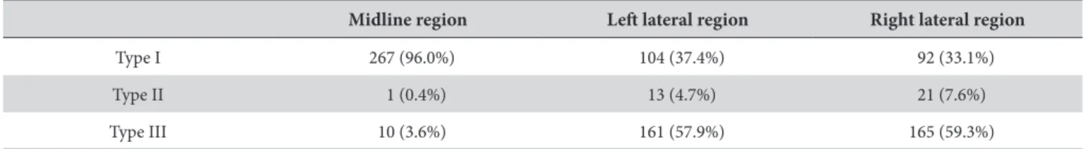
In the midline region, the predominant mandible shape was type I (96%). In the right and left lateral regions, the predominant shape was type III (57.9% and 59.4%), respectively. In the midline region, only 1 patient (0.4%) presented type II, while 13 (4.7%) and 21 (7.6%) patients had this shape on the left and right side, respectively. The distribution of different types of mandible shape at the midline region and in the lateral regions is shown in Table 3.
The assessment of the presence of lingual foramina in the midline and in the lateral regions revealed a negative statistical correlation (r=-0,149; p=0,013). There was no statistically significant correlation between age and vascular vessels, or for age and lingual foramina (p> 0.05). In addition, no correlation was detected regarding to the presence of lingual foramina and dentate or edentulous patients (p> 0.05).
DISCUSSION
Vascular injuries have been associated with surgical complications in the anterior region of the mandible21-23. The objective of this study
was to evaluate the frequency and location of the lingual foramina and the morphology of the mandible.
In the sample analyzed, 246 (88%) patients presented midline lingual foramina and 210 (75%) patients presented lingual foramina in the lateral regions. Previous studies have shown that the prevalence of lingual foramina varied between 73%10, 81%16,
90.35%24 and 100%11,12,17.
In our study, 408 midline lingual foramina were identified with a mean diameter of 0.93 mm. In fact, the mean diameter has
Table 1. Distribution of lateral lingual foramina according to dental region
Dental region
31 32 33 34 35 41 42 43 44 45
Number of lingual foramina 25 55 33 50 33 27 32 38 42 51
Percentile distribution 6.47 14.24 8.54 12.95 8.54 6.99 8.29 9.88 10.88 13.21
Table 2. Diameter and height of the lingual foramina in relation to the base of the mandible at the midline and lateral regions of the mandible
Diameter Height
Mean SD Max Min Mean SD Max Min
(n=408)
Midline lingual foramina 0.9 0.3 0.1 2.5 9.7 5.7 0 30.7
(n=386)
Lateral lingual foramina 0.8 0.3 0.17 2.6 8.7 6.7 0.6 34.8
SD: standard deviation; Max: Maximum value; Min: minimum value.
Table 3. Distribution of different shapes of mandible in the midline and lateral regions
Midline region Left lateral region Right lateral region
Type I 267 (96.0%) 104 (37.4%) 92 (33.1%)
Type II 1 (0.4%) 13 (4.7%) 21 (7.6%)
previously been reported as 0.70 mm12 and 0.80 mm13,24. However,
other studies have reported a smaller diameter (0.31 mm)10.
Our data also revealed that the mean height of the base of the mandible to the lingual foramen was 9.72 mm. In line with this study, Loukas et al.13 reported a mean height value of 10.30 mm
while Rosano et al.11 found a mean height of 12.50 mm. However,
other studies10 found lower values (0.5 mm). In the lateral regions,
386 lingual foramina were found in 75% of the sample evaluated in this study. Corroborating our data, another study17 found lingual
foramina in 80% of the sample, with a mean diameter of 0.8 mm. Regarding the shape of the mandible at the midline, most of the sample presented the type I format (96%), and there was no significant difference between dentate and edentulous patients. Other authors25 found that 69% of the sample presented mandibular
morphology with the base wider than the alveolar crest, which corresponds to the type I format of this study. The type II shape (wider alveolar crest than the base) is the most likely to be affected by surgical complications due to the difficulty of directly visualizing the base of the mandible. Thus, the mandibular shape is of great
importance, because lingual concavity or declivity of the lingual cortex can lead to higher risks of lingual perforation during the installation of implants9.
There was no positive correlation between the presence of vascular vessels and lingual foramina, and age did not influence the frequency of these structures. Moreover, no correlation was found related to presence of lingual foramina in dentate or edentulous patients. Thus, aging as well as presence of teeth does not seem to influence the prevalence and location of these structures.
This study shows a high prevalence of lingual foramina in the midline and the lateral regions. Moreover, the anterior region of the mandible, located between the mentual foramina, is often accessed for the installation of implants. Because severe surgical complications may occur more frequently due to the presence of these anatomical structures7,9, it is very important that a thorough
analysis is carried out during surgical planning. Therefore, cone beam computerized tomography is an excellent tool, which helps to reduce the incidence of surgical complications in the anterior region of the mandible.
REFERENCES
1. Woo BM, Al-Bustani S, Ueeck BA. Floor of mouth haemorrhage and life-threatening airway obstruction during immediate implant placement in the anterior mandible. Int J Oral Maxillofac Surg. 2006 Oct;35(10):961-4. http://dx.doi.org/10.1016/j.ijom.2006.03.020. PMid:16829038. 2. Kalpidis CD, Setayesh RM. Hemorrhaging associated with endosseous implant placement in the anterior mandible: a review of the literature.
J Periodontol. 2004 May;75(5):631-45. http://dx.doi.org/10.1902/jop.2004.75.5.631. PMid:15212344.
3. Greenstein G, Cavallaro J, Tarnow D. Practical application of anatomy for the dental implant surgeon. J Periodontol. 2008 Oct;79(10):1833-46. http://dx.doi.org/10.1902/jop.2008.080086. PMid:18834237.
4. Lamas Pelayo J, Peñarrocha Diago M, Martí Bowen E, Peñarrocha Diago M. Intraoperative complications during oral implantology. Med Oral Patol Oral Cir Bucal. 2008 Apr;13(4):E239-43. PMid:18379448.
5. Longoni S, Sartori M, Braun M, Bravetti P, Lapi A, Baldoni M, et al. Lingual vascular canals of the mandible: the risk of bleeding complications during implants procedures. Implant Dent. 2007 Jun;16(2):131-8. http://dx.doi.org/10.1097/ID.0b013e31805009d5. PMid:17563503. 6. Hofschneider U, Tepper G, Gahleitner A, Ulm C. Assessment of the blood supply to the mental region for reduction of bleeding complications
during implant surgery in the interforaminal region. Int J Oral Maxillofac Implants. 1999 May-Jun;14(3):379-83. PMid:10379111. 7. Niamtu J 3rd. Near-fatal airway obstruction after routine implant placement. Oral Surg Oral Med Oral Pathol Oral Radiol Endod. 2001
Dec;92(6):597-600. http://dx.doi.org/10.1067/moe.2001.116503. PMid:11740473.
8. Mraiwa N, Jacobs R, Steenberghe D, Quirynen M. Clinical assessment and surgical implications of anatomic challenges in the anterior mandible. Clin Implant Dent Relat Res. 2003 Dec;5(4):219-25. http://dx.doi.org/10.1111/j.1708-8208.2003.tb00204.x. PMid:15127992. 9. Pigadas N, Simoes P, Tuffin JR. Massive sublingual haematoma following osseo-integrated implant placement in the anterior mandible. Br
Dent J. 2009 Jan;206(2):67-8. http://dx.doi.org/10.1038/sj.bdj.2009.2. PMid:19165260.
10. Jaju P, Jaju S. Lingual vascular canal assessment by dental computed tomography: a retrospective study. Indian J Dent Res. 2011 Mar-Apr;22(2):232-6. http://dx.doi.org/10.4103/0970-9290.84293. PMid:21891891.
11. Rosano G, Taschieri S, Gaudy JF, Testori T, Del Fabbro M. Anatomic assessment of the anterior mandible and relative hemorrhage risk in implant dentistry: a cadaveric study. Clin Oral Implants Res. 2009 Aug;20(8):791-5. http://dx.doi.org/10.1111/j.1600-0501.2009.01713.x. PMid:19489928.
12. Gahleitner A, Hofschneider U, Tepper G, Pretterklieber M, Schick S, Zauza K, et al. Lingual vascular canals of the mandible: evaluation with dental CT. Radiology. 2001 Jul;220(1):186-9. http://dx.doi.org/10.1148/radiology.220.1.r01jl05186. PMid:11425994.
13. Loukas M, Kinsella CR Jr, Kapos T, Tubbs RS, Ramachandra S. Anatomical variation in arterial supply of the mandible with special regard to implant placement. Int J Oral Maxillofac Surg. 2008 Apr;37(4):367-71. http://dx.doi.org/10.1016/j.ijom.2007.11.007. PMid:18262766. 14. Neves FS, Torres MG, Oliveira C, Campos PS, Crusoé-Rebello I. Lingual accessory mental foramen: a report of an extremely rare anatomical
variation. J Oral Sci. 2010 Sep;52(3):501-3. http://dx.doi.org/10.2334/josnusd.52.501. PMid:20881347.
16. Katakami K, Mishima A, Kuribayashi A, Shimoda S, Hamada Y, Kobayashi K. Anatomical characteristics of the mandibular lingual foramina observed on limited cone-beam CT images. Clin Oral Implants Res. 2009 Apr;20(4):386-90. http://dx.doi.org/10.1111/j.1600-0501.2008.01632.x. PMid:19298292.
17. Tagaya A, Matsuda Y, Nakajima K, Seki K, Okano T. Assessment of the blood supply to the lingual surface of the mandible for reduction of bleeding during implant surgery. Clin Oral Implants Res. 2009 Apr;20(4):351-5. http://dx.doi.org/10.1111/j.1600-0501.2008.01668.x. PMid:19298289.
18. Babiuc I, Tărlungeanu I, Păuna M. Cone beam computed tomography observations of the lingual foramina and their bony canals in the median region of the mandible. Rom J Morphol Embryol. 2011;52(3):827-9. PMid:21892525.
19. Tepper G, Hofschneider UB, Gahleitner A, Ulm C. Computed tomographic diagnosis and localization of bone canals in the mandibular interforaminal region for prevention of bleeding complications during implant surgery. Int J Oral Maxillofac Implants. 2001 Jan-Feb;16(1):68-72. PMid:11280364.
20. von Arx T, Matter D, Buser D, Bornstein MM. Evaluation of location and dimensions of lingual foramina using limited cone-beam computed tomography. J Oral Maxillofac Surg. 2011 Nov;69(11):2777-85. http://dx.doi.org/10.1016/j.joms.2011.06.198. PMid:21855197.
21. Choi DY, Woo YJ, Won SY, Kim DH, Kim HJ, Hu KS. Topography of the lingual foramen using micro-computed tomography for improving safety during implant placement of anterior mandibular region. J Craniofac Surg. 2013 Jul;24(4):1403-7. http://dx.doi.org/10.1097/ SCS.0b013e31828b75da. PMid:23851818.
22. Kilic E, Doganay S, Ulu M, Çelebi N, Yikilmaz A, Alkan A. Determination of lingual vascular canals in the interforaminal region before implant surgery to prevent life-threatening bleeding complications. Clin Oral Implants Res. 2014 Feb;25(2):e90-3. http://dx.doi.org/10.1111/ clr.12065. PMid:23176525.
23. Yildirim YD, Güncü GN, Galindo-Moreno P, Velasco-Torres M, Juodzbalys G, Kubilius M, et al. Evaluation of mandibular lingual foramina related to dental implant treatment with computerized tomography: a multicenter clinical study. Implant Dent. 2014 Feb;23(1):57-63. http:// dx.doi.org/10.1097/ID.0000000000000012. PMid:24394340.
24. Scaravilli MS, Mariniello M, Sammartino G. Mandibular lingual vascular canals (MLVC): evaluation on dental CTs of a case series. Eur J Radiol. 2010 Nov;76(2):173-6. http://dx.doi.org/10.1016/j.ejrad.2009.06.002. PMid:19596533.
25. Quirynen M, Mraiwa N, van Steenberghe D, Jacobs R. Morphology and dimension of the mandibular jaw bone in the interforaminal region in patients requiring implants in the distal areas. Clin Oral Implants Res. 2003 Jun;14(3):280-5. http://dx.doi.org/10.1034/j.1600-0501.2003.140305.x. PMid:12755778.
CONFLICTS OF INTERESTS
The authors declare no conflicts of interest.
*CORRESPONDING AUTHOR
Marcela Claudino, Departamento de Odontologia, UEPG – Universidade Estadual de Ponta Grossa, Av. General Carlos Cavalcanti, 4748, Uvaranas, 84030-900 Ponta Grossa - PR, Brasil, e-mail: marcelaclaudino@hotmail.com