Ultrasensitive Colorimetric and Ratiometric Detection of Cu
2+
:
Acid
−Base Properties, Complexation, and Binding Studies
Daniel J. Fanna,
†,#Luís M. P. Lima,
‡Alexander R. Craze,
†Adrian Trinchi,
§Richard Wuhrer,
∥Leonard F. Lindoy,
⊥Gang Wei,
#Jason K. Reynolds,
*
,†and Feng Li
*
,††School of Science and Health, Western Sydney University, Locked Bag 1797, Penrith, New South Wales 2751, Australia ‡Instituto de Tecnologia Química e Biológica António Xavier, Universidade Nova de Lisboa, Av. da República, 2780-157 Oeiras,
Portugal
§CSIRO Manufacturing, Private Bag 33, Clayton, Victoria 3169, Australia
∥Advanced Materials Characterisation Facility, Western Sydney University, Locked Bag 1797, Penrith, New South Wales 2751,
Australia
⊥School of Chemistry, University of Sydney, Sydney, New South Wales 2006, Australia #CSIRO Manufacturing, P.O. Box 218, Lindfield, New South Wales 2070, Australia
*
S Supporting InformationABSTRACT: Herein, we report the synthesis and character-ization of a chemosensor, 5-(diethylamino)-2-(2,3-dihydro-1H-perimidin-2-yl)phenol (HL), synthesized from a condensation between 4-(diethylamino)salicylaldehyde and 1,8-diamino-naphthalene. Upon investigation of the sensing properties of HL, it was found that this sensor may be employed for simple yet efficient detection of Cu2+in aqueous methanol solutions. The selective and ratiometric response to Cu2+ yielded an outstandingly low limit of detection of 3.7 nM by spectro-photometry and is also useful as a naked-eye sensor from 2.5 μM. The system was studied by spectrophotometric pH
titrations to determine Cu2+binding constants and complex speciation. Binding of Cu2+to HL occurs in 1:1 stoichiometry, in good agreement with high-resolution electrospray ionization mass spectrometry (ESI-HRMS) results, Cu2+titrations, and Job’s plot experiments, while the coordination geometry was tentatively assigned as square pyramidal by spectroscopic studies.
■
INTRODUCTIONThe design and synthesis of new chemosensors for the detection of trace metals, anions, and small molecules continue to be strongly pursued due to their potential for use in medical, biological, industrial, and environmental applications.1−16 Currently, the detection of biologically active metals such as Fe2+/3+,17,18
Cd2+,19,20
Hg2+,21,22
and Cu2+23−26
is receiving considerable attention due to their possible adverse effects on human health.27 Of these metals, copper is the third most abundant transition metal found in humans and plays an essen-tial role in several cuproenzymes.28However, free copper is also able to oxidize cellular components through its redox activity, damaging nucleic acids, proteins, and lipids.29Hence, an imbal-ance of copper may be detrimental to human health, causing pathogenesis such as Parkinson’s disease, Alzheimer’s disease, Wilson’s disease, Menkes disease, and Amyotrophic Lateral Sclerosis (ALS).30−32In particular, copper is a common pollut-ant due to its widespread usage in industry, agriculture and drinking water systems. The World Health Organization (WHO) has advised that 2 ppm (31.5μM) is the recommended upper level for copper in drinking water.33Although a consid-erable number of chemosensor systems have been developed for
copper(II) ions (Table 1), the design and successful construc-tion of ultrasensitive and highly selective systems, particularly those with naked-eye detection ability, still represents a signif-icant challenge.
Herein, we report a potential chemosensor, HL (Scheme 1), capable of extremely fast colorimetric and ratiometric detection of Cu2+ with an impressively low detection limit of 3.7 nM. Detection of copper at low micromolar levels is also possible in the absence of a spectrophotometer, as the visible response of HL to Cu2+is significant. Spectrophotometric acid−base studies were carried out in addition to several UV−vis-based metal detection assays, ESI-HRMS, and electron paramagnetic reso-nance (EPR) studies in order to probe the nature of the binding of HL to Cu2+in terms of its sensing ability for this metal ion.
■
RESULTS AND DISCUSSIONSynthesis and Structure of HL. HL was synthesized from the condensation of 4-(diethylamino)salicylaldehyde and Received: June 28, 2018
Accepted: August 20, 2018 Published: September 4, 2018
Article
http://pubs.acs.org/journal/acsodf
Cite This:ACS Omega 2018, 3, 10471−10480
Derivative Works (CC-BY-NC-ND) Attribution License, which permits copying and redistribution of the article, and creation of adaptations, all for non-commercial purposes.
Downloaded via UNIV NOVA DE LISBOA 00900 on April 26, 2019 at 16:19:33 (UTC).
1,8-diaminonaphthalene in isopropanol in the presence of a cata-lytic amount of p-toluenesulfonic acid, as shown inScheme 1. It was characterized via 1H and 13C NMR, ESI-HRMS,
pendicular (Figure 1). Both edge-to-face and face-to-face π-contacts are present in the lattice between the naphthalene moieties (Figure S6). Crystallographic data is summarized in
Table S1, and detailed crystallographic information can also be found in theSupporting Information.
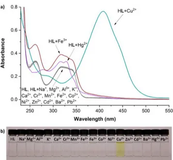
Screening of Metal Cations. The interaction with common metal cations was investigated using 20μM solutions of HL in a MeOH/H2O (1:1, v/v; HEPES, 20 mM; pH = 7.00) mixture by addition of two equivalents (40μM) of the cations. In the presence of Cu2+, the absorbance spectrum develops imme-diately a strong absorption at 408 nm (ε = 3.815 × 10⁴M−1cm−1) associated with generation of a yellow color (Figure 2). Small peak changes were also observed for Hg2+and Fe3+, but impor-tantly no other cations gave rise to an intense band centered at 408 nm as observed for Cu2+. The sensing mechanism likely involves a dual ligand-to-metal charge transfer (LMCT) trans-ition (an intense band at 408 nm) characteristic of a Cu2+center coordinated to a phenoxyl58−60and naphthalene amine based61,62 ligand such as HL, which also incorporates an electron donor diethylamino group.
Acid−Base Equilibrium Studies. To characterize the acid−base properties of HL and investigate its interaction with Cu2+, pH titrations monitored by UV−vis spectrophotometry were performed for HL alone and in the presence of Cu2+. The main spectral features of HL varying with a change of pH are the composite band at 310−350 nm assigned to a intraligand π−π* charge transfer (CT) transition from phenol and phenolate,58 the shifting band at 260−280 nm assigned to a intraligand π−π* charge transfer (CT) from naphthalene amine,61,62 and the increasing absorption band at 230 nm (Figure S7a). The spectral variations were fitted to a speciation model containing three protonation equilibria (Figure S7b), and the protonation equilibrium constants obtained are presented inTable 2. The highest protonation detected occurs around pH = 11 and is associated with moderate spectral change, being assigned to protonation of the phenoxyl group as found in similar
10 mM tris-HCl, pH 7.0)
UV−vis DMSO/Bis-tris buffer (1:1, v/v) 360 38
UV−vis DMSO/Bis-tris buffer (3:2, v/v, 10 mM bis-tris, pH 7.0)
200 39
fluorescence MeOH/H2O (2:8, v/v) 314 and
184
40
UV−vis MeOH/HEPES buffer (1:1, v/v, pH 7.4)
140 41
fluorescence HEPES buffer/CH3CN (3:2, v/v,
100 mM HEPES, pH 7.2)
110 42
UV−vis DMSO/HEPES buffer (9:1, v/v, pH 7.0)
100 43
UV−vis CH3CN 61.9 44
fluorescence EtOH/HEPES buffer (8:2, v/v, 20 mM HEPES, pH 7.2)
40 45
fluorescence CH3CN/tris-HCl (1:1, v/v, 10 mM
tris-HCl, pH 7)
40 46
UV−vis CH3CN/H2O (varying ratio) 40 47
fluorescence DMF/HEPES buffer (3:7, v/v, 20 mM HEPES, pH 7.4)
15 48
fluorescence Tris buffer (25 mM, pH 7.4) 11.2 49
UV−vis CH3CN 7.8 and
5.7
50
UV−vis CH3CN/Tris buffer (1:1, v/v, 10 mM
Tris, pH = 7.0) 5.2, 4.9, and 4.5 51 fluorescence CH3CN/MeOH/H2O (1:9:10, v/v/v) 4 and 3 52
UV−vis MeOH/HEPES buffer (1:1, v/v, 20 mM HEPES, pH 7.00) 3.7 This work fluorescence CH3CN 2.3 53 UV−vis/ fluorescence CH3CN 1.9, 1.8,and 1.7 54 fluorescence H2O/CH3CN (2:1, v/v) 1.45 55
fluorescence HEPES buffer (20 mM, pH 7.0) 1.1 56
fluorescence Britton−Robinson buffer/CH3CN
(9:1, v/v, pH = 7.02)
0.739 57
functions.63Around pH = 5.5, there is an intense spectral change likely associated with protonation of the tertiary amine attached to the phenoxyl group. Below pH = 3.5, there is a very small spectral change possibly arising from protonation of one of the secondary amines of the dihydroperimidine group.
In the presence of an equimolar amount of Cu2+, the UV−vis spectrum of HL changes significantly on moving from acidic to basic pH with a new band developing centered at ca. 408 nm (Figure S8). The spectral variations were in this case successfully fitted to a speciation model containing three complex species corresponding to different protonation states (Figure 3), and the complexation equilibrium constants obtained are presented in
Table 2. It should be stressed that all complex species found are of 1:1 metal-to-ligand ratio. At lower pH, the complex exists mainly as the species [CuHL]2+, while around and above neutral pH, there are two hydroxo species formed likely from depro-tonation of water molecules coordinated to the metal center. It is worth noticing that the [CuL]+species was not obtained in the data fitting, suggesting that it may only exist in very low abundance in equilibrium, if at all. Thus, what is observed
corresponds to a double deprotonation step of the complex from [CuHL]2+to [CuL(OH)] around pH = 4−5. The complexation constants obtained for this system evidence a high thermody-namic stability of the complex formed between HL and Cu2+, also resulting in the almost complete absence of free Cu2+above pH = 5 (Figure 3) under the conditions employed.
Cu2+ Detection Performance. The stoichiometry of the interaction between HL and Cu2+was also probed in buffered pH by the continuous variation method (Job’s plot). The molar ratios between HL and Cu2+in a MeOH/H
2O (1:1, v/v; HEPES, 20 mM; pH = 7.00) medium were varied with the combined Figure 1.Thermal ellipsoid structure of HL drawn at 50% probability: (a) side view; (b) top view. Bifurcated hydrogen H-bonding represented by dotted lines.
Figure 2.(a) UV−vis absorbance spectra of HL (20 μM) in a MeOH/ H2O (1:1, v/v; HEPES, 20 mM; pH = 7.00) mixture in the presence of
two equivalents (40μM) of common metal ions. (b) Color changes observed by the naked-eye, colorimetric detection for Cu2+(yellow),
while solutions of HL and HL plus other cations are all colorless.
Table 2. Overall (logβ) and Stepwise (log K) Thermodynamic Equilibrium Constantsafor the
Protonation of HL and Its Complexation with Cu2+at 25.0± 0.1° C in MeOH/H2O (1:1, v/v) Medium at 0.10± 0.01 M in KCl
equilibrium logβ equilibrium log K L−+ H+⇄ HL 10.93(1) L−+ H+⇄ HL 10.93(1) L−+ 2H+⇄ (H 2L)+ 16.50(1) HL + H+⇄ (H2L)+ 5.57(1) L−+ 3H+⇄ (H 3L)2+ 19.85(4) (H2L)++ H+⇄ (H3L)2+ 3.35(4) Cu2++ L−+ H+⇄ [CuHL]2+ 19.11(1) [CuL] ++ H+⇄ [CuHL]2+ − Cu2++ L−⇄ [CuL]+ b Cu2++ L−⇄ [CuL]+ − Cu2++ L−⇄ [CuLH−1] + H+ 10.52(1) CuL(OH) + H+⇄ [CuL]+ − Cu2++ L−⇄ [CuLH−2]−+ 2 H+ 1.01(3) [CuL(OH)2]−+ H+⇄ [CuL(OH)] 9.51(3)
aValues in parentheses are standard deviations in the last significant
figures.bNot determined.
Figure 3.Species distribution diagram for HL in the presence of 1 equiv of CuCl2versus pH (at 25μM in MeOH/H2O 1:1).
addition of Cu2+up to 20μM, a directly proportionate increase in the band at 408 nm was observed. An isosbestic point was observed at 346 nm, occurring between the peak maximum for HL (∼320 nm) and that for the complex (408 nm), indicating a formation via a single equilibrium step. Furthermore, after 20μM had been added, no further spectrophotometric change was observed, indicating saturation of HL with 1 equiv of Cu2+. This is in good agreement with the 1:1 stoichiometry found above. A linear relationship was observed on plotting the ratio between the complex peak maximum and the isosbestic point (408 nm/346 nm) absorptions versus the effective Cu2+ concentration, indicating that this spectrophotometric change is ratiometric and thus can be used to estimate the concentration of Cu2+independently of the HL concentration. The limit of detection (LoD) and limit of quantification (LoQ) were calculated from this ratiometric change of 408/346 nm, yielding
Cu Detection Selectivity and Reversibility. The
selectivity of HL for Cu2+ was also probed in the presence of competing cations; UV−vis experiments were conducted in which Cu2+ complexation was investigated in solutions con-taining an excess of a competing metal ion. This was carried out in a MeOH/H2O (1:1, v/v; HEPES, 20 mM; pH = 7.00) medium, where HL was present at 20μM, Cu2+at 1 equiv (20μM), and a competing cation in a 4 equiv (80μM) excess (see UV−vis spectraFigure S11). As shown inFigure 6a, the selectivity of HL for Cu2+is unaffected by the presence of any of the competing cations employed. In each case, the solution color observed by the naked-eye is in good agreement with the UV−vis measure-ments, with all solutions exhibiting a yellow color associated with formation of the Cu2+complex (Figure 6b).
Reversibility studies employing ethylenediaminetetraacetic acid disodium salt (H2Na2EDTA) were undertaken in order to probe the robustness of HL with respect to its repetitive binding to Cu2+. As with previous experiments, HL was prepared as a 20μM solution in a MeOH/H2O (1:1, v/v; HEPES, 20 mM; pH = 7.00) mixture. Initially Cu2+was added at 1 equiv (20μM), followed by 4 equiv (80μM) of EDTA4−. Subsequent cycles were undertaken by adding a further 80μM of Cu2+followed by 80μM of EDTA4−(Figure 7). The addition of EDTA4−results in the disappearance of the band at 408 nm, while the further addition of an excess of Cu2+results in the reformation of this band. As the absorbance peak (λmax= 408 nm) was not signi fi-cantly affected after several cycling events, this result indicates that HL is suitable as a reversible sensor.
Structural Study of the Cu2+Complex. In order to probe the binding mode of HL to Cu2+, we focused on the visible and EPR spectra of the complex. The EPR spectra of frozen complex solutions in buffered MeOH/H2O (1:1, v/v; HEPES, 20 mM; pH = 7.00) media, in the absence and also in the presence of additional O- or N-donor atoms (respectively, DMSO or pyridine,Figure 8), all unambiguously display a single species of rhombic symmetry and a dx2−y2 ground state, consistent with elongated rhombic-octahedral or distorted square-based pyr-amidal stereochemistry.64The A hyperfine coupling constants and g factors of these complex samples obtained by spectral simulation (Table 3) are indicative of either N3O or N2O2 equatorial coordination donor sets as seen from the values of g3 and A3,65pointing to involvement of one O and 2 N donor atoms from HL on the equatorial positions of the complex. Additionally, the visible spectra obtained for samples under similar conditions to those used for the EPR study (Figure S12) display two distinguishable absorptions (as shoulders of the higher energy one) centered at the 610−620 and 730−750 nm regions, a feature that has been assigned to Cu2+complexes of square pyra-midal geometry featuring strong axial bonding interaction.66,67 Thus, we propose that these spectroscopic results are in accord with HL forming a distorted square pyramidal complex in solu-tion by coordinasolu-tion of Cu2+ to the phenoxyl O and both Figure 4.(a) Absorption spectral change of HL (20μM) upon titration
with CuCl2in a MeOH/H2O (1:1, v/v; HEPES, 20 mM; pH = 7.00)
mixture. The peak associated with formation of the complex is atλmax=
408 nm. (b) The linear plot used for LoD and LoQ calculations was obtained by plotting the absorbance ratio of the complex band over the isosbestic peak.
secondary amine N atoms to define an approximate equatorial coordination plane, with additional donors from the media com-pleting the coordination sphere at the remaining equatorial and axial positions of the metal center.
Detection of Cu2+in Tap Water. To evaluate the sensing potential of HL in real world samples, two tap waters suitable for human consumption were taken from the Sydney area in New South Wales, Australia. A third sample of known concentration
(5 μM of CuCl2) was prepared using Milli-Q water and a standardized Cu2+solution. These samples were prepared in a MeOH/H2O (1:1, v/v; HEPES, 20 mM; pH = 7.00) buffered media with HL at 20 μM. The samples were analyzed using UV−vis, and the ratio of their absorbance at 408/346 nm plotted against a linear calibration plot (Figures S13 and S14). As shown inTable 4, the measured 5μM spike of each tap water, and the 5μM laboratory prepared water sample are in good agreement with the expected values. Thus, HL is suitable for the quantitative detection of Cu2+far below the WHO guidelines of 2 mg/L (31.5μM).
Figure 5.Detection of Cu2+by the naked eye at 20μM of HL in MeOH/H
2O (1:1, v/v; HEPES, 20 mM; pH = 7.00) with increasing concentrations of CuCl2.
Figure 6.(a) Competing ion study with HL (20μM) and 1 equiv (20μM) of Cu2+in the presence of 4 equiv (80μM) of other metal ions in a MeOH/H2O (1:1, v/v; HEPES, 20 mM; pH = 7.00) medium at
λmax= 408 nm. (b) Competing ion solutions as visible to the naked-eye;
the detection of Cu2+by the observed color change to yellow remains unaffected by other metal ions.
Figure 7.Reversibility of the response of HL (20μM) in a MeOH/ H2O (1:1, v/v; HEPES, 20 mM; pH = 7.00) mixture atλmax= 408 nm
after cycling with additions of Cu2+(20μM initially, 80 μM thereafter)
and EDTA4−(80μM).
Figure 8.EPR spectra of the complex of HL with Cu2+in different
buffered media (HEPES, 20 mM; pH = 7.00); dotted lines are the corresponding simulated spectra.
■
CONCLUSIONSIn summary, we report an efficient colorimetric Cu2+ chemo-sensor for use in aqueous methanol media. The chemo-sensor HL exhibits an excellent ratiometric response, also visible by the naked-eye, corresponding to a clear color change from colorless to yellow. In addition, Cu2+sensing is selective, being unaffected by the presence of the wide range of other metal cations tested. In buffered medium at pH = 7, an outstanding 3.7 nM limit of detection for Cu2+was obtained in the low nanomolar range, one of the most sensitive reported in aqueous media. Furthermore, the reaction stoichiometry between HL and Cu2+was unambig-uously established as 1:1, and the complexation is reversible employing EDTA4−/Cu2+cycling. The coordination geometry of the formed complex was also tentatively assigned as square pyramidal. The complexation constants determined from the pH titrations point to the formation of a thermodynamically stable complex that exists almost exclusively as [CuL(OH)] in the range of pH = 5−8. According to the preliminary results, the simplicity and effectiveness of this sensor show great potential for real word applications in Cu2+detection via qualitative (visible color change) and quantitative (spectrophotometric) methods.
■
MATERIALS AND METHODSChemicals and Reagents. All chemicals and solvents were of commercial grade and used without further purification. All aqueous solutions were prepared with type I water from a Milli-Q water purification system. Metal standards were prepared from chloride (Na+, Mg2+, Al3+, K+, Ca2+, Cr3+, Mn2+, Fe2+, Fe3+, Co2+, Ni2+, Cu2+, Zn2+, Cd2+, Ba2+and Hg2+) or nitrate salts (Pb2+) of analytical grade, then standardized using literature complexo-metric titration methods with H2Na2EDTA.68
Synthesis of HL. The preparation of HL was optimized from a literature procedure (Scheme 1).69In a 50 mL round-bottom flask, 4-(diethylamino)salicylaldehyde (1000 mg, 5.2 mmol), 1,8-diaminonaphthalene (825 mg, 5.2 mmol), and a catalytic amount of p-toluenesulfonic acid (PTSA) were combined and dissolved in isopropanol (30 mL). This mixture was heated at 65°C for 24 h, and the resulting precipitate was filtered and washed with 50% isopropanol (H2O/isopropanol, v/v). The product was then dried under vacuum at 40°C, yielding 1.4 g (82%) of HL as a white crystalline powder. Crystals suitable for SC-XRD were grown by slow evaporation of HL in an ethanol/acetone mixture (1:4 v/v).1H NMR (DMSO-d
6with TMS, 300 MHz) δ (ppm) 9.16 (s. 1H), 7.22 (d. 1H), 7.13 (t. 2H), 6.98 (d. 2H),
TMS, 75.5 MHz) δ (ppm) 156.36, 148.37, 143.71, 134.34, 129.24, 126.63, 115.07,113.57, 112.50, 104.36, 102.99, 98.31, 61.22, 43.76, 12.39. ESI-HRMS (positive-ion detection, MeOH, m/z): calculated for [HL + H]+, 334.1919; found, 334.1884. FT-IR (ATR νmax/cm−1): 3333, 2966, 1596, 1562, 1411, 1216, 1118, 818, 761. Melting point: 156.7°C. UV−vis (1 cm optical path length, MeOH−H2O, 1/1, v/v, HEPES 20 mM, pH = 7.00): 20μM HL, λmax= 320 nm (abs = 0.277,ε = 13 850), 262 nm (abs = 0.324,ε = 16 200).
General Methods. Characterization of HL was carried out using a range of techniques. 1H and 13C NMR spectra were measured on a Bruker DRX-300 NMR spectrometer. FT-IR measurements were conducted on a Bruker Vertex 70 spectro-meter. High resolution electrospray ionization mass spectra (ESI-HRMS) were collected using a Waters Xevo QToF quad-rupole mass spectrometer with samples dissolved in methanol. Simultaneous thermal analysis (STA) was undertaken on a Netzsch STA449 C Jupiter thermo-microbalance. A PerkinElmer Lambda 45 UV−vis spectrophotometer was used to conduct the Job’s plot and titration experiments, while all other UV−vis works were performed using a Varian Cary 100 spectropho-tometer. Quartz cuvettes with a 1 cm optical path length were used for all UV−vis measurements. Single crystal X-ray diffraction (SC-XRD) was undertaken at the Australian Syn-chrotron on the MX1 beamline at 100(2) K with silicon double crystal monochromated radiation.70 Data collection was through Blu-Ice71software, with data integration and reduction undertaken using XDS;72following this, an empirical absorption correction was applied using SADABS software.73The crystal structure was solved by direct methods, and the full-matrix least-squares refinements were conducted using the SHELX74,75suite of programs via Olex2 interface.76
UV−Vis Spectroscopy. A 2.00 mM stock solution of HL was prepared in MeOH; this solution was used for all spectro-scopic experiments. Metal interaction studies were conducted using 20μM HL in a MeOH/H2O (1:1, v/v, HEPES 20 mM, pH = 7.00) buffered mixture with 2 equiv (40 μM) of various chloride and nitrate salts. Competing ion studies were prepared with a similar mixture, 20μM HL in a MeOH/H2O (1:1, v/v, HEPES 20 mM, pH = 7.00) mixture. CuCl2was added as 1 equiv (20μM), with competing metal ions added in a higher excess (80 μM). Job’s plot measurements were undertaken using literature protocols,77,78where HL and Cu2+were prepared in various molar ratios, with the combined concentration of both at 50 μM in all analytical solutions. These molar ratio mixtures were prepared in a MeOH/H2O (1:1, v/v, HEPES 20 mM, pH = 7.00) buffered mixture.
The complex formed by HL and Cu2+was studied in more detail in the visible range using higher concentration samples in both the absence and the presence of additional coordinating compounds, namely, DMSO as a good O-donor and pyridine as a good N-donor. These were prepared in buffered aqueous
sample (μM) (μM) (%) (%)
tap 1 0.00 8.57 0.85
5.00 13.62 100.34 0.82
tap 2 0.00 4.19 0.26
5.00 9.13 99.38 1.14
medium (HEPES 20 mM, pH = 7.00) containing both HL and Cu2+at a 1:1 concentration ratio in the 0.78−1.0 mM range in the following solvent mixtures (v/v): (a) MeOH/H2O 1:1, (b) MeOH/H2O/DMSO 2:2:1, and (c) MeOH/H2O/DMSO/ pyridine 50:50:25:1. In these spectra, the relevant bands (600− 800 nm) appear as shoulders on the more energetic band at ∼410 nm, so the maxima of these absorptions were determined employing the second-derivative method.
EPR Spectroscopy Studies. The electron paramagnetic resonance spectra were measured on a Bruker EMX 300 spec-trometer operating at the X-band and equipped with a continuous-flow cryostat for liquid nitrogen. Here, the complex formed by HL with Cu2+was also studied in the absence and in the presence of additional coordinating compounds, similar to the procedure used for visible absorption studies. The samples were prepared in buffered aqueous mixtures (HEPES 20 mM, pH = 7.00) containing both HL and Cu2+at a 1:1 concentration ratio in the 0.20−0.48 mM range in the following solvent mixtures (v/v): (a) MeOH/H2O 1:1, (b) MeOH/H2O/DMSO 2:2:1, and
(c) MeOH/H2O/DMSO/pyridine 50:50:25:1. The EPR
spectra of the frozen sample solutions were acquired at 130 K at a microwave power of 2.0 mW and a frequency of 9.5 GHz. The experimental spectra were then simulated using the SpinCount software79 in order to obtain the corresponding g factors and A hyperfine coupling constants. All spectra were successfully simulated considering the presence of a single paramagnetic complex species.
Equilibrium Studies. The equilibrium constants for the protonation of HL and the formation of its Cu2+complexes were determined by UV−vis pH titrations in the range of pH = 3−11. The experimental setup used in these titrations consisted of a closed titration vessel of 5−70 mL volume with thermostat jacket set on top of a 801 magnetic stirrer and connected to a Dosimat Plus 865 buret, all from Metrohm, as well as being coupled to a Haake F3 thermostatic water circulator for temper-ature control. All experiments were performed at 25± 0.1 °C in MeOH/H2O (1:1, v/v) medium at 0.10± 0.01 M in KCl, using standardized KOH solutions in MeOH/H2O (1:1, v/v) as titrant (at ca. 0.10 M for protonation and 0.02 M for complexa-tion); a nitrogen atmosphere was maintained in the titration vessel. The KOH titrant solutions were prepared from dilution of a commercial ampule and standardized using Gran’s method.80The starting solutions for titrations contained KCl at 0.10 M, HCl at 1.0× 10⁻³M (for pH≈ 3.0), HL at 2.5 × 10⁻⁵M (and CuCl2in equimolar amount in the case of complexation), all in a starting volume of 20 mL. After each titrant addition, a sample of ca. 1 mL was taken from the titration solution, placed in a 1 cm Hellma 114-QS cuvette and measured on a PerkinElmer lambda 45 UV−vis spectrometer, after which the sample was returned to the titration solution. The pH was not measured experimentally; instead the proton concentration was calculated from the addition volumes and concentrations of the titrant solutions. The titration data were fitted using the HypSpec software,81in the range of 240−370 nm in the absence of Cu2+ or of 370−500 nm in the presence of Cu2+. Species distribution diagrams were plotted with the HySS software at 25μM of HL.82 Sensitivity Studies of HL with Cu2+. To determine the sensitivity of HL to Cu2+, a metal titration approach was used in buffered pH, where 5 μL additions of CuCl2aqueous solution were added to a cuvette with 20 μM of HL dissolved in a MeOH/H2O (1:1, v/v; HEPES, 20 mM; pH = 7.00) mixture, until no further spectral shift was observed. The limit of detec-tion (LoD) and limit of quantification (LoQ) were calculated
using 3σ/m and 10σ/m, respectively,83whereσ is the standard deviation of a blank solution and m is the slope of the linear plot obtained. For the standard deviation (σ) determination, we took 10 repeated spectra measurements of a blank solution (HL solution in the same conditions used for measurements but without any addition of Cu2+) and calculated the standard deviation for the absorbance values at 408 nm, obtaining a value ofσ = 2.34 × 10⁻⁴.
Detection of Cu2+in Tap Water. Tap water samples were taken from residential drinking taps in southeast (tap 1) and northwest (tap 2) Sydney, New South Wales, Australia. An addi-tional sample of a known Cu2+concentration (5μM) was pre-pared in Milli-Q water. To probe the accuracy and precision of HL, tap water solutions were also spiked with 5μM of Cu2+, and all samples were measured with 5 replicates.
Quantitative detection of Cu2+in these samples was carried out by UV−vis spectroscopy with HL (20 μM) in a MeOH/ H2O (1:1, v/v; HEPES, 20 mM; pH = 7.00) buffered media. The ratiometric response taken from 408/346 nm was plotted against a calibration curve in the same media. As the water sample was 40% of the total in cuvette volume, a correction for sample dilution was applied.
■
ASSOCIATED CONTENT*
S Supporting InformationThe Supporting Information is available free of charge on the
ACS Publications websiteat DOI:10.1021/acsomega.8b01483. Crystallographic structure of HL (CIF)
Additional characterization details of HL and binding studies including 1H and 13C NMR, ESI-HRMS, STA, FT-IR, various UV−vis experiments, and crystallographic data (PDF)
■
AUTHOR INFORMATION Corresponding Authors *E-mail:feng.li@westernsydney.edu.au(F.L.). *E-mai:j.reynolds@westernsydney.edu.au(J.K.R.). ORCID Luís M. P. Lima:0000-0002-9729-330X Feng Li:0000-0001-8465-9678 NotesThe authors declare no competingfinancial interest.
CCDC-1820800 contains details of the crystallographic data for this article; these data can be obtained free of charge from the Cambridge Crystallographic Data Centre viawww.ccdc.cam.ac. uk/data_request/cif.
■
ACKNOWLEDGMENTSThe authors thank the Western Sydney University (WSU) Advance Material Characterisation Facility and the WSU Mass Spectrometry Facility for instrumental access. D.J.F. thanks WSU for a Postgraduate Research Award and CSIRO for a Ph.D. top-up award. L.M.P.L. is grateful for financial support from project LISBOA-01-0145-FEDER-007660 (Microbiologia Molec-ular, Estrutural e Celular) funded by FEDER funds through COMPETE2020-Programa Operacional Competitividade e Internacionalização (POCI) and by national funds through Fundação para a Ciência e a Tecnologia (FCT) and also for a postdoctoral fellowship (SFRH/BPD/73361/2010) from FCT. The crystallographic data acquisition was undertaken on the MX1 beamline at the Australian Synchrotron, Clayton, Victoria,
Phenomena. In Ion Exchange and Solvent Extraction; Moyer, B. A., Ed.; CRC Press: Boca Raton, FL, 2013; Vol. 21, pp 1−48.
(3) Gale, P. A.; Caltagirone, C. Fluorescent and Colorimetric Sensors for Anionic Species. Coord. Chem. Rev. 2018, 354, 2−27.
(4) Kaur, B.; Kaur, N.; Kumar, S. Colorimetric Metal Ion Sensors− A Comprehensive Review of the Years 2011−2016. Coord. Chem. Rev. 2018, 358, 13−69.
(5) Aulsebrook, M. L.; Graham, B.; Grace, M. R.; Tuck, K. L. Lanthanide Complexes for Luminescence-Based Sensing of Low Molecular Weight Analytes. Coord. Chem. Rev. 2017,DOI: 10.1016/ j.ccr.2017.11.018.
(6) Wu, D.; Sedgwick, A. C.; Gunnlaugsson, T.; Akkaya, E. U.; Yoon, J.; James, T. D. Fluorescent Chemosensors: The Past, Present and Future. Chem. Soc. Rev. 2017, 46 (23), 7105−7123.
(7) Sakai, R.; Satoh, T.; Kakuchi, T. Polyacetylenes as Colorimetric and Fluorescent Chemosensor for Anions. Polym. Rev. 2017, 57 (1), 159−174.
(8) Pan, J.; Tang, F.; Ding, A.; Kong, L.; Yang, L.; Tao, X.; Tian, Y.; Yang, J. A Small-Molecule Chemosensor for the Selective Detection of 2,4,6-Trinitrophenol (TNP). RSC Adv. 2015, 5 (1), 191−195.
(9) Shanmugaraju, S.; Mukherjee, P. S. π-Electron Rich Small Molecule Sensors for the Recognition of Nitroaromatics. Chem. Commun. 2015, 51 (89), 16014−16032.
(10) Lin, V. S.; Chen, W.; Xian, M.; Chang, C. J. Chemical Probes for Molecular Imaging and Detection of Hydrogen Sulfide and Reactive Sulfur Species in Biological Systems. Chem. Soc. Rev. 2015, 44 (14), 4596−4618.
(11) Butler, S. J.; Parker, D. Anion Binding in Water at Lanthanide Centres: From Structure and Selectivity to Signalling and Sensing. Chem. Soc. Rev. 2013, 42 (4), 1652−1666.
(12) Esipenko, N. A.; Koutnik, P.; Minami, T.; Mosca, L.; Lynch, V. M.; Zyryanov, G. V.; Anzenbacher, P. First Supramolecular Sensors for Phosphonate Anions. Chem. Sci. 2013, 4 (9), 3617−3623.
(13) Bernier, N.; Carvalho, S.; Li, F.; Delgado, R.; Félix, V. Anion Recognition by a Macrobicycle Based on a Tetraoxadiaza Macrocycle and an Isophthalamide Head Unit. J. Org. Chem. 2009, 74 (13), 4819− 4827.
(14) Li, F.; Delgado, R.; Drew, M. G. B.; Felix, V. Dioxadiaza- and Trioxadiaza-Macrocycles Containing One Dibenzofuran Unit Selective for Cadmium. Dalton Trans. 2006, No. 45, 5396−5403.
(15) Vázquez, M.; Fabbrizzi, L.; Taglietti, A.; Pedrido, R. M.; González-Noya, A. M.; Bermejo, M. R. A Colorimetric Approach to Anion Sensing: A Selective Chemosensor of Fluoride Ions, in Which Color Is Generated by Anion-Enhancedπ Delocalization. Angew. Chem. 2004, 116 (15), 1996−1999.
(16) Wallace, K. J.; Belcher, W. J.; Turner, D. R.; Syed, K. F.; Steed, J. W. Slow Anion Exchange, Conformational Equilibria, and Fluorescent Sensing in Venus Flytrap Aminopyridinium-Based Anion Hosts. J. Am. Chem. Soc. 2003, 125 (32), 9699−9715.
(17) Xuan, W.; Pan, R.; Wei, Y.; Cao, Y.; Li, H.; Liang, F.-S.; Liu, K.-J.; Wang, W. Reaction-Based “Off−On” Fluorescent Probe Enabling Detection of Endogenous Labile Fe2+ and Imaging of Zn2+-Induced
Fe2+ Flux in Living Cells and Elevated Fe2+ in Ischemic Stroke.
Bioconjugate Chem. 2016, 27 (2), 302−308.
(18) Wang, C.; Liu, Y.; Cheng, J.; Song, J.; Zhao, Y.; Ye, Y. Efficient FRET-Based Fuorescent Ratiometric Chemosensors for Fe3+and Its
Application in Living Cells. J. Lumin. 2015, 157, 143−148.
Living Cells. RSC Adv. 2016, 6 (9), 7668−7673.
(22) Song, J.; Huai, M.; Wang, C.; Xu, Z.; Zhao, Y.; Ye, Y. A New FRET Ratiometric Fluorescent Chemosensor for Hg2+ and Its
Application in Living EC 109 Cells. Spectrochim. Acta, Part A 2015, 139, 549−554.
(23) Li, D.; Sun, X.; Huang, J.; Wang, Q.; Feng, Y.; Chen, M.; Meng, X.; Zhu, M.; Wang, X. A Carbazole-Based “Turn-on” Two-Photon Fluorescent Probe for Biological Cu2+Detection Vis Cu2+-Promoted Hydrolysis. Dyes Pigm. 2016, 125, 185−191.
(24) Li, M.; Sun, Y.; Dong, L.; Feng, Q.-C.; Xu, H.; Zang, S.-Q.; Mak, T. C. Colorimetric Recognition of Cu2+and Fluorescent Detection of
Hg2+ in Aqueous Media by a Dual Chemosensor Derived from
Rhodamine B Dye with a NS 2 Receptor. Sens. Actuators, B 2016, 226, 332−341.
(25) Yin, S.; Leen, V.; Van Snick, S.; Boens, N.; Dehaen, W. A Highly Sensitive, Selective, Colorimetric and near-Infrared Fluorescent Turn-on Chemosensor for Cu2+Based on BODIPY. Chem. Commun. 2010,
46 (34), 6329−6331.
(26) He, X.; Zhang, J.; Liu, X.; Dong, L.; Li, D.; Qiu, H.; Yin, S. A Novel BODIPY-Based Colorimetric and Fluorometric Dual-Mode Chemosensor for Hg2+and Cu2+. Sens. Actuators, B 2014, 192, 29−35.
(27) Trace Elements in Human Health and Disease; Prasad, A. S., Oberleas, D., Eds.; Academic Press: New York, 1976.
(28) Uauy, R.; Olivares, M.; Gonzalez, M. Essentiality of Copper in Humans. Am. J. Clin. Nutr. 1998, 67 (5), 952S−959S.
(29) Aust, S. D.; Morehouse, L. A.; Thomas, C. E. Role of Metals in Oxygen Radical Reactions. J. Free Radicals Biol. Med. 1985, 1 (1), 3−25. (30) Gaggelli, E.; Kozlowski, H.; Valensin, D.; Valensin, G. Copper Homeostasis and Neurodegenerative Disorders (Alzheimer’s, Prion, and Parkinson’s Diseases and Amyotrophic Lateral Sclerosis). Chem. Rev. 2006, 106 (6), 1995−2044.
(31) Bull, P. C.; Thomas, G. R.; Rommens, J. M.; Forbes, J. R.; Cox, D. W. The Wilson Disease Gene Is a Putative Copper Transporting P− Type ATPase Similar to the Menkes Gene. Nat. Genet. 1993, 5 (4), 327−337.
(32) de Bie, P.; Muller, P.; Wijmenga, C.; Klomp, L. W. J. Molecular Pathogenesis of Wilson and Menkes Disease: Correlation of Mutations with Molecular Defects and Disease Phenotypes. J. Med. Genet. 2007, 44 (11), 673−688.
(33) World Health Organization. Guidelines for Drinking-Water Quality, 4th ed.; World Health Organization: Geneva, 2011.
(34) You, G. R.; Jang, H. J.; Jo, T. G.; Kim, C. A Novel Displacement-Type Colorimetric Chemosensor for the Detection of Cu2+and GSH
in Aqueous Solution. RSC Adv. 2016, 6 (78), 74400−74408. (35) Noh, J. Y.; Park, G. J.; Na, Y. J.; Jo, H. Y.; Lee, S. A.; Kim, C. A Colorimetric“Naked-Eye” Cu(II) Chemosensor and pH Indicator in 100% Aqueous Solution. Dalton Trans. 2014, 43 (15), 5652−5656.
(36) Kang, J. H.; Lee, S. Y.; Ahn, H. M.; Kim, C. Sequential Detection of Copper(II) and Cyanide by a Simple Colorimetric Chemosensor. Inorg. Chem. Commun. 2016, 74, 62−65.
(37) Elmorsi, T. M.; Aysha, T. S.; Machalický, O.; Mohamed, M. B. I.; Bedair, A. H. A Dual Functional Colorimetric and Fluorescence Chemosensor Based on Benzo[f]Fluorescein Dye Derivatives for Copper Ions and PH; Kinetics and Thermodynamic Study. Sens. Actuators, B 2017, 253, 437−450.
(38) Lee, S. Y.; Bok, K. H.; Kim, J. A.; Kim, S. Y.; Kim, C. Simultaneous Detection of Cu2+ and Cr3+ by a Simple Schiff-Base Colorimetric
Chemosensor Bearing NBD (7-Nitrobenzo-2-Oxa-1, 3-Diazolyl) and Julolidine Moieties. Tetrahedron 2016, 72 (35), 5563−5570.
(39) Kim, M. S.; Jo, T. G.; Ahn, H. M.; Kim, C. A Colorimetric and Fluorescent Chemosensor for the Selective Detection of Cu2+and Zn2+
Ions. J. Fluoresc. 2017, 27 (1), 357−367.
(40) Kumawat, L. K.; Kumar, M.; Bhatt, P.; Sharma, A.; Asif, M.; Gupta, V. K. An Easily Accessible Optical Chemosensor for Cu2+Based
on Novel Imidazoazine Framework, Its Performance Characteristics and Potential Applications. Sens. Actuators, B 2017, 240, 365−375.
(41) Qu, L.; Yin, C.; Huo, F.; Chao, J.; Zhang, Y.; Cheng, F. A Pyridoxal-Based Dual Chemosensor for Visual Detection of Copper Ion and Ratiometric Fluorescent Detection of Zinc Ion. Sens. Actuators, B 2014, 191, 158−164.
(42) Zhang, B.; Diao, Q.; Ma, P.; Liu, X.; Song, D.; Wang, X. A Sensitive Fluorescent Probe for Cu2+ Based on Rhodamine B Derivatives and Its Application to Drinking Water Examination and Living Cells Imaging. Sens. Actuators, B 2016, 225, 579−585.
(43) Lin, Q.; Chen, P.; Liu, J.; Fu, Y.-P.; Zhang, Y.-M.; Wei, T.-B. Colorimetric Chemosensor and Test Kit for Detection Copper (II) Cations in Aqueous Solution with Specific Selectivity and High Sensitivity. Dyes Pigm. 2013, 98 (1), 100−105.
(44) Roy, D.; Chakraborty, A.; Ghosh, R. Perimidine Based Selective Colorimetric and Fluorescent Turn-off Chemosensor of Aqueous Cu
2+: Studies on Its Antioxidant Property along with Its Interaction with
Calf Thymus-DNA. RSC Adv. 2017, 7 (64), 40563−40570.
(45) Wang, Z.; Yang, J.; Yang, Y.; Fang, H.; Xu, X.; Rui, J.; Su, F.; Xu, H.; Wang, S. A Novel Hexahydroquinazolin-2-Amine-Based Fluo-rescence Sensor for Cu2+ from Isolongifolanone and Its Biological
Applications. RSC Adv. 2017, 7 (53), 33263−33272.
(46) Sarkar, S.; Roy, S.; Sikdar, A.; Saha, R.; Panja, S. S. A Pyrene-Based Simple but Highly Selective Fluorescence Sensor for Cu2+Ions
via a Static Excimer Mechanism. Analyst 2013, 138 (23), 7119−7126. (47) Paul, S.; Ghosh, P.; Bhuyan, S.; Mukhopadhyay, S. K.; Banerjee, P. Nanomolar-Level Selective Dual Channel Sensing of Cu2+and CN−
from an Aqueous Medium by an Opto-Electronic Chemoreceptor. Dalton Trans. 2018, 47 (4), 1082−1091.
(48) Jia, H.; Yang, M.; Meng, Q.; He, G.; Wang, Y.; Hu, Z.; Zhang, R.; Zhang, Z. Synthesis and Application of an Aldazine-Based Fluorescence Chemosensor for the Sequential Detection of Cu2+ and Biological
Thiols in Aqueous Solution and Living Cells. Sensors 2016, 16 (1), 79. (49) Naskar, B.; Modak, R.; Maiti, D. K.; Bauzá, A.; Frontera, A.; Maiti, P. K.; Mandal, S.; Goswami, S. A Highly Selective“ON−OFF” Probe for Colorimetric and Fluorometric Sensing of Cu2+in Water.
RSC Adv. 2017, 7 (19), 11312−11321.
(50) Shan, Y.; Liu, Z.; Cao, D.; Liu, G.; Guan, R.; Sun, N.; Wang, C.; Wang, K. Coumarinic Chalcone Derivatives as Chemosensors for Cyanide Anions and Copper Ions. Sens. Actuators, B 2015, 221, 463− 469.
(51) Wang, M.; Leung, K.-H.; Lin, S.; Chan, D. S.-H.; Kwong, D. W. J.; Leung, C.-H.; Ma, D.-L. A Colorimetric Chemosensor for Cu2+Ion
Detection Based on an Iridium(III) Complex. Sci. Rep. 2015, 4, 6794. (52) Zhao, Y.-C.; Chen, C.; Chen, B.; Wang, P.-Y.; Wu, Z.-B.; Li, H.; Song, B.-A.; Yang, S. Tunable Fluorescent Probes for Selective Detection of Copper (II) and Sulphide with Completely Different Modes. Sens. Actuators, B 2017, 241, 168−173.
(53) Udhayakumari, D.; Velmathi, S. Naphthalene Thiourea Derivative Based Colorimetric and Fluorescent Dual Chemosensor for F−and Cu2+/Hg2+Ions. Supramol. Chem. 2015, 27 (7−8), 539− 544.
(54) Udhayakumari, D.; Velmathi, S.; Chen, W.-C.; Wu, S.-P. A Dual-Mode Chemosensor: Highly Selective Colorimetric Fluorescent Probe for Cu2+and F−Ions. Sens. Actuators, B 2014, 204, 375−381.
(55) Liu, C.; Jiao, X.; He, S.; Zhao, L.; Zeng, X. A Highly Selective and Sensitive Fluorescent Probe for Cu2+ Based on a Novel Naphthali-mide−Rhodamine Platform and Its Application in Live Cell Imaging. Org. Biomol. Chem. 2017, 15 (18), 3947−3954.
(56) Zhu, D.; Ren, A.; He, X.; Luo, Y.; Duan, Z.; Yan, X.; Xiong, Y.; Zhong, X. A Novel Ratiometric Fluorescent Probe for Selective and
Sensitive Detection of Cu2+ in Complete Aqueous Solution. Sens.
Actuators, B 2017, 252, 134−141.
(57) Yuan, Y.; Sun, S.; Liu, S.; Song, X.; Peng, X. Highly Sensitive and Selective Turn-on Fluorescent Probes for Cu2+Based on Rhodamine B.
J. Mater. Chem. B 2015, 3 (26), 5261−5265.
(58) Sokolowski, A.; Leutbecher, H.; Weyhermüller, T.; Schnepf, R.; Bothe, E.; Bill, E.; Hildebrandt, P.; Wieghardt, K. Phenoxyl-Copper(II) Complexes: Models for the Active Site of Galactose Oxidase. JBIC, J. Biol. Inorg. Chem. 1997, 2 (4), 444−453.
(59) Thomas, F.; Jarjayes, O.; Duboc, C.; Philouze, C.; Saint-Aman, E.; Pierre, J.-L. Intramolecularly Hydrogen-Bonded versus copper(II) Coordinated Mono- and Bis-Phenoxyl Radicals. Dalton Trans. 2004, No. 17, 2662−2669.
(60) Storr, T.; Verma, P.; Pratt, R. C.; Wasinger, E. C.; Shimazaki, Y.; Stack, T. D. P. Defining the Electronic and Geometric Structure of One-Electron Oxidized Copper−Bis-Phenoxide Complexes. J. Am. Chem. Soc. 2008, 130 (46), 15448−15459.
(61) Sahoo, S. K.; Sharma, D.; Moirangthem, A.; Kuba, A.; Thomas, R.; Kumar, R.; Kuwar, A.; Choi, H.-J.; Basu, A. Pyridoxal Derived Chemosensor for Chromogenic Sensing of Cu2+ and Fluorogenic
Sensing of Fe3+in Semi-Aqueous Medium. J. Lumin. 2016, 172, 297−
303.
(62) Qu, L.; Yin, C.; Huo, F.; Zhang, Y.; Li, Y. A Commercially Available Fluorescence Chemosensor for Copper Ion and Its Application in Bioimaging. Sens. Actuators, B 2013, 183, 636−640.
(63) Parween, A.; Mandal, T. K.; Guillot, R.; Naskar, S. Acid−Base Behavior, Electrochemical Properties and DFT Study of Redox Non-Innocent Phenol−Imidazole Ligands and Their Cu Complexes. Polyhedron 2015, 99, 34−46.
(64) Hathaway, B. J.; Billing, D. E. The Electronic Properties and Stereochemistry of Mono-Nuclear Complexes of the Copper(II) Ion. Coord. Chem. Rev. 1970, 5 (2), 143−207.
(65) Peisach, J.; Blumberg, W. E. Structural Implications Derived from the Analysis of Electron Paramagnetic Resonance Spectra of Natural and Artificial Copper Proteins. Arch. Biochem. Biophys. 1974, 165 (2), 691−708.
(66) Addison, A. W.; Rao, T. N.; Reedijk, J.; van Rijn, J.; Verschoor, G. C. Synthesis, Structure, and Spectroscopic Properties of Copper(II) Compounds Containing Nitrogen-Sulphur Donor Ligands; the Crystal and Molecular Structure of Aqua[1,7-Bis(N-Methylbenzimidazol-2[Prime or Minute]-Yl)-2,6-Dithiaheptane]Copper(II) Perchlorate. J. Chem. Soc., Dalton Trans. 1984, No. 7, 1349−1356.
(67) Miyoshi, K.; Tanaka, H.; Kimura, E.; Tsuboyama, S.; Murata, S.; Shimizu, H.; Ishizu, K. Electrochemical and Spectroscopic Studies on Copper(II) Complexes of Macrocyclic Ligands as Models for Square-Pyramidal Metal Active Sites of Copper(II) Complexes of Bleomycin and Glutathione. Inorg. Chim. Acta 1983, 78, 23−30.
(68) Schwarzenbach, G.; Flaschka, H. Complexometric Titrations, 2nd ed.; Translated by Irving, H. M. N. H.; Methuen: London, 1969; pp 3− 295.
(69) Latif, N.; Mishriky, N.; Assad, F. M.; Meguid, S. A. Reaction of Biologically Activeβ-Nitrostyrenes with o-Phenylenediamine: A New Route to the Synthesis of 2-Substituted Benzimidazoles. Indian J. Chem. Sect. B Org. Chem. Med. Chem. 1982, 21B (9), 872−874.
(70) Cowieson, N. P.; Aragao, D.; Clift, M.; Ericsson, D. J.; Gee, C.; Harrop, S. J.; Mudie, N.; Panjikar, S.; Price, J. R.; Riboldi-Tunnicliffe, A.; et al. MX1: A Bending-Magnet Crystallography Beamline Serving Both Chemical and Macromolecular Crystallography Communities at the Australian Synchrotron. J. Synchrotron Radiat. 2015, 22 (1), 187− 190.
(71) McPhillips, T. M.; McPhillips, S. E.; Chiu, H.-J.; Cohen, A. E.; Deacon, A. M.; Ellis, P. J.; Garman, E.; Gonzalez, A.; Sauter, N. K.; Phizackerley, R. P.; et al. Blu-Ice and the Distributed Control System: Software for Data Acquisition and Instrument Control at Macro-molecular Crystallography Beamlines. J. Synchrotron Radiat. 2002, 9 (6), 401−406.
(72) Kabsch, W. Automatic Processing of Rotation Diffraction Data from Crystals of Initially Unknown Symmetry and Cell Constants. J. Appl. Crystallogr. 1993, 26 (6), 795−800.
(78) Renny, J. S.; Tomasevich, L. L.; Tallmadge, E. H.; Collum, D. B. Method of Continuous Variations: Applications of Job Plots to the Study of Molecular Associations in Organometallic Chemistry. Angew. Chem., Int. Ed. 2013, 52 (46), 11998−12013.
(79) Petasis, D. T.; Hendrich, M. P. Quantitative Interpretation of Multifrequency Multimode EPR Spectra of Metal Containing Proteins, Enzymes, and Biomimetic Complexes. In Methods in Enzymology; Qin, P. Z., Warncke, K., Eds.; Academic Press, 2015; Vol. 563, Chapter 8, pp 171−208.
(80) Rossotti, F. J. C.; Rossotti, H. Potentiometric Titrations Using Gran Plots: A Textbook Omission. J. Chem. Educ. 1965, 42 (7), 375.
(81) Gans, P.; Sabatini, A.; Vacca, A. Determination of Equilibrium Constants from Spectrophometric Data Obtained from Solutions of Known PH: The Program pHab. Ann. Chim. 1999, 89 (1−2), 45−49. (82) Alderighi, L.; Gans, P.; Ienco, A.; Peters, D.; Sabatini, A.; Vacca, A. Hyperquad Simulation and Speciation (HySS): A Utility Program for the Investigation of Equilibria Involving Soluble and Partially Soluble Species. Coord. Chem. Rev. 1999, 184 (1), 311−318.
(83) Magnusson, B. The Fitness for Purpose of Analytical Methods: A Laboratory Guide to Method Validation and Related Topics; Eurachem, 2014.