5 1 4
Lopes et al
Double outlet left ventricle
Arq Bras Cardiol 2001; 76: 514-6.
Real e Benemérita Sociedade Portuguesa de Beneficência de São Paulo Mailing address: Lilian M. Lopes – Rua Batista Caetano, 79/111 – 04108-130 – São Paulo, SP - Brazil
Lilian Maria Lopes, Paula Iamac N omura de Rangel, Angela Maria Broglio Soraggi, Beatriz Helena Sanches Furlanetto, Gláucio Furlanetto
São Paulo, SP - Brazil
Double-Outlet Left Ventricle. Echocardiographic Diagnosis
Case Report
This is a case report of a double-outlet left ventricle associated with tricuspid atresia and hypoplasia of the right ventricle, diagnosed during echocardiography with color-flow imaging, in a three-month-old child who presented with fatigue and cyanosis. The child underwent palliative pulmonary arterial banding without an invasive procedure, and showed sustained improvement during follow-up.
Double-outlet left ventricle is a rare anomaly in which the aorta and pulmonary arteries arise completely or mostly from the morphologically left ventricle. In 1819,1 Marchal
provided the first description in the literature of this ano-maly. It was not until 1967 that Sakakibara et al 2 described it
in detail however. Recent studies report that the incidence is lower than 1 per 200,000 live births 3.
Case Report
A three-month-old female child, weighing 2.8kg, was admitted to the hospital with fatigue on light effort. On examination, she was found to be in regular clinical con-dition, cyanotic, tachypneic, had a respiratory rate of 45 ipm, and a heart rate of 130bpm. Pulmonary auscultatory fin-dings were characterized by diffuse rales, and cardiac aus-cultation was characterized by a systolic murmur at the medium left sternal border. The second heart sound was split and fixed. Liver enlargement was identified at 2.5cm below the right costal margin, and the pulses were symme-tric and palpable. The chest X-ray showed cardiomegaly, and a diffuse pulmonary infiltrate in the right mid-lung zone was revealed on bronchogram and was treated as broncho-pneumonia. Doppler echocardiography with color flow ima-ging showed situs solitus, levocardia, interatrial communi-cation of the ostium secundum type, tricuspid atresia, and a
severely hypoplastic right ventricle (fig. 1). Both arteries arose exclusively from the left ventricle. The pulmonary ar-tery was posterior and left positioned, the aorta was anteri-or and right positioned, as in D-transposition of great arte-ries. Subaortic ventricular communication was identified to-gether with mitral-pulmonary Fibrous continuity (fig. 2). A continuous-wave Doppler examination showed increased pulmonary flow, with a 2.12 m/s velocity. Color-flow imaging showed mild mitral and pulmonary regurgitation.
Pulmonary artery banding was indicated, and the pos-toperative period was uneventful. The patient was sent back to the rejeral center for follow-up examination and a fontan operation was planned for the future.
Discussion
Double-outlet left ventricle is a rare anomaly in which the aorta and pulmonary arteries arise completely or mostly from the morphologically left ventricle. According to this definition, double-outlet left ventricle is a very heteroge-nous entity, presenting wide variations in morphology. Thus, Van Praagh et al 1 described 14 different types, and
Bharati et al 4 report that it is not a precise entity, due to the
heterogeneity of the morphological findings. Otero Coto et al 5 stressed the need for a precise description of
intra-cardiac morphology for treatment guidance.
Gouton et al 6,in a review of 126 cases, reported a
grea-ter incidence of levocardia (96%), atrial situs solitus (92.8%), subaortic interventricular communication (70%), and D-transposition (65%). Atrioventricular valve ano-malies were observed in 30% of cases, mostly in the tricus-pid valve (atresia, 16 cases; hypoplasia, 3 cases; stenosis, 7 cases; dysplasia, 8 cases; Ebstein, 3 cases). In this eventua-lity, as in this study, right ventricle hypoplasia is commonly associated.
Khanolkar et al 7 reported for the first time the
asso-ciation of double-outlet left ventricle, tricuspid atresia, and
cor triatriatum dexter, which is very rare due to persistence
of the embryonic right valve of sinus venosus.
Due to the great variability of associated malforma-tions, each case has a specific surgical approach, through
Arq Bras Cardiol 2001; 76: 514-6.
Lopes et al Double outlet left ventricle
5 1 5 the use of several techniques that are classified according
to the presence or absence of hypoplasia of the right ven-tricle, pulmonary stenosis, and a relationship of interven-tricular communication to the great arteries 8,9. In the
pre-sence of a normal right ventricle, the best approach is biven-tricular repair, and in the presence of hypoplasia of the right ventricle, cavo pulmonary derivation of the Fontan type would be the procedure of choice. Tubes are commonly used to translocate the pulmonary artery to the right ven-tricle in biventricular repair, because pulmonary valve ste-nosis is a common association.
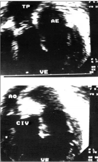
Before the advent of 2-D echocardiography, the diag-nosis of double-outlet left ventricle was only possible through cardiac catheterization, postmortem examination, or even as surgical findings. Currently, 2-D echocardiogra-phy together with color-flow imaging enables fast diagno-sis, showing both arteries arising from the morphologic left ventricle. No further difficulties occur in identifying the rela-Fig. 1 - Upper panel: Four chamber view depicting. Tricuspid atresia (arrow) and the hypoplastic right ventricle (VD).
Lower panel: Aorta (Ao) and pulmonary artery (TP) longitudinally related to the left
ventricle. AD - Right atrium; AE - Left Atrium; VE Left ventricle. Fig. 2 - Upper panel: Pulmonary artery (TP) totally related to the left ventricle (VE) and mitral-pulmonary continuity, in anterior position.
Lower panel: The aorta (Ao) is related to the left ventricle (VE) overiding a ventricu-lar septal (CIV). AE: Left atrium.
tionship between sigmoid valves and atrioventricular valves; presence or absence of infundibulum; common as-sociated anomalies, such as aortic coarctation, aortic ste-nosis, pulmonary steste-nosis, tricuspid atresia, right ventricle hypoplasia, and interventricular communication.
It is important to highlight the rarity of the case des-cribed here because it did not involve pulmonary stenosis (17% of cases). Eighty-three percent of cases described by Donald and William 10 of double-outlet left ventricle
asso-ciated with subaortic interventricular communication and with anterior and right aorta were also associated with pul-monary stenosis. This case is in agreement with data found in the literature 11 where an improvement in
5 1 6
Lopes et al
Double outlet left ventricle
Arq Bras Cardiol 2001; 76: 514-6.
1. Van Praagh R, Weinberg PM, Srebro JP. Double outlet left ventricle, In: Adams FH, Emmanouilides GL, Riemen-Schneider TA, eds. Heart Diseases in Infants Children and Adolescents. Baltimore: Williams and Wilkins, 1989: 461-73.
2. Sakakibara S, Takao A, Arai T, Hashimoto A, Nogi M. Both great vessels arising from the left ventricle (Double outlet left ventricle) (Origin of both great vessels from the left ventricle) Bull Heart Institute. Japan, 1967: 66.
3. Wilkinson JL. Double outlet left ventricle. In: Anderson RH, Macartney FJ, Shinebourne EA, Tynan M, eds. Paediatric cardiology, vol 2. Edinburgh: Churchill Living-Stone, 1987: 889-911.
4. Bharati S, Lev M, Stewart R, McAllister HA, Kirklin JW. The morphologic spec-trum of double outlet left ventricle and its surgical significance. Circulation 1978; 58: 558-65.
5. Otero Coto E, Quero Jimenez M, Castaneda AR, Rufilanchas JJ, Deverall PB. Double outlet from chambers of left ventricular morphology. Br Heart J 1979, 42: 15-21.
References
6. Gouton M, Bozio A, Rey C, Sassolas F, Vaksmann G, Filippo S. Le ventricule gauche à double issue: une cardiopathie rare et étonnante de diversité. Arch Mal Coeur Vaiss 1996; 89: 553-9.
7. Khanolkar UB, Deshpande JR, Kinare SG. Double outlet left ventricle with cor triatriatum. Indian Heart J 1990; 42: 393-5.
8. McElhinneyD, Reddy V, Hanley F. Pulmonary root translocation for biventricu-lar repair of double-outlet left ventricle with absent subpulmonic conus. J Thorac Cardiovasc Surg 1997; 114: 501-3.
9. DeLeon SY, Ow EP, Chiemmongkoltip P, et al. Alternatives in biventricular repair of double-outlet left ventricle. Ann Thorac Surg 1995; 60: 213-6. 10. Donald Jr H, William E. Double outlet left ventricle, In: Adams FH,
Emmanoui-lides GL, Riemen-schneider TA, eds. Heart Diseases in Infants Children and Adolescents. 5th ed. Baltimore: Williams and Wilkins, 1995, 1270-6.