2015/2016
João Luís Aragão Rodrigues
Ectropion
Mestrado Integrado em Medicina
Área: Oftalmologia Tipologia: Monografia
Trabalho efetuado sob a Orientação de: Doutora Sara Filipa Teixeira Ribeiro
Trabalho organizado de acordo com as normas da revista: Ophtalmic Plastic and Reconstructive Surgery
João Luis Aragão Rodrigues
Ectropion
ECTROPION 1
2
Sara F T Ribeiro, MD, PhD1,3, João L A Rodrigues2, Maria Shekhovtsova, MD2,
3
Antonio A V Cruz, MD, PhD3
4 5
1
Department of Ophthalmology, Hospital de Braga, School of Health Sciences,
6
University of Minho, Braga, Portugal.
7
2
School of Medicine of Porto, University of Porto, Porto, Portugal.
8
3
Department of Ophthalmology, Otorhinolaryngology and Head and Neck Surgery,
9
Hospital das Clínicas-Campus, School of Medicine of Ribeirão Preto, University of
10
São Paulo, Ribeirão Preto, São Paulo, Brazil.
11
12
Runing head: Ectropion. 13
14
Corresponding author: Sara Filipa Teixeira Ribeiro 15
Postal address: Department of Ophthalmology, Hospital de Braga, School of Health
16
Sciences, University of Minho, Rua Sete Fontes – São Vítor 4710-243 Braga,
17 Portugal. 18 Email: sara_ribeiro25@hotmail.com 19 Telephone: 00351-919589546 20
Précis 21
Ectropion is an abnormal eversion of the eyelid margin. The understanding of the
22
underlying pathophysiological mechanism is the key to a successful surgical
23
treatment.
ABSTRACT 25
26
Purpose: Ectropion is one of the most common eyelid malpositions. The treatment 27
of ectropion is still a subject of controversy. The authors reviewed the literature about
28
ectropion, including definition, diagnosis, pathophysiological mechanisms,
29
classification and surgical treatments.
30
Methods: A literature search was performed on the MEDLINE database using the 31
keywords ectropion, eyelid malposition, cicatricial ectropion, congenital ectropion,
32
involucional ectropion, mechanical ectropion, tarsal strip; lazy-T; canthoplasty,
33
canthopexy, skin flaps, skin grafts. Only articles in english were included.
34
Results: There is no consensus about the best surgical treatment for this eyelid 35
malposition, however, we know that the identification of the anatomic abnormality
36
associated is the most important point to chose the correct surgical treatment. The
37
literature on the treatment of ectropion mainly includes descriptions of surgical
38
techniques without objective measurements of the results, and uncontrolled studies.
39
Conclusions: The scientific literature on ectropion is vast, however there is a need 40
for quantitative studies on the effects of ectropion correction using different surgical
41
techniques.
Ectropion is an eversion (outward turning) of the eyelid margin. This lid
43
malposition may affect the lower or upper lid and leads to cosmetic and functional
44
deficits, such as conjunctival hyperemia, corneal exposure, eye irritation, epiphora,
45
chronic conjunctivitis, and in rare cases visual loss. It can be medial or lacrimal,
46
lateral or complete (involving the entire eyelid).1,2
47 48
METHODS OF LITERATURE SEARCH 49
A literature search was performed on the MEDLINE database using the
50
keywords ectropion, eyelid malposition, cicatricial ectropion, congenital ectropion,
51
involucional ectropion, mechanical ectropion, tarsal strip; lazy-T; canthoplasty,
52
canthopexy, skin flaps, skin grafts. Only articles in english were included.
53
The research adhered to the tenets of the Declaration of Helsinki and was
54
approved by the Ethics Committee of the Hospital de Braga, Braga, Portugal. An
55
informed consent was obtained from patients whose photos were used in this article.
56
57
CLASSIFICATION OF ECTROPION 58
59
Ectropion is usually classified as congenital, involutional, cicatricial,
60
mechanical and paralytic.1,2 These categories are not absolute and some cases fit in
61
more than one type. For instance, a four-lid ectropion in a newborn with lamellar
62
ichthyosis can be considered as a congenital or a cicatricial ectropion. Similarly, the
63
ectropion of a child born with an orbital cyst, which mechanically pushes the lid
64
margin, may be seen as a congenital mechanical ectropion, and congenital facial
65
palsies may cause a congenital paralytic ectropion.
From a therapeutic perspective, rather than focusing on the clinical categories
67
it is more useful to look at the underlying pathophysiological mechanism (Table 1)
68
because the correct identification of the anatomic abnormality associated with the lid
69
margin malposition is the key to a successful surgical procedure.
70 71
CONGENITAL ECTROPION 72
The designation “congenital ectropion” is used to name a variety of conditions
73
with distinct pathophysiological mechanisms. A typical example is the so-called
74
congenital upper eyelid eversion. This rare lid abnormality, usually present at birth, is
75
characterized by eversion of both upper lid margins with prolapsed chemotic
76
conjunctiva. It is typically symmetrical but some degree of asymmetry is not
77
uncommon. Several factors have been implicated in its pathophysiology including
78
trauma at birth, orbicularis hypotonia, posterior lamella vertical elongation and failure
79
of the orbital septum to fuse with the levator aponeurosis with adipose tissue
80
interposition. Most cases are not associated with ocular or general abnormalities.
81
However, the incidence appears to be higher in Down syndrome. This peculiar form
82
of upper lid ectropion responds well to conservative treatment such as topical
83
ointments and lubricants, patching or temporary tarsorrhaphy.3-6
84
A totally different situation is represented by newborns with severe forms of
85
lamellar ichthyosis. In these patients eyelid eversion is due to severe skin
86
contraction. All four lids may undergo a severe form of cicatricial ectropion that might
87
require early skin grafting in order to prevent corneal ulceration and eye perforation.
88
Shortening of the anterior lamella is also implicated in other syndromes associated
89
with congenital ectropion such as blepharophimosis (lower lid laterally)7,
blepharocheilodontic syndrome(lower eyelids)8-10 and Down syndrome (upper and
91
lower lids)11, as well a variety of sporadic congenital anomalies including craniofacial
92 clefting.12 93 94 INVOLUTIONAL ECTROPION 95
Involutional ectropion is the most frequent form of lower eyelid eversion,
96
commonly found in aged patients. The Blue Mountain Eye Study assessed the
97
prevalence and associations of ectropion in a large cohort of residents of Sydney,
98
Australia (3654 people aged 49-97 years). Ectropion prevalence was higher in men
99
(5.1%) than women (3.0%). The prevalence of lower eyelid ectropion increased with
100
age, reaching 16.7% among patients above 80 years old.13 Damasceno studied a
101
Brazilian population of 24 565 elderly people and found similar results, with a
102
prevalence of 17.7% in subjects aged 80 years or more.14
103
Several factors have been implicated in the pathogenesis of this eyelid
104
deformity, all related to an abnormal laxity of the lid support system including tarsal
105
and orbital septum atrophy, thinning of the skin and subcutaneous tissues,
106
elongation of the tarsus and pretarsal orbicularis, medial and lateral canthal tendon
107
laxity and dehiscence, and elongation or desinsertion of the lower lid retractors.2
108
Heimmel suggested that when these predisposing factors are present, the key to
109
final lower eyelid position is the globe axial projection, with relatively exophthalmic
110
eyes being more likely to develop tarsal ectropion.15
111
Histopathological studies have confirmed the role of age-related changes in
112
the tarsal plate, inferior retractors, orbicularis oculi and lateral canthal tendon in the
lids with involutional ectropion.16,17 The affected lid shows the presence of collagen
114
degeneration and elastosis of the tarsal plate and canthal tendons, an increased
115
amount of adipose tissue in the distal tarsus, and focal degeneration, fibrosis, and
116
elastosis of the pretarsal orbicularis.18-20 In chronic sun exposure, actinic damage on
117
the anterior lamella contributes as an additional factor to lower eyelid eversion in
118
patients with involutional ectropion.21
119
The evaluation of a patient with involutional ectropion should start with the
120
inspection of the lid margin and the position of the puncta since one the first
121
manifestations of lower eyelid ectropion is epiphora secondary to lacrimal punctum
122
eversion. The patency of the puncta and lacrimal drainage system should always be
123
examined.
124
The lower eyelid laxity can be detected using the “pinch test” and the “snap
125
back test”. The result is abnormal if the lid can be distended more than 6 mm from
126
the globe or does not briskly return to its natural position. The lateral canthus should
127
form an acute angle and a rounded shape is indicative of lateral tendon elongation. If
128
during traction of the lower lid there is lateral displacement of the lacrimal punctum
129
towards the limbus, there is medial tendon laxity.2, 22
130
The condition of the lower lid skin is assessed by pulling the lower lid margin
131
upwards. With traction the lid margin should reach a point at least 2 mm above the
132
limbus. A relatively immobile margin indicates vertical shortening of the anterior
133
lamella. This cicatricial component of the involutional ectropion is a common finding
134
when the ectropion is present for a long period of time or can be the result of mild
135
actinic changes of the skin. 2, 22
136
CICATRICIAL ECTROPION 138
Cicatricial ectropion is caused by anterior lamella shortening. Depending on
139
the etiologic mechanism, cicatricial entropion can affect the upper lids, the lower lids
140
or both (as observed in same cases of ichthyosis). It usuallyinvolves secondary
141
eyelid scars resulting from trauma23, 24, burns25-27 or from a large contingent of skin
142
diseases such as ichthyoses28, 29, discoid lupus erythematosus30, inherited
143
epidermolysis bullosa31, generalized eruptive keratoacanthoma32-34, cutaneous
144
leishmaniasis35, pityriasis rubra pilaris36, and pyoderma gangrenosum37, 38.
145
Postoperative complications of eyelid tumors39, 40, blepharoplasty41-44 and skin
146
resurfacing45, 46 are also common causes of lower lid cicatricial ectropion. Other
147
sources of skin damage resulting in ectropion are radiotherapy47, 48 and the use of
148
drugs such as docetaxel49, fluorouracil50-52, anthrax53, prostaglandins54, 55,
149
brimonidine55, betaxolol55, dorzolamide55, timolol55, and iopidine56.
150
151
PARALYTIC ECTROPION 152
Paralytic ectropion is caused by the lack of normal innervation of the
153
orbicularis muscle. Failure of normal lid closure with lower lid laxity and ectropion,
154
upper eyelid retraction and brow ptosis are the clinical signs. The causes of seventh
155
nerve palsy are myriad, but can be broadly divided, in the order of frequency, into
156
idiopathic (Bell’s palsy), traumatic (birth canal trauma or forceps delivery, surgical
157
trauma, facial and temporal bone fractures), infectious (herpes zoster, Lyme disease,
158
HIV infection, polio, mumps, cytomegalovirus, mononucleosis, leprosy, cat scratch
159
fever), and neoplastic (acoustic neuroma, cerebellopontine angle tumors).57
In paralytic ectropion due to surgical trauma, the timing for facial reanimation
161
surgery is under debate. Watts et al advocate that rehabilitation surgery with gold
162
weight implantation and lateral tarsal strip should be performed immediately, at the
163
time of facial nerve sacrifice.58
164 165
MECHANICAL ECTROPION 166
Mechanical ectropion is caused by eyelid tumors that evert the lower lid or
167
inflammatory disorders that cause orbicularis spasm. Large tumors or cysts near the
168
lid margin, acute proptosis with chemosis, eyelid and periocular edema, significant
169
herniated orbital fat and traction on the lower eyelid skin from spectacles can
170
mechanically cause ectropion. The treatment is directed at the cause.2, 59-62
171
172
TREATMENT 173
Lower or upper eyelid ectropions are usually managed with surgery. As
174
mentioned before, the key for a successful surgical procedure is the correct
175
preoperative identification of the underlying etiologic factor. Depending on the
176
mechanism provoking the lid margin rotation, a variety of procedures are used to
177
stabilize the lid margin. For instance, lower eyelid horizontal laxity with normal
178
canthal tendon tonus can be addressed by pentagonal full thickness eyelid resection.
179
If the lateral canthal tendonis abnormally lax tarsal strip procedures are indicated.1, 2,
180
63, 64 This useful procedure is a variant of the old Bick’s lateral resection65
and
181
consists of a lateral canthotomy and cantholysis; excision of skin and conjunctiva,
182
leaving a free strip of tarsus; fixation of the tarsal strip to the periosteum of the lateral
orbital wall; and reconstruction of the lateral canthus to create the appropriate height
184
and tension of the lower eyelid.63, 66 Usually monofilament non-absorbable sutures
185
are used 2, 64, 67, 68, but long-acting absorbable sutures will also work without
186
recurrence of horizontal laxity. 66, 69, 70
187
The repair of medial tendon laxity is more complicated than that of its lateral
188
counterpart because of the intimate relation of the tendon with the canaliculi. The
189
high rate of postoperative lacrimal problems following surgery on the medial canthus
190
leads some surgeons to delay correction of the medial canthal tendon until laxity is
191
advanced. Medial canthal tendon correction has been attempted by resection or
192
plication of the tendon medial to the lower punctum, anchoring the tissue to either
193
the anterior periosteum or posterior lacrimal crest.71-75
194
Lacrimal ectropion without horizontal laxity is classically treated with an
195
excision of a diamond of conjunctiva and retractors below the punctum. If a
196
horizontal laxity is present, a full-thickness wedge excision horizontally tight of the lid
197
margin can be associated with the pentagon excision creating a lazy-T procedure.
198
The wedge excision can be displaced laterally or be replaced by a tarsal strip. 76, 77
199
In both procedures the role of lower lid retractor plication has been
200
emphasized. Advanced tarsal ectropions require a large excision of conjunctiva and
201
retractors by a posterior approach combined with everting sutures and horizontal
202
tightening.78-80
203
Since cicatricial ectropion is due to an anterior lamella deficiency, various
204
surgical techniques can be used to lengthen the underlying deformity. Z-plasties can
205
be used to manage linear scars. 1, 2, 81 Large shortages of skin require local flaps 26,
206
82-85
or skin grafts 27, 48, 86. There are a large variety of flaps that can be used to
correct lower and upper ectropions depending on the location and extension of the
208
cicatricial process. Free skin grafts may be obtained from the upper lid, retroauricular
209
region, supraclavicular area or inner aspect of the upper arm.1, 2, 81
210
In severe lower eyelid ectropion, there is often inadequate muscular support
211
for the pretarsal lower eyelid. When the pretarsal orbicularis muscle is damaged, a
212
fascia lata sling is an option for supporting the lower lid.84
213
214
CONCLUSION 215
A successful functional and cosmetic correction of ectropion depends on the
216
understanding that ectropion is a group of eyelid malpositions that may have
217
different etiological factors and may affect lateral, central or medial portion of the lids,
218
alone or in combination. Surgery techniques must address the anatomic
219
abnormalities responsible for the ectropion. The choice of the right procedure leads
220
to a successful outcome for the patient which is the main goal.
REFERENCES 222
1. Hintschich C. Correction of entropion and ectropion. Dev Ophthalmol 2008;41:85-102.
223
2. Bedran EG, Pereira MV, Bernardes TF. Ectropion. Semin Ophthalmol 2010;25:59-65.
224
3. Dohvoma VA, Nchifor A, Ngwanou AN, et al. Conservative management in congenital
225
bilateral upper eyelid eversion. Case Rep Ophthalmol Med 2015;2015:389289.
226
4. Krishnappa NC, Deb AK, Poddar C. Congenital total eversion of upper eyelids in a newborn
227
with Down's syndrome. Oman J Ophthalmol 2014;7:98-9.
228
5. Ibraheem WA. Bilateral congenital upper eyelid eversion: the clinical course and outcome
229
of conservative management. Pan Afr Med J 2014;17:215.
230
6. Fasina O. Management of bilateral congenital upper eyelid eversion with severe
231
chemosis. J Ophthalmic Vis Res 2013;8:175-8.
232
7. Sandramouli S, Betharia SM. Blepharophimosis syndrome: an atypical case. Eye (Lond)
233
1994;8:482-4.
234
8. Gorlin RJ, Wiedemann HR. Blepharo-cheilo-dontic (BCD) syndrome. Acta Ophthalmol
235
Scand Suppl 1996;219:22.
236
9. Guion-Almeida ML, Rodini ES, Kokitsu-Nakata NM, Bologna-Amantini D.
Blepharo-Cheilo-237
Dontic (BCD) syndrome: report on four new patients. Am J Med Genet 1998;76:133-6.
238
10. Yen MT, Lucci LM, Anderson RL. Management of eyelid anomalies associated with
239
Blepharo-cheilo-dontic syndrome. Am J Ophthalmol 2001;132:279-80.
240
11. Sellar PW, Bryars JH, Archer DB. Late presentation of congenital ectropion of the eyelids
241
in a child with Down syndrome: a case report and review of the literature. J Pediatr
242
Ophthalmol Strabismus 1992;29:64-7.
243
12. Iida A, Narai S, Takagi R, et al. Blepharo-cheilo-dontic (BCD) syndrome: case report. Cleft
244
Palate Craniofac J 2006;43:237-43.
13. Mitchell P, Hinchcliffe P, Wang JJ, et al. Prevalence and associations with ectropion in an
246
older population: the Blue Mountains Eye Study. Clin Experiment Ophthalmol
2001;29:108-247
10.
248
14. Damasceno RW, Osaki MH, Dantas PE, Belfort R, Jr. Involutional entropion and ectropion
249
of the lower eyelid: prevalence and associated risk factors in the elderly population. Ophthal
250
Plast Reconstr Surg 2011;27:317-20.
251
15. Heimmel MR, Enzer YR, Hofmann RJ. Entropion-ectropion: the influence of axial globe
252
projection on lower eyelid malposition. Ophthal Plast Reconstr Surg 2009;25:7-9.
253
16. Chua J, Choo CT, Seah LL, et al. A 5-year retrospective review of Asian ectropion: how
254
does it compare to ectropion amongst non-Asians? Ann Acad Med Singapore 2011;40:84-9.
255
17. Bashour M, Harvey J. Causes of involutional ectropion and entropion--age-related tarsal
256
changes are the key. Ophthal Plast Reconstr Surg 2000;16:131-41.
257
18. Kocaoglu FA, Katircioglu YA, Tok OY, et al. The histopathology of involutional ectropion
258
and entropion. Can J Ophthalmol 2009;44:677-9.
259
19. Damasceno RW, Osaki MH, Dantas PE, Belfort R, Jr. Involutional ectropion and
260
entropion: clinicopathologic correlation between horizontal eyelid laxity and eyelid
261
extracellular matrix. Ophthal Plast Reconstr Surg 2011;27:321-6.
262
20. Damasceno RW, Heindl LM, Hofmann-Rummelt C, et al. Pathogenesis of involutional
263
ectropion and entropion: the involvement of matrix metalloproteinases in elastic fiber
264
degradation. Orbit 2011;30:132-9.
265
21. Marshall JA, Valenzuela AA, Strutton GM, Sullivan TJ. Anterior lamella actinic changes as
266
a factor in involutional eyelid malposition. Ophthal Plast Reconstr Surg 2006;22:192-4.
267
22. Frueh BR, Schoengarth LD. Evaluation and treatment of the patient with ectropion.
268
Ophthalmology 1982;89:1049-54.
23. Desciak EB, Eliezri YD. Surgical Pearl: Temporary suspension suture (Frost suture) to help
270
prevent ectropion after infraorbital reconstruction. J Am Acad Dermatol 2003;49:1107-8.
271
24. Salgarelli AC, Bellini P, Multinu A, Landini B, Consolo U. Tarsal strip technique for
272
correction of malposition of the lower eyelid after treatment of orbital trauma. Br J Oral
273
Maxillofac Surg 2009;47:298-301.
274
25. Astori IP, Muller MJ, Pegg SP. Cicatricial, postburn ectropion and exposure keratitis.
275
Burns 1998;24:64-7.
276
26. Kostakoglu N, Ozcan G. Orbicularis oculi myocutaneous flap in reconstruction of
277
postburn lower eyelid ectropion. Burns 1999;25:553-7.
278
27. Mandrekas AD, Zambacos GJ, Anastasopoulos A. Treatment of bilateral severe eyelid
279
burns with skin grafts: an odyssey. Burns 2002;28:80-6.
280
28. Cruz AA, Menezes FA, Chaves R, et al. Eyelid abnormalities in lamellar ichthyoses.
281
Ophthalmology 2000;107:1895-8.
282
29. Craiglow BG, Choate KA, Milstone LM. Topical tazarotene for the treatment of ectropion
283
in ichthyosis. JAMA Dermatol 2013;149:598-600.
284
30. Kopsachilis N, Tsaousis KT, Tourtas T, Tsinopoulos IT. Severe chronic blepharitis and
285
scarring ectropion associated with discoid lupus erythematosus. Clin Exp Optom
286
2013;96:124-5.
287
31. Fine JD, Johnson LB, Weiner M, et al. Eye involvement in inherited epidermolysis bullosa:
288
experience of the National Epidermolysis Bullosa Registry. Am J Ophthalmol
2004;138:254-289
62.
290
32. Consigli JE, Gonzalez ME, Morsino R, et al. Generalized eruptive keratoacanthoma
291
(Grzybowski variant). Br J Dermatol 2000;142:800-3.
33. Oakley A, Ng S. Grzybowski's generalized eruptive keratoacanthoma: remission with
293
cyclophosphamide. Australas J Dermatol 2005;46:118-23.
294
34. Anzalone CL, Cohen PR. Generalized eruptive keratoacanthomas of Grzybowski. Int J
295
Dermatol 2014;53:131-6.
296
35. Chaudhry IA, Hylton C, DesMarchais B. Bilateral ptosis and lower eyelid ectropion
297
secondary to cutaneous leishmaniasis. Arch Ophthalmol 1998;116:1244-5.
298
36. Durairaj VD, Horsley MB. Resolution of pityriasis rubra pilaris-induced cicatricial
299
ectropion with systemic low-dose methotrexate. Am J Ophthalmol 2007;143:709-10.
300
37. Procianoy F, Barbato MT, Osowski LE, Bocaccio FJ, Bakos L. Cicatricial ectropion
301
correction in a patient with pyoderma gangrenosum: case report. Arq Bras Oftalmol
302
2009;72:384-6.
303
38. Thampy RS, Al-Niaimi F, Lyon C, Duff CG, Leatherbarrow B. Management of cicatricial
304
ectropion secondary to pyoderma gangrenosum. Orbit 2014;33:129-31.
305
39. Rubin P, Mykula R, Griffiths RW. Ectropion following excision of lower eyelid tumours
306
and full thickness skin graft repair. Br J Plast Surg 2005;58:353-60.
307
40. Salgarelli AC, Francomano M, Magnoni C, Bellini P. Cicatricial iatrogenic lower eyelid
308
malposition in skin cancer surgery: results of a combined approach. J Craniomaxillofac Surg
309
2012;40:579-83.
310
41. Botti G. Blepharoplasty: A classification of selected techniques in the treatment and
311
prevention of lower lid margin distortions. Aesthetic Plast Surg 1998;22:341-8.
312
42. Morax S, Touitou V. Complications of blepharoplasty. Orbit 2006;25:303-18.
313
43. Patrocinio TG, Loredo BA, Arevalo CE, et al. Complications in blepharoplasty: how to
314
avoid and manage them. Braz J Otorhinolaryngol 2011;77:322-7.
44. Mack WP. Complications in periocular rejuvenation. Facial Plast Surg Clin North Am
316
2010;18:435-56.
317
45. Demas PN, Bridenstine JB. Diagnosis and treatment of postoperative complications after
318
skin resurfacing. J Oral Maxillofac Surg 1999;57:837-41.
319
46. Fife DJ, Fitzpatrick RE, Zachary CB. Complications of fractional CO2 laser resurfacing: four
320
cases. Lasers Surg Med 2009;41:179-84.
321
47. Tarallo M, Rizzo MI, Monarca C, et al. Optimal care for eyelid contraction after
322
radiotherapy: case report and literature review. J Oral Maxillofac Surg 2012;70:2459-65.
323
48. Kim HJ, Hayek B, Nasser Q, Esmaeli B. Viability of full-thickness skin grafts used for
324
correction of cicatricial ectropion of lower eyelid in previously irradiated field in the
325
periocular region. Head Neck 2013;35:103-8.
326
49. Kaya AO, Buyukberber S, Coskun U, et al. Acute erythema and edematous skin reaction
327
and ectropion following docetaxel in a patient with non-small cell lung cancer. Cutan Ocul
328
Toxicol 2008;27:327-31.
329
50. Lewis JE. Temporary ectropion due to topical fluorouracil. Int J Dermatol 1997;36:79.
330
51. Obi EE, McDonald A, Kemp E. A bilateral cicatricial ectropion and bilateral upper lid
331
shortening caused by 5-fluorouracil toxicity in a patient with dihydropyrimidine
332
dehydrogenase deficiency. Cutan Ocul Toxicol 2011;30:157-9.
333
52. Nikkhah D, Abood A, Watt D. Cicatricial ectropion: a complication of topical
5-334
fluorouracil. J Plast Reconstr Aesthet Surg 2012;65:e9-10.
335
53. Soysal HG, Kiratli H, Recep OF. Anthrax as the cause of preseptal cellulitis and cicatricial
336
ectropion. Acta Ophthalmol Scand 2001;79:208-9.
337
54. Altieri M, Ferrari E. Do prostaglandin analogs affect eyelid position and motility? J Ocul
338
Pharmacol Ther 2011;27:511-7.
55. Hegde V, Robinson R, Dean F, Mulvihill HA, Ahluwalia H. Drug-induced ectropion: what is
340
best practice? Ophthalmology 2007;114:362-6.
341
56. Britt MT, Burnstine MA. Iopidine allergy causing lower eyelid ectropion progressing to
342
cicatricial entropion. Br J Ophthalmol 1999;83:992-3.
343
57. Mavrikakis I. Facial nerve palsy: anatomy, etiology, evaluation, and management. Orbit
344
2008;27:466-74.
345
58. Watts TL, Chard R, Weber SM, Wax MK. Immediate eye rehabilitation at the time of
346
facial nerve sacrifice. Otolaryngol Head Neck Surg 2011;144:353-6.
347
59. Ashfaq I, Kyprianou I, Ahluwalia H. A large kissing (divided) naevus presenting with
348
complete mechanical ptosis and lower lid ectropion. J Plast Reconstr Aesthet Surg
349
2009;62:e87-8.
350
60. Kampp JT, Kouba DJ, Fincher EF, Moy RL. Basal cell carcinoma masquerading as the
351
chronic ectropion of lamellar ichthyosis. Dermatol Surg 2008;34:963-7.
352
61. Baker HE, Berry-Brincat A, Zaki I, Cheung D. Three different consecutive manifestations
353
of morphoeic BCC in the same patient: presenting first as ectropion, then entropion, and
354
finally medial canthal dystopia with epicanthus inversus. Orbit 2008;27:183-5.
355
62. Evans M, Chang E, Yu DL, Rao NA. Granular cell tumour: a rare caruncle lesion. Br J
356
Ophthalmol 2006;90:246-7.
357
63. Anderson RL, Gordy DD. The tarsal strip procedure. Arch Ophthalmol 1979;97:2192-6.
358
64. Della Rocca DA. The lateral tarsal strip: illustrated pearls. Facial Plast Surg
2007;23:200-359
2.
360
65. Bick MW. Surgical management of orbital tarsal disparity. Arch Ophthalmol
1966;75:386-361
9.
66. Jordan DR, Anderson RL. The lateral tarsal strip revisited. The enhanced tarsal strip. Arch
363
Ophthalmol 1989;107:604-6.
364
67. Kam KY, Cole CJ, Bunce C, Watson MP, Kamal D, Olver JM. The lateral tarsal strip in
365
ectropion surgery: is it effective when performed in isolation? Eye (Lond) 2012;26:827-32.
366
68. Tenzel RR, Buffam FV, Miller GR. The use of the "lateral canthal sling" in ectropion
367
repair. Can J Ophthalmol 1977;12:199-202.
368
69. Hsuan J, Selva D. The use of a polyglactin suture in the lateral tarsal strip procedure. Am
369
J Ophthalmol 2004;138:588-91.
370
70. Meyer DR. The use of a polyglactin suture in the lateral tarsal strip procedure. Am J
371
Ophthalmol 2005;139:758-9.
372
71. O'Donnell BA, Anderson RL, Collin JR, et al. Repair of the lax medial canthal tendon. Br J
373
Ophthalmol 2003;87:220-4.
374
72. Moe KS, Kao CH. Precaruncular medial canthopexy. Arch Facial Plast Surg
2005;7:244-375
50.
376
73. Sullivan TJ, Collin JR. Medical canthal resection: an effective long-term cure for medial
377
ectropion. Br J Ophthalmol 1991;75:288-91.
378
74. Francis IC, Wilcsek GA, Sharma S, Coroneo MT. Transcaruncular medial orbitotomy for
379
stabilization of the posterior limb of the medial canthal tendon. Clin Experiment Ophthalmol
380
2001;29:85-9.
381
75. Edelstein JP, Dryden RM. Medial palpebral tendon repair for medial ectropion of the
382
lower eyelid. Ophthal Plast Reconstr Surg 1990;6:28-37.
383
76. Manners RM. Surgical repair of medial ectropion. Eye (Lond) 1995;9:365-7.
384
77. Smith B. The "lazy-T" correction of ectropion of the lower punctum. Arch Ophthalmol
385
1976;94:1149-50.
78. Ferguson AW, Chadha V, Kearns PP. The not-so-lazy-T: a modification of medial
387
ectropion repair. Surgeon 2006;4:87-9.
388
79. O'Donnell B. Age-related medial ectropion of the lower eyelid. Aust N Z J Ophthalmol
389
1994;22:183-6.
390
80. Fong KC, Mavrikakis I, Sagili S, Malhotra R. Correction of involutional lower eyelid medial
391
ectropion with transconjunctival approach retractor plication and lateral tarsal strip. Acta
392
Ophthalmol Scand 2006;84:246-9.
393
81. Miletic D, Elabjer BK, Bosnar D, Busic M. Our approach to operative treatment of lower
394
lid ectropion. Acta Clin Croat 2010;49:283-7.
395
82. Xue CY, Dai HY, Li L, et al. Reconstruction of lower eyelid retraction or ectropion using a
396
paranasal flap. Aesthetic Plast Surg 2012;36:611-7.
397
83. Xu JH, Tan WQ, Yao JM. Bipedicle orbicularis oculi flap in the reconstruction of the lower
398
eyelid ectropion. Aesthetic Plast Surg 2007;31:161-6.
399
84. Qian JG, Wang XJ, Wu Y. Severe cicatrical ectropion: repair with a large advancement
400
flap and autologous fascia sling. J Plast Reconstr Aesthet Surg 2006;59:878-81.
401
85. Manku K, Leong JK, Ghabrial R. Cicatricial ectropion: repair with myocutaneous flaps and
402
canthopexy. Clin Experiment Ophthalmol 2006;34:677-81.
403
86. Lau CK, Huang S, Cormack G. Minimising the risk of ectropion when full thickness skin
404
grafting lower eyelid defects. J Plast Reconstr Aesthet Surg 2008;61:1562-4.
Table 1. Type of ectropion, affected lids and pathophysiologic mechanism.
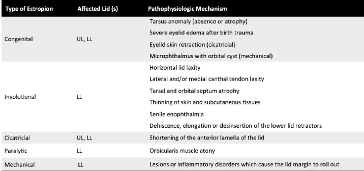
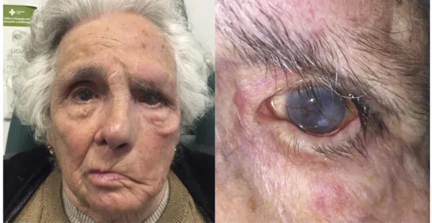
Figure 1. Lamellar ichthyosis. Congenital cicatricial ectropion of the lower
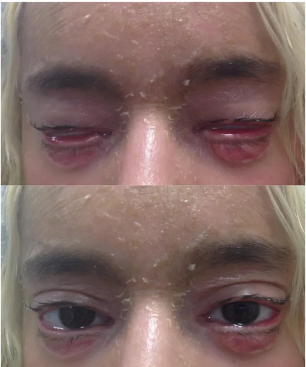
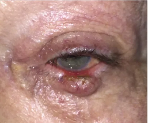
Figure 2. Cicatricial ectropion caused by an excision of basal cell carcinoma of the
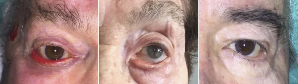
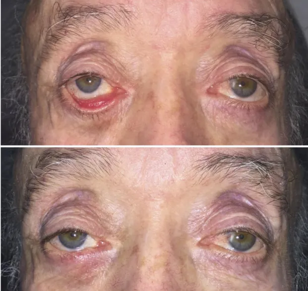
Figure 4. Left lower lid paralytic ectropion. Note lower lid laxity and ectropion, brow
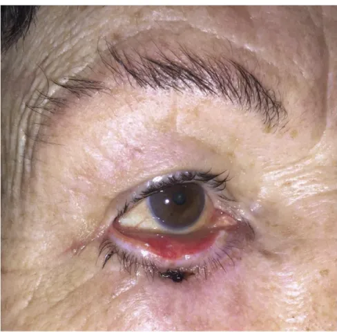
Figure 6. Lacrimal ectropion of the right lower eyelid (top) corrected with a Lazy-T
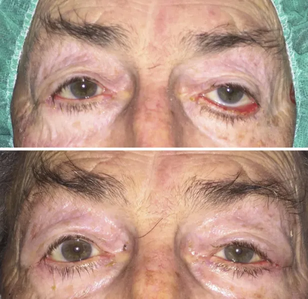
Figure 7. Left involutional lower lid ectropion. Top: Preoperative appearence.
Um agradecimento muito especial à minha orientadora, a Doutora Sara
Filipa Ribeiro Teixeira. Agradecimento também muito especial à família e
aos amigos que sempre me acompanharam nesta jornada.
Ophthalmic Plastic and Reconstructive Surgery
INFORMATION FOR AUTHORSOphthalmic Plastic and Reconstructive Surgery (OPRS) publishes Original
Investigations describing clinical and laboratory investigations, Case Reports, Surgical Techniques, Anatomy & Physiology, and Reviews, and by invitation, Commentaries,
Perspectives, and Editorials. The journal is owned and sponsored by the American Society of Ophthalmic Plastic and Reconstructive Surgery (ASOPRS) but welcomes submissions from authors who are not members of the Society.
GENERAL INFORMATION AND POLICIES
OPRS subscribes to the policies outlined in the “Uniform Requirements for Manuscripts
Submitted to Biomedical Journals” written by the International Committee of Medical Journal Editors (www.ICMJE.org).
Authorship. OPRS expects each author to have made a significant intellectual contribution
to the design or execution of the project, to the writing of the manuscript, or both. Each author must take full responsibility for his or her contribution and must have approved the final manuscript. (Lundberg GD, Glass RM. What does authorship mean in a peer-reviewed medical journal? [editorial] JAMA 1996;276:75.)
Papers from the ASOPRS Annual Scientific Symposia. OPRS holds copyright and has
the right of first refusal for all manuscripts derived from papers presented at the Annual Fall and Spring Symposia of the American Society of Ophthalmic Plastic and Reconstructive Surgery. Permission to submit such work elsewhere must be sought from the Editor. Presenters who violate this policy will be excluded by the Program Committee from participation in the Annual Scientific Symposia for 2 years.
Prior and Repetitive Publication. OPRS will not consider manuscripts that have appeared
in part or in total in other publications, except in special circumstances by approval of the Editor. Updates of previously published studies that add little data to an existing publication will not be considered. Overlap between patient groups described in serial manuscripts must be acknowledged, and references to previous publications that include the same patients must be provided. Authors uncertain as to whether specific data represent prior or repetitive publication should alert the Editor in the transmittal letter and include copies of the
publication(s) in question.
Institutional Review Board Approval
For all manuscripts reporting data from studies involving human subjects, human-derived material, human medical records, or animals, formal ethical standards review and approval, or formal review and waiver, by an appropriate institutional review board (IRB) or ethics committee is required and must be described in the Methods section. Case reports presenting the clinical or surgical results on three patients or less, are generally not
considered research, and therefore do not require IRB review. However, discussion or chart reviews of four or more patients, especially when informed consent is obtained, or any form of analysis or comparison with other studies is performed, may be considered research, and should be reviewed by an IRB board for approval or waiver. The IRB decision should be included in the introduction. For those investigators who do not have access to a formal institutional ethics review committee, a non-institutional regional IRB should be utilized. For non-USA investigators, who do not have access to any formal review board, please include a
statement to this effect in the Methods, and include a statement that the study adhered to the principles outlined in the Declaration of Helsinki
(http://www.wma.net/en/30publications/10policies/b3/). Editors may request that authors provide documentation of the formal review and recommendation from the institutional review board or ethics committee responsible for oversight of the study.
Patient Consent
For investigations on human subjects, state in the Methods section the manner in which informed consent was obtained from the study participants (ie, oral or written) and for US authors, that the study was HIPPA-compliant (http://www.hhs.gov/ocr/privacy/index.html).
Clinical Photographs
All clinical and radiographic photographs that permit identification of the patient must have a signed consent by the patient or guardian, which is to be archived by the authors. This statement does not have to be submitted with the manuscript. A statement that this consent was obtained and is on file must be included in the Methods or Acknowledgement section. It is not acceptable to place bars over the patient's features, but in cases where permissions are unobtainable, the photographs must be very tightly cropped to the feature being
displayed. If identification is still possible after cropping, OPRS cannot use the photograph. In the case of a patient who is deceased, written permission must be provided by the patient's next of kin.
Animal Studies. If animals were used in a study, the manuscript should include the
appropriate IRB approval statement.
Statistical Analysis. OPRS strongly advises statistical consultation about data collection and
analysis. Statistical methods must be identified in table footnotes, illustration legends, or text explanations. Software programs used for complex statistical analyses must be identified to enable reviewers to verify calculations.
MANUSCRIPT PREPARATION
Manuscripts should be prepared using Microsoft Word. The manuscript should be paginated and double-spaced, with 1-inch margins and left-justified text. Please activate line
numbering: File/pagesetup/page layout: Show line numbers, Line numbering:
Continuous/Begin with line 1. Article length can be estimated according to the following formula: 1 journal page = 3 double-spaced type-written pages, OR 4 single-image figures, OR 2 tables. Submissions should include the intended article type as follows.
Original Investigation: These articles discuss original clinical or basic research with a detailed
review of the literature that the research supports, expands or modifies. Submissions should be limited to six journal pages.
Major Review: These articles present a comprehensive review of the current literature
summarizing our state of knowledge on a specific subject. Submissions are limited to 10 journal pages.
Surgical Technique: This article describes a new or modified surgical procedure, with or
without case descriptions to illustrate the technique. Limited to 2 journal pages. Authors are encouraged to submit a video to accompany Surgical Technique articles to enhance the article content. Please refer to the Supplemental Digital Content section of this document for
instructions on submitting videos as Supplemental Digital Content, including directions on necessary file formats and required call-outs within the article.
Anatomy & Physiology: Includes original anatomical or physiological descriptions related to
the eyelids, orbit, or lacrimal systems. Limited to 3 journal pages.
Case Report: These are clinical descriptions of one or more patients with a brief discussion of
the relevant literature. Case reports will be published on-line only. Limited to 2 journal pages and 12 references. For all case reports originating in the USA, the introductory paragraph should state that collection and evaluation of protected patient health information was HIPAA-compliant.
Images in Ophthalmic Plastic and Reconstructive Surgery: Submissions should contain a
maximum of 2 figures with accompanying figure legend. Video segments may also be included but submissions must include a high-resolution still image. Limited to ½ journal page. Please see Images in OPRS for more details.
Letter to the Editor: Letters are brief non-peer reviewed comments that relate to recently
published papers in the journal; they may also present a brief discussion of the authors' clinical or research experience. Limited to 1 journal page. Letter titles should be “Re:” or “Reply re:” followed by the title of the article to be discussed, and the discussed article should be listed as a reference. If the letter pertains only to your own research experience, just an appropriate title is needed. Letters should be addressed to “To the Editor:”.
Title Page. The title page should include the following information:
1. Title–no longer than 135 characters. Declarative titles should not be used.
2. Names of authors–provide first name, middle initial, last name, and advanced degrees or professional certification.
3. Institutional affiliation–indicate each author’s affiliation during the course of the study in footnotes on the title page using superscript numbers, not symbols (e.g., John Doe1).
4. Meeting presentation–if the material has been presented previously, supply the name, place, and date of the meeting.
5. Financial support–identify all sources, public and private. Provide the agency name and city, company name and city, fellowship name, and grant number.
6. Proprietary interest statement–each author is expected to disclose any type of financial interest that is related to the manuscript, including stock or ownership of a business entity connected to a product described in the paper, paid consulting for the company or competing companies, or patent rights to a drug or piece of equipment. Authors must disclose personal or family ownership or potential rights to more than 1% of the company or competing company and whether they have any interest in marketing any product, drug, instrument, or piece of equipment discussed in the manuscript.
7. Running head–no longer than 60 characters.
8. Corresponding author–contact information for reprints.
Précis. On a separate page (page 2), include a one-sentence précis (35 words or fewer)
summarizing the main finding or outcome of the study. The précis will appear under the title in the Table of Contents and should not duplicate the abstract conclusion.
Structured Abstract. Each manuscript must include a structured abstract of no more than
250 words (except for Case Reports; see below). The abstract must appear on a separate page (page 3) and should include four separate sections:
Purpose: Provide a concise statement of the study goal (e.g., the question to be answered
or the hypothesis to be tested).
Methods: Identify the study design using a phrase such as randomized or nonrandomized
clinical trial, case-controlled study, cross-sectional study, cohort study, case series, case report, meta-analysis, review, experimental study, or historical manuscript. Patient selection, interventions, and outcome measures must be defined. For studies involving human
subjects, human-derived materials, or human medical records, include a statement declaring IRB approval, Helsinki adherence or HIPAA compliance as appropriate.
Results: Briefly summarize the principal measurements (data) obtained and relevant
statistical analysis.
Conclusions: State the specific conclusions derived from the data analysis and their clinical
significance.
Abstract for Case Reports. Provide an unstructured summary not to exceed 150 words on
a separate page following the précis.
Text. All manuscripts must follow generally recognized standards for presenting scientific
material. See the CBE Manual for Authors, Editors, and Publishers, 6th ed. (Council of Biology Editors; 1995). Type size (True Type fonts) should be 12 point. The Introduction, without a heading, should refer only to the most pertinent past publications and should not be an extensive review of the literature. Methods should be written with sufficient detail to permit others to duplicate the work. Results must be concise and not simply a reiteration of data presented in Tables. Discussion should be restricted to the significant findings
presented. Digressions and speculation are not appropriate.
References. References should follow text and begin on a separate page. They must be
double spaced and numbered consecutively in order of appearance in the text. References should be designated by superscript numbers following all punctuation (except semicolons and colons).
1. List only references that you have read and that are pertinent to the manuscript. 2. Cite only published studies as references. You may acknowledge “unpublished data” or “submitted” articles within parentheses in the text. Reference to a “personal communication” within parentheses in the text must be accompanied by a signed permission letter from the individual being cited.
3. Abstracts such as those published in the annual meeting programs of the American Society of Ophthalmic Plastic and Reconstructive Surgery (ASOPRS), the American Academy of Ophthalmology (AAO), or the Association for Research in Vision and Ophthalmology (ARVO) are considered “unpublished” and should be cited in parentheses in the text. For example:-by Smith et al. (Invest Ophthalmol Vis Sci 28 (Suppl):54, 1989).
4. Oral or poster presentations are similarly unpublished and may be cited only in parentheses in the text. Platform and poster presentations at annual meetings are customarily indicated: (Smith AB, presented at the AAO Annual Meeting, Atlanta, 1995). 5. Books or articles “in press” may be cited as numbered references. Such citations should be updated before publication, if possible.
Journal abbreviations should be those used by the National Library of Medicine, as found in Index Medicus. If in doubt as to the correct abbreviation, cite the complete journal name.
Do not underline journal titles, and do not use periods in abbreviations of journal titles or in author initials. Please follow precisely the format and punctuation shown in the following examples.
Journal Article–(If four or fewer authors, list all) Anderson RL, Beard C. The levator
aponeurosis: attachments and their clinical significance. Arch Ophthalmol1977;95:1437– 41.
Journal Article–(If five or more authors, list only the first three and add et al) Meyer
DR, Bui HX, Carlson JA, et al. Silicon granulomas and dermatomyositis-like changes associated with chronic eyelid edema after silicone breast implant. Ophthal Plast Reconstr
Surg 1998;14:182–8.
Chapter in a Book–Kaltreider SA, Sherman DD, McGetrick JJ. Eyelid trauma. In: Dortzbach
RK, ed. Ophthalmic plastic surgery: prevention and management of complications. New York: Raven Press, 1994:157–74.
Book-Miller NR. Walsh and Hoyt’s clinical neuro-ophthalmology. 4th ed. Vol. 4. Baltimore:
Williams & Wilkins; 1991; 2102–14.
Letter to the Editor-Sneed SR, Blodi CF, Berger BB, et al. Pneumocystis carinii choroiditis
in patients receiving inhaled pentamidine [letter]. N Engl J Med 1990;322:936 –7.
Online Journal Article-LaPorte RE, Marler E, Akazawa S, Sauer F. The death of biomedical
journals. JAMA [serial online]. 1996;310:1387–90. Available
athttp://www.jama.com/jama/archive/6991ed2.htm. Accessed June 16, 1997.
Web Site–Health Care Financing Administration. 1996 statistics at a glance. Available
at: http://www.hcfa.gov/stats/stathili.htm. Accessed December 2, 1997.
Tables. Do not embed tables within the body of the manuscript. Each table must be
numbered consecutively using Arabic numbers, be mentioned in the text, and be titled. Each column must have a heading. All abbreviations must be explained in the legend. Please do not place more than one table on a page.
Legends. Figure legends (photos, drawings, graphs) should be placed at the end of the
manuscript. Do not embed figures within the body of the manuscript. Figures must be numbered consecutively as they appear in the text. For histologic figures, stains and magnifications should be noted in the legend. Any figure that has been published elsewhere should have an acknowledgment to the original source; a copy of the release to publish the figure, signed by the copyright holder, must also be submitted. Legends must identify all symbols or letters that appear on the prints.
Abbreviations. Restrict abbreviations to those that are widely used and understood. Avoid
abbreviations that have meaning only in the context of your specific manuscript. An abbreviation should appear first in parentheses immediately after the term or phrase to which it refers. Abbreviations may only be used if the term appears five or more times in the text. Abbreviations are not used in abstracts.
Drug/Manufacturer Names. Use generic names only in the text body. Include the trade
after the first use of the generic name. In the case of equipment, include manufacturer’s name, city, state, and/or country.
Illustrations. Illustrations should be prepared according to the image guidelines for online
manuscript submission.
Copyright. Consideration of manuscripts for publication in Ophthalmic Plastic and
Reconstructive Surgery is dependent on the assurance that the material (in whole or part) is
not under consideration by another journal, is not in press in any other format, and has not been previously published. Each author must sign a statement transferring copyright ownership to the American Society of Ophthalmic Plastic and Reconstructive Surgery. Copyright transfer forms are printed in each issue of the journal and are available online at the journal’s Web site (www.op-rs.com).
Conflicts of Interest. Authors must state all possible conflicts of interest in the manuscript,
including financial, consultant, institutional and other relationships that might lead to bias or a conflict of interest. If there is no conflict of interest, this should also be explicitly stated as none declared. All sources of funding should be acknowledged in the manuscript. All relevant conflicts of interest and sources of funding should be included on the title page of the
manuscript with the heading “Conflicts of Interest and Source of Funding:”.
For example: Conflicts of Interest and Source of Funding: A has received honoraria from Company Z. B is currently receiving a grant (#12345) from Organization Y, and is on the speaker’s bureau for Organization X – the CME organizers for Company A. For the remaining authors none were declared.
Each author must complete and submit the journal's copyright transfer agreement, which includes a section on the disclosure of potential conflicts of interest based on the
recommendations of the International Committee of Medical Journal Editors, "Uniform Requirements for Manuscripts Submitted to Biomedical Journals"
(www.icmje.org/update.html).
A copy of the form is made available to the submitting author within the Editorial Manager submission process. Co-authors will automatically receive an Email with instructions on completing the form upon submission.
Open access
LWW's hybrid open access option is offered to authors whose articles have been accepted for publication. With this choice, articles are made freely available online immediately upon publication. Authors may take advantage of the open access option at the point of
acceptance to ensure that this choice has no influence on the peer review and acceptance process. These articles are subject to the journal's standard peer-review process and will be accepted or rejected based on their own merit.
Authors of accepted peer-reviewed articles have the choice to pay a fee to allow perpetual unrestricted online access to their published article to readers globally, immediately upon publication. The article processing charge for Ophthalmic Plastic and Reconstructive Surgery is $2,100. The article processing charge for authors funded by the Research Councils UK (RCUK) is $2,640. The publication fee is charged on acceptance of the article and should be paid within 30 days by credit card by the author, funding agency or institution. Payment
must be received in full for the article to be published open access. Any additional standard publication charges, such as for color images, will also apply.
Authors retain copyright
Authors retain their copyright for all articles they opt to publish open access. Authors grant LWW a license to publish the article and identify itself as the original publisher.
Creative Commons license
Articles opting for open access will be freely available to read, download and share from the time of publication. Articles are published under the terms of the Creative Commons License Attribution-NonCommerical No Derivative 3.0 which allows readers to disseminate and reuse the article, as well as share and reuse of the scientific material. It does not permit commercial exploitation or the creation of derivative works without specific permission. To view a copy of this license
visit:http://creativecommons.org/licenses/by-nc-nd/3.0.
Compliance with NIH, RCUK, Wellcome Trust and other research funding agency accessibility requirements
A number of research funding agencies now require or request authors to submit the post-print (the article after peer review and acceptance but not the final published article) to a repository that is accessible online by all without charge. As a service to our authors, LWW identifies to the National Library of Medicine (NLM) articles that require deposit and transmits the post-print of an article based on research funded in whole or in part by the National Institutes of Health, Howard Hughes Medical
Institute, or other funding agencies to PubMed Central. The revised Copyright Transfer Agreement provides the mechanism. LWW ensures that authors can fully comply with the public access requirements of major funding bodies worldwide. Additionally, all authors who choose the open access option will have their final published article deposited into PubMed Central.
RCUK and Wellcome funded authors can choose to publish their paper as open access with the payment of an article process charge (gold route), or opt for their accepted manuscript to be deposited (green route) into PMC with an embargo.
With both the gold and green open access options, the author will continue to sign the Copyright Transfer Agreement (CTA) as it provides the mechanism for LWW to ensure that the author is fully compliant with the requirements. After signature of the CTA, the author will then sign a License to Publish where they will then own the copyright. Those authors who wish to publish their article via the gold route will be able to publish under the terms of the Attribution 3.0 (CCBY) License. To view of a copy of this license visit: http://creativecommons.org/licenses/by/2.0/. Those authors who wish to publish their article via the green route will be able to publish under the rights of the Attribution Non-commercial 3.0 (CCBY NC) license
(http://creativecommons.org/licenses/by-nc/2.0/).
It is the responsibility of the author to inform the Editorial Office and/or LWW that they have RCUK funding. LWW will not be held responsible for retroactive deposits to PMC if the author has not completed the proper forms.
FAQ for open access
http://links.lww.com/LWW-ES/A48
Cancer Classification Scheme. Authors should use the American Joint Commission on
Cancer classification scheme when describing patients with ophthalmic malignancies; see American Joint Committee on Cancer. AJCC Cancer Staging Manual, 7th Edition, New York, Springer, 2010.
Corresponding Author. The editorial office must be supplied with phone and fax numbers,
and the e-mail address for the corresponding author. Please do not chose a corresponding author who will not be available to respond to editorial queries during the evaluation and publication process. The editorial office must be notified of any changes in the order of authorship, author name change, or address or phone number of the corresponding author. Always indicate the manuscript number in subsequent communications or correspondence.
Receipt of Manuscript, Review Process, and Revisions. Each manuscript will be
acknowledged via e-mail in the order received. The acknowledgment will note the number assigned to the manuscript; this number should be referenced during all subsequent
communications about the manuscript. OPRS does not reveal the identity of its reviewers but does provide pertinent comments to the corresponding author. Usually two and sometimes several reviewers will participate in the review of a manuscript. Re-review may be required after revision if, in the judgment of the Editor, sufficient modification of the manuscript or data justifies another review cycle.
ONLINE MANUSCRIPT SUBMISSION
Ophthalmic Plastic and Reconstructive Surgery accepts online submission of manuscripts
through Editorial Manager™, which is linked to www.op-rs.com. The site contains instructions and advice on how to use the system, guidance on the creation/scanning and saving of electronic art, and supporting documentation.
Although Editorial Manager presently accepts many file formats, authors are advised to use only those that are acceptable to Lippincott Williams & Wilkins, the publisher, in order to ensure proper publication in the print issues. Please see individual sections below for specific file requirements for text, tables, and figures. Please review the files as they upload to ensure each file name has a corresponding file extension (i.e., .doc, .tif). Adherence to the guidelines is essential if efficient and expeditious processing of your manuscript is to be achieved. Manuscripts not submitted in the correct format will be returned to authors for revision before peer review. Authors who submit their manuscripts through Editorial Manager are asked not to send e-mailed or hard copies of the manuscript to the Journal’s editorial office.
File Formats. Following the guidelines for manuscript preparation, text and tables should be
prepared using Microsoft Word. Figures should be submitted in TIFF format.
File Size. Manuscripts are distributed to reviewers via the Web. However, reviewers who use
telephone modems may experience unacceptable download delays if the files are too large. Some simple measures can avoid unnecessarily large files. Do not scan pages of text. Do not scan printed figures unless no original digital or film image exists. If a scanned figure is unavoidable, please use Adobe PhotoShop or a similar program to reduce the file size (not
necessarily the image size). For example, crop the picture to exclude surrounding “white space.” Black-and-white line drawings or grayscale figures should not be saved as color documents; this will increase file size without increasing the information content of the file.
SUPPLEMENTAL DIGITAL CONTENT
Supplemental Digital Content (SDC). Authors may submit SDC that enhances their
article’s text via Editorial Manager to LWW journals to be considered for online posting. SDC may include standard media such as text documents, graphs, audio, video, etc. On the Attach Files page of the submission process, please select Supplemental Audio, Video, or Data for your uploaded file as the Submission Item. If an article with SDC is accepted, our production staff will create a URL with the SDC file. The URL will be placed in the call-out within the article. SDC files are not copyedited by LWW staff; they will be presented digitally as submitted. For a list of all available file types and detailed instructions, please
visit http://links.lww.com/A142.
SDC Call-outs. SDC must be cited consecutively in the text of the submitted manuscript.
Citations should include the type of material submitted (Audio, Figure, Table, etc.), be clearly labeled as “Supplemental Digital Content,” include the sequential list number, and provide a description of the supplemental content. All descriptive text should be included in the call-out, as it will not appear elsewhere in the article. Example: We performed many tests on the degrees of flexibility in the elbow (see Video, Supplemental Digital Content 1, which
demonstrates elbow flexibility) and found our results inconclusive.
List of SDC. A listing of SDC must be submitted at the end of the manuscript file. Include
the SDC number and file type of the SDC. This text will be removed by our production staff and not be published. Example: Supplemental Digital Content 1.wmv.
SDC File Requirements. All acceptable file types are permissible up to 10 MBs. For audio or
video files greater than 10 MBs, authors should first query the journal office for approval. For a list of all available file types and detailed instructions, please
visit http://links.lww.com/A142.
IMAGES
Photomicrographs of histopathologic sections must be submitted in color. Authors are encouraged to submit other figures in color, when appropriate. The cost to authors for color reproduction is $100 per figure. A figure can comprise one or more images/figure parts. Color costs can be reduced by judicious grouping of color images into composite figures. If figures are submitted in color, it is assumed the author intends to pay for color reproduction. Do not paste figures into word processing documents; submit them as separate files, without their captions. Label each file with its figure number and upload the figures in numerical order.
The following summarizes our printer’s guidelines for image preparation. Additional details can be found at http://cpc.cadmus.com/da/guidelines.asp. Please follow these guidelines carefully. Image manipulation by the printer will substantially increase the cost to authors.
Please note: Images should be submitted in TIFF format. JPEG, GIFF, PowerPoint, Excel,
CorelDRAW, Quattro Pro, MS Word, and downloaded Internet image files are not acceptable at this time.
Resolution: The minimum requirements for resolution are:
● 1200 DPI/PPI for monochrome.
● For purely black and white images, such as line graphs and artistic renderings. ● 300 DPI/PPI for halftones (CMYK/grayscale).
For color or black-and-white images containing pictures only, including photographs not containing text labeling or thin lines.
● 600 DPI/PPI for combination halftones (CMYK/grayscale)
For color or black-and-white images containing pictures and text labeling and/or thin lines. Lower resolutions may compromise print quality; higher resolutions will not improve output quality and will only increase file size.
Color Mode. Color images must be submitted in CMYK (cyan, magenta, yellow, black)
mode, not RGB (red, green, blue) mode. Radiographs should be submitted in grayscale mode. Black-and-white line art can be submitted in grayscale or bitmap mode.
Cropping and sizing. All graphics should be submitted at their actual size; that is, they
should be 100% of their print dimensions so that no scaling is necessary. Images should fit a column width of 3.5 inches. Crop figures (or change the pagesize of your document) so that no unnecessary white space is left bordering the figure.
Line Art. Line art must be submitted as a TIFF file at a resolution of 1200 dpi. If prepared in
PowerPoint or Excel, line art can be imported into Photoshop and converted to TIFF format or can be printed on a photoquality printer and then rescanned at 1200 dpi.