Community-based transconjunctival
marginal rotation for cicatricial
trachoma in Indians from the
Upper Rio Negro basin
1Federação das Organizações dos Índios do Rio Negro, São Gabriel da Cachoeira,
AM, Brasil
2Departamento de Oftalmologia, Otorrinolaringologia e Cirurgia de Cabeça e Pescoço,
Faculdade de Medicina de Ribeirão Preto, Universidade de São Paulo, Ribeirão Preto, SP, Brasil
O.E. Soares1 and
A.A.V. Cruz2
Abstract
The objective of the present study was to describe, for the first time in Brazil, the use by a non-ophthalmologist of a community-based marginal rotation procedure by a posterior approach in the indigenous population from the Upper Rio Negro basin. Seventy-three upper eyelids of 46 Indians (11 males and 35 females) with cicatricial upper eyelid entropion and trichiasis were operated in the Indian communi-ties using a marginal rotational procedure by a posterior approach by a non-ophthalmologist physician who had general surgery experience but only an extremely short period (one week) of ophthalmic training. Subjects were reevaluated 6 months after surgery. Results were clas-sified according to the presence and location of residual trichiasis and symptoms were assessed according to a three-level subjective scale (better, worse or no change). Fifty-six eyelids (76.7%) were free from trichiasis, whereas residual trichiasis was observed in 17 eyelids (23.3%) of 10 subjects. In these cases, trichiasis was either lateral or medial to the central portion of the lid. Of these 10 patients, only 4 reported that the surgery did not improve the irritative symptoms. We conclude that marginal rotation by a posterior approach is an effective and simple procedure with few complications, even when performed by non-specialists. Due to its simplicity the posterior approach is an excellent option for community-based upper eyelid entropion surgery.
Correspondence
A.A.V. Cruz
Departamento de Oftalmologia, Otorrinolaringologia e Cirurgia de Cabeça e Pescoço, FMRP, USP Av. Bandeirantes, 3900 14049-900 Ribeirão Preto, SP Brasil
E-mail: [email protected] Publication supported by FAPESP.
Received May 27, 2003 Accepted January 9, 2004
Key words
•Trachoma
•Community-based surgery
•Marginal rotation
•Trichiasis
•Entropion
Introduction
Trachoma continues to be a leading cause of preventable blindness in some regions of developing countries. Since blindness results from corneal damage caused by chronic en-tropion and trichiasis, eyelid surgery is an essential measure in programs for the
elimi-nation of trachoma (1).
procedure manual was published advocating the use of marginal rotation by an anterior approach as the standard procedure to be performed in community-based programs (2). We report our experience in correcting upper eyelid trachomatous entropion with a marginal rotational procedure by a posterior approach by an non-ophthalmologist physi-cian in two Indian communities in the Upper Rio Negro basin, and discuss the use of this technique as an option for community-based surgeries.
Material and Methods
From October 2000 to September 2002, 73 upper eyelids of 46 Indians (11 males and 35 females) with cicatricial upper eyelid en-tropion and trichiasis were operated in the communities along the Rivers Tiquié and Papuri. Both rivers belong to the Upper Rio Negro basin, an extremely hot and humid region, located between 70º 20' and 64º 40' W longitude and 1º 45' S and 2º and 15' N latitude in the northwest of the Amazonas State in Brazil. The hygiene level of the communities was poor, with a high insect population. Thirty-nine Indians belonged to the Hüpde (N = 36) and Dãw (N = 3) groups of the Maku language division and only 7 belonged to the Tukano division. Since age is not measured by the Indians of this study, they were classified into three age levels: children (N = 1), adults (N = 25) and elderly subjects (N = 20). Fifteen Indians (32.6%) had corneal opacities.
All surgeries were performed by the same non-ophthalmologist physician (O.E.S.) who had previous experience with general sur-gery but virtually none with ophthalmic pro-cedures. He had been working with the Maku Indians for three years and was well ac-cepted by them. His only training in eyelid surgery consisted of a period of one week attendance at the Oculoplastic Service at the University Hospital of the Faculty of Medi-cine of Ribeirão Preto, University of São
Paulo. During this period he performed 6 transconjunctival marginal rotations under supervision.
Surgical technique
The operation is essentially a marginal rotation by a posterior approach. This sur-gery is an old and effective procedure that has been well described by several surgeons (3-6).
Briefly, the eyelid is infiltrated in the subcutaneous pretarsal area with adequate amounts of local anesthetic. The lid is then everted over a cotton-tipped applicator and held in position with a lid traction suture. Using a number 15 scalpel blade, a curvilin-ear incision parallel to the lid margin is made 2 to 3 mm from the lid margin through the conjunctiva and tarsal plate, transecting the tarsal plate into a marginal portion and a distal portion. During this incision, care is take to avoid injuring the overlying orbicu-laris muscle. As the tarsus is avascular, the only sources of bleeding are the conjunctival vessels. Usually, the amount of bleeding is minimal and easily controlled with compres-sion maneuvers. Next, the two portions of the incised tarsus are carefully freed from the overlying orbicularis muscle with Westcott scissors.
direction (Figure 1D). As the sutures are tied, the marginal tarsal portion is rotated outwards causing the lashes to evert. The amount of correction is adjusted when tying the knots. A small amount of overcorrection exposing the entire posterior border of the margin is desirable. Due to the local working conditions, the sutures were removed only 30 days after surgery. Compliance with topi-cal medication was assured only for 48 h after surgery.
During the procedures, which typically lasted 10 min per eyelid, the operating field was protected with a mosquito net. An elec-trocautery was not available and thus never used in the surgeries. Sedation was applied only to the only child operated using keta-mine (5 mg/kg, im).
All subjects were reevaluated 6 months after surgery. Results were classified ac-cording to the presence and location of re-sidual trichiasis. Symptoms were assessed according to a three-level subjective scale (better, worse or no change). Any complica-tion was also recorded. The study was ap-proved by the Ethics Committee of the Fede-ração das Organizações dos Indios do Rio Negro.
Results
The postoperative evaluation revealed that 56 eyelids (76.7%) were free from tri-chiasis, whereas residual trichiasis was ob-served in 17 eyelids (23.3%) of 10 patients. In these cases, trichiasis was either lateral or medial to the central portion of the lid. Of these 10 patients, only 4 reported that the surgery did not improve the irritative symp-toms. All the remaining 42 patients reported that the symptoms improved after surgery. Figure 2 shows a typical successful marginal rotation achieved with the procedure. Figure 3 displays the dramatic change in the behav-ior of a subject after surgery with the elimi-nation of the blepharospasm induced by the upper eyelid entropion. Complications such
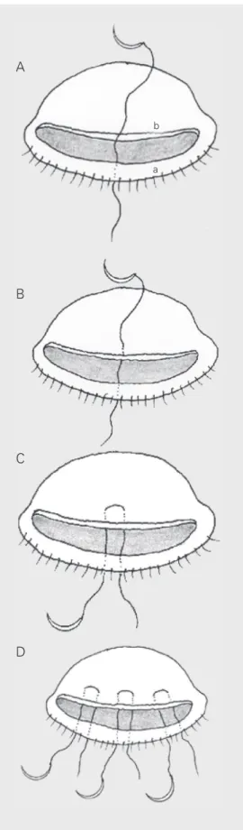
b
a
Figure 1. A, Surgeon’s view: a curvilinear incision has divided the tarsal plate into two por-tions: marginal (a) and distal (b). A single-armed suture was in-serted at the center of the lid through the anterior lamella, just behind the lash line, passed un-der the marginal tarsal portion and emerged in the defect cre-ated by the incision. B, The needle was next passed through the distal tarsal portion and passed back under the mar-ginal portion emerging at the anterior lamella. C, Additional sutures are passed in the same manner laterally and medially. D, Final result: the distal tarsal por-tion (dotted line) is now over the marginal portion.
A
B
C
is the replacement of the loose stroma of the tarsal conjunctiva with a subepithelial thick and fibrous membrane (7). This membrane is avascular, adherent to the tarsal plate and composed of vertically oriented fibers. The presence of this abnormal cicatricial tissue is the key factor causing the buckling of the tarsal plate and inward rotation of the lid margin towards the globe (entropion). Tra-chomatous trichiasis is, thus, essentially re-lated to the entropion caused by cicatricial changes of the posterior eyelid lamella.
A wide variety of surgical procedures for outwards rotation of the upper eyelid margin have been described. In the setting of a spe-cialized oculoplastic center, complex opera-tions involving lid splitting, anterior lamella recession and grafting can be used (8-11). However, in community-based surgeries so-phisticated procedures are not feasible and the focus must be directed at the simplest surgery capable of rotating the upper eyelid margin.
In order to overcome the rotational forces induced by the cicatricial changes of the posterior lamella, most rotational margin pro-cedures combine a tarsal plate fracture with the use of sutures to reposition the tarsal fragment containing the lid margin away from the globe. These surgeries can be done by an anterior or posterior approach. In both cases, a full-thickness tarsotomy is the es-sential step.
The anterior approach was described in 1964 by Ballen (12) who applied it to the upper eyelid, but the procedure was origi-nally described by Wies for the lower eyelid. A complete blepharotomy (incision of all layers of the lid: skin, orbicularis, tarsus and conjunctiva) needs to be made. This tech-nique is recommended by the WHO and described in detail in the 1993 WHO manual (2).
The posterior approach is much simpler than the anterior approach because the inci-sion is limited to the conjunctiva and tarsal plate. As the skin and orbicularis muscle are
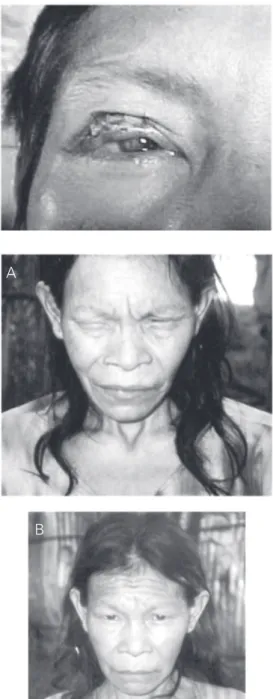
Figure 2. Successful marginal rotation.
Figure 3. A, Blepharospasm in-duced by trachomatous entro-pion. B, Relief of blepharospasm after marginal rotation by a pos-terior approach.
as defective closure or eyelid notching were not observed. One patient developed a bac-terial conjunctivitis that was controlled with antibiotic drops.
Discussion
The main histopathologic finding in bi-opsies of tarsal plates of upper eyelids af-fected by cicatricial trachoma and trichiasis
A
not disturbed, bleeding is minimal. Accord-ing to Kersten et al. (5), marginal rotation by a posterior approach has being used since the sixth century and different authors have already attested to the efficacy of this modal-ity of marginal rotation for the treatment of trachomatous entropion and trichiasis (3,4,6). It is not clear why the anterior approach was recommended by the WHO as the pre-ferred technique to be used in community-based surgeries. First, as pointed out in the 1993 manual, surgery is directed towards the eye and care should be taken to avoid globe injury. Second, when the anterior lamella is opened, the lid may bleed profusely (2). These problems are minimized with the pos-terior approach. As the incision is placed on an everted lid, the surgery is done away from the globe and bleeding is not an issue be-cause the posterior lamella is relatively avas-cular. We are aware of only one randomized trial comparing the anterior and posterior approaches for the treatment of trachoma-tous trichiasis in which it was concluded that both techniques had the same efficacy but fewer complications occurred when rotation was done by the posterior approach (13).
Our experience with tarsotomy by poste-rior approach was extremely positive. We have used this surgery in the hospital to manage any degree of trachomatous entro-pion. The results of the present study indi-cate that even with a minimal amount of training the surgeon was able to alleviate the symptoms of 91.3% of the subjects and 76.7% of the eyelids were free of trichiasis after a 6-month follow-up period.
Two points deserve comment when tri-chiasis recurrence is taken as a parameter of tarsal rotation effectiveness. The first is the location of the trichiasis. In our case, the recurrent misdirected eyelashes were always lateral or medial to the limbus and thus did not touch the cornea. Normally, this finding indicates that tarsotomy was not done across the entire extension of the tarsal plate. An-other possibility is that the tarsal incision
was not parallel to the margin. In order to achieve a uniform degree of margin rotation, the tarsotomy must be performed in a curvi-linear fashion, maintaining the same dis-tance from the margin. If the incision is placed more posteriorly, the margin will not rotate properly. Any surgery has its own learning curve, and the presence of segmen-tal trichiasis might suggest that the surgeon still needs some training with the tarsotomy. In any case, secondary surgery can then be performed in the segments (lateral, medial or both) that were not incised.
The second aspect is related to the differ-entiation between entropion and trichiasis. The WHO grading system is excellent for the epidemiological assessment of trachoma but does not measure the degree of eyelid in-ward rotation. Trichiasis is not necessarily associated with entropion. A typical example is leprosy, a disease that does not induce cicatricial changes in the eyelid posterior lamella. We have shown that a high percent-age of leprosy patients with paralytic la-gophthalmos and no conjunctival scarring have trichiasis (14). Trichiasis may also have undetermined causes, appearing as an idio-pathic disorder (9,15). Marginal rotation pro-cedures are aimed at rotating the lid margin. It is entirely possible that the surgery achieves its goal by producing a normal margin posi-tion even though trichiasis is still present.
chosen for the least invasive procedure (tar-sotomy by a posterior approach) was of para-mount importance for the acceptance of sur-gery. Only when the community saw that the
first subjects being operated upon had virtu-ally no complaints or complications was sur-gery well accepted.
References
1. Bailey R & Lietman T (2001). The SAFE strategy for elimination of trachoma by 2020: will it work? Bulletin of the World Health Organ-ization, 79: 233-236.
2. Reacher M, Foster A & Huber J (1993). Trichiasis Surgery for Tra-choma. The Bilamellar Tarsal Rotation Procedure. World Health Organization and the Edna McConnell Clark Foundation, New York. 3. Carre JB (1972). Tratamiento del entropion y de la triquíasis por via conjunctival (técnica personal). Archivos de la Sociedad Española de Oftalmología, 32: 683-698.
4. Nasr AM (1989). Eyelid complications in trachoma. I. Cicatricial entropion. Ophthalmic Surgery, 20: 800-807.
5. Kersten RC, Kleiner FP & Kulvin DR (1992). Tarsotomy for the treatment of cicatricial entropion with trichiasis. Archives of Oph-thalmology, 110: 714-717.
6. Halasa AH & Jarudi N (1974). Tarsotomy for the correction of cicatricial entropion. Annals of Ophthalmology, 6: 837-840. 7. Al-Rajhi AA, Hidayat A, Nasr A & Al-Faran M (1993). The
histopathol-ogy and the mechanism of entropion in patients with trachoma.
Ophthalmology, 100: 1293-1296.
8. Lyon DB & Dortzbach RK (1994). Entropion, trichiasis, and distichiasis. In: Dortzbach RK (Editor), Ophthalmic Plastic Surgery: Prevention and Management of Complications. Raven Press, New York.
9. Martin RT, Nunery WR & Tanenbaum M (1995). Entropion, trichi-asis, and distichiasis. In: McCord Jr CD, Tanenbaum M & Nunery WR (Editors), Oculoplastic Surgery. 3rd edn. Raven Press, New York.
10. Kemp EG & Collin JRO (1986). Surgical management of upper lid entropion. British Journal of Ophthalmology, 70: 575-579. 11. Goldberg RA, Joshi AR, McCann JD & Shorr N (1999). Management
of severe cicatricial entropion using shared mucosal grafts.
Ar-chives of Ophthalmology, 117: 1255-1259.
12. Ballen PH (1964). A simple procedure for the relief of trichiasis and entropion of the upper lid. Archives of Ophthalmology, 72: 239-240. 13. Adamu Y & Alemayehy W (2002). A randomized clinical trial of the success rates of bilamellar tarsal rotation and tarsotomy for upper eyelid trachomatous trichiasis. Ethiopian Medical Journal, 40: 107-114.
14. Guimaraes FC & Cruz AAV (1998). Eyelid changes in long-standing leprosy. Ophthalmic Plastic and Reconstructive Surgery, 14: 239-243.
15. Figueiredo ARP & Soares EJC (1992). Trichiasis: diagnosis and management. Orbit, 11: 137-146.
16. West S, Lynch M, Munoz B, Katala S, Tobin S & Mmbaga BB (1994). Predicting surgical compliance in a cohort of women with trichiasis.
International Ophthalmology, 18: 105-109.
17. Rabiu MM & Abiose A (2001). Magnitude of trachoma and barriers to uptake of lid surgery in a rural community of northern Nigeria.
Ophthalmic Epidemiology, 8: 181-190.
18. Bowman RJ, Faal H, Jatta B, Myatt M, Foster A, Johnson GJ & Bailey RL (2002). Longitudinal study of trachomatous trichiasis in the Gambia: barriers to acceptance of surgery. Investigative Oph-thalmology and Visual Science, 43: 936-940.
19. Oliva MS, Munoz B, Lynch M, Mkocha H & West SK (1997-1998). Evaluation of barriers to surgical compliance in the treatment of trichiasis. International Ophthalmology, 21: 235-241.
20. Courtright P (1994). Acceptance of surgery for trichiasis among rural Malawian women. East African Medical Journal, 71: 803-804. 21. Alves APX, Medina NH & Cruz AAV (2002). Trachoma and ethnic
diversity in the Upper Rio Negro Basin of Amazonas State, Brazil.