UNIVERSIDADE DE LISBOA Faculdade de Medicina Veterinária
RECOMBINANT FELINE INTERFERON OMEGA THERAPY IN CATS NATURALLY INFECTED WITH FELINE IMMUNODEFICIENCY VIRUS: CLINICAL, VIRAL AND
IMMUNOLOGICAL RELEVANCE
RODOLFO ASSIS OLIVEIRA LEAL
CONSTITUIÇÃO DO JURI
PRESIDENTE
Reitor da Universidade de Lisboa
VOGAIS
Doutora Aura Antunes Colaço
Doutor Miguel Agostinho Sousa Pinto Torres Fevereiro
Doutor Carlos Manuel Lopes Vieira Martins
Doutora Berta Maria Fernandes Ferreira São Braz
Doutora Maria Manuela Grave Rodeia Espada Niza
Doutora Solange Judite Coelho Alves Gil
ORIENTADORA
Doutora Solange Judite Coelho Alves Gil
CO-ORIENTADORES
Doutora Maria Manuela Grave Rodeia Espada Niza
Doutor Luís Manuel Morgado Tavares
2014
UNIVERSIDADE DE LISBOA Faculdade de Medicina Veterinária
RECOMBINANT FELINE INTERFERON OMEGA THERAPY IN CATS NATURALLY INFECTED WITH FELINE IMMUNODEFICIENCY VIRUS: CLINICAL, VIRAL AND
IMMUNOLOGICAL RELEVANCE
RODOLFO ASSIS OLIVEIRA LEAL
TESE DE DOUTORAMENTO EM CIÊNCIAS VETERINÁRIAS ESPECIALIDADE DE CLÍNICA
CONSTITUIÇÃO DO JURI
PRESIDENTE
Reitor da Universidade de Lisboa
VOGAIS
Doutora Aura Antunes Colaço
Doutor Miguel Agostinho Sousa Pinto Torres Fevereiro
Doutor Carlos Manuel Lopes Vieira Martins
Doutora Berta Maria Fernandes Ferreira São Braz
Doutora Maria Manuela Grave Rodeia Espada Niza
Doutora Solange Judite Coelho Alves Gil
ORIENTADORA
Doutora Solange Judite Coelho Alves Gil
CO-ORIENTADORES
Doutora Maria Manuela Grave Rodeia Espada Niza
Doutor Luís Manuel Morgado Tavares
2014
i
Porque expressar sentimentos numa outra língua, nunca nos permite transmitir convenientemente o que sentimos, decidi escrever os agradecimentos e a dedicatória em “bom português”.
Já lá vão cinco anos desde o verão de 2009…! Estava a acabar o internato em Paris e numa das muitas incursões a Portugal, bati à porta da Professora Rodeia! O futuro era incerto e a minha vontade de ir mais longe também…! Depois de uma conversa em género de desabafo, expressei-lhe o meu interesse pela carreira académica, sobretudo motivada pelo ensino. Sou filho de dois professores e partilhar conhecimento é algo que sempre achei que gostava de fazer na vida. Ali começou esta aventura…
Nunca gostei de investigação pura… ou melhor…sempre fui mais clínico que investigador. Todos o sabem e não o escondo. Acredito que tal como não nasci para ser cirurgião, nasci para dar asas à minha impulsividade e abraçar a Medicina Interna…! Investigação requer paciência, meticulosidade e insistência…qualidades que não colam bem com a minha impulsividade e rapidez nata…! Mas se para fazer o doutoramento, a investigação é um requisito…fiz-me a essa longa estrada! Cedo percebi que não era fácil, muito mais para mim, que comecei bem longe, com mãos grandes demais para pipetar 2 microlitros…! Também cedo me familiarizei com contaminações, primers e tive que aprender a lidar com o fracasso, com o voltar atrás, com resultados longe do espectável…! Percebi que podia juntar a investigação ao quotidiano clínico e a mudança para um projecto de investigação clínica permitiu-me continuar o contacto com a minha formação base! Hoje digo-o…sabe bem olhar para trás, ver resultados e ver que os mesmos podem ser aplicados! Hoje sei que descobri a investigação e que essa área me mudou! Vejo que fiz parte de uma equipa e essa equipa testou, demonstrou e concluiu, contribuindo para o avanço da medicina veterinária. Nem tudo foi fácil…ou melhor…não foi mesmo nada fácil! Não nego que vacilei muitas vezes, que me questionei, que quase fracassei em obstáculos menores…mas foram esses problemas diários, que também me moldaram como pessoa. Hoje, sinto que cheguei ao fim dessa estrada! Foi uma viagem longa, percorrida nos últimos 5 anos…com curvas e contra-curvas…! Hoje sou uma pessoa diferente…!! Consegui tirar partido de áreas que não conhecia, aproveitei o melhor das dificuldades e testei os meus limites! Hoje, olho para trás e vejo que aprendi tanto…! Das metodologias de topo (em especial o Real-time!) aos truques básicos de bancada, dos resultados inesperados, aos papers publicados…hoje sou uma pessoa diferente!...e estas 200 páginas reflectem isso…! Como diria a professora Rodeia…esta é…”a obra da minha vida!”
iii
À minha mulher, aos meus pais e a todos os gatos FIV positivos que colaboraram nesta odisseia, contribuindo para o avanço da ciência…
v
AGRADECIMENTOS
Hoje acho que o trabalho solitário é algo passado. Só em equipa, conseguimos ir longe e “mover montanhas”. Por isso, esta tese não é fruto só do meu esforço. Engloba nas linhas e entrelinhas, pessoas e personalidades que guardo comigo para vida. Por isso, o meu Obrigado:
Aos meus orientadores e co-orientadores. Em detalhe…obrigado Professora Manuela Rodeia, minha co-orientadora, por me ter feito o convite e aberto a porta para que tudo isto fosse possível. É uma referência para mim! Fez-me descobrir a Medicina Interna e o seu carácter assertivo e directo, fez (e tem feito) de mim grande parte do que sou hoje. Obrigado pela amizade e pela força que me dá.
À Professora Solange Gil, a minha orientadora, por me ter guiado neste projecto e por, aliando a amizade ao profissionalismo, me ter conduzido e amparado nas curvas e contra-curvas destes anos. Obrigado pela confiança e pela partilha de fracassos, de êxitos, de arranhadelas e de dificuldades.
Ao Professor Luís Tavares, meu co-orientador, presidente da FMV e coordenador do CIISA. Obrigado por acreditar e confiar no meu potencial, acolhendo-me na sua equipa com tamanha amizade e profissionalismo.
À Professora Cristina Vilela, pelas palavras e pela motivação que sempre me deu. Relembro-me frequentemente do seu sorriso, das nossas conversas e da força inigualável de uma professora única, que partiu cedo demais. Esta tese, é também para si, que sempre acreditou em mim e que isto era possível.
À Professora Ana Duarte, pela paciência de me moldar nos caminhos da investigação, pela ajuda de bancada e pela disponibilidade constante.
À Clara Cartaxeiro, pela amizade, pela ajuda, pelo sentido de humor e pelas gargalhadas que alegravam os meus dias.
Ao Nuno Félix e à Joana Dias, meus companheiros de saga nesta estrada, cada um a seu tempo. Obrigado pela partilha, pela força e pela cumplicidade científica que desenvolvemos. Ao David McGahie e à Virbac. Obrigado pela colaboração e pelo espírito de equipa desenvolvido.
Aos colegas e amigos do CIISA, pelo acolhimento e pela inter-ajuda constante. Obrigado por estarem sempre prontos a esclarecer-me a mínima dúvida.
vi
À União Zoófila de Lisboa, à Associação de Amigos dos Animais Abandonados da Moita, aos donos dos gatos FIV positivos que colaboraram no estudo e aos veterinários referentes, obrigado pela confiança e pela colaboração no desenvolvimento deste trabalho.
Aos estagiários que colaboraram no laboratório de Virologia em especial a Inês Siborro, a Joana Cravo, a Filipa Vassallo e Silva e a Sofia Sirage. Obrigado pela disponibilidade e pelos bons momentos que fizeram o meu dia-a-dia na bancada.
Ao Professor António Ferreira, director do Hospital Escolar, obrigado pela possibilidade de continuar a exercer clínica, que tanto alento me dava em períodos árduos desta etapa. À Professora Teresa Villa de Brito, pela amizade, pela parceria e pela partilha de conhecimentos de endocrinologia que encheram os meus dias e complementaram este período da minha vida. Obrigado pela confiança e por termos montado em conjunto o serviço de Endocrinologia.
A todos os colegas do Hospital Escolar da FMV-UTL, auxiliares, recepcionistas, enfermeiros e estagiários, que de uma forma ou de outra, colaboraram no projecto e contribuíram para o meu crescimento profissional e pessoal nos últimos anos. Á Joana Pontes um obrigado especial pela amizade, pelo suporte em tantos momentos difíceis. Ao Pedro Lourenço, pela cumplicidade, amizade e auxílio em colheitas de campo que incluíram arranhadelas e unhas partidas!
À Natacha Couto e à Professora Constança, pela oportunidade de explorar o gosto pelo ensino e pela partilha de momentos únicos nessa experiência de dar aulas, que tanto me gratificou.
Às pessoas que fizeram o meu quotidiano na FMV. Um obrigado especial à tia Salomé. Pela amizade e por aturar o “pépino” de uma forma tão particular, tranquilizando as minhas manhãs durante a escrita da tese.
À Ana Vieira e Inês Ajuda, minhas colegas de gabinete e cúmplices desta saga de doutoramento, pela amizade, pelas pausas para café e pela inter-ajuda do C3.10.
E porque este projecto profissional não seria exequível sem um suporte pessoal estável, obrigado à minha mulher, Ana Rute Vilar, pela cumplicidade, pela tranquilidade que me transmite quando faço dos problemas mais do que eles são, pela dedicação, pela tolerância e paciência para estas minhas aventuras… ! Obrigado Xi por me fazeres tão feliz dia após dia e por me fazeres acreditar que com calma e ponderação, consigo ir longe.
vii
Aos meus pais, por me terem feito deste cimento de querer sempre progredir e ser melhor amanhã do que sou hoje. Obrigado por me terem incutido valores de persistência e me terem demonstrado que pelo facilitismo não chegamos longe.
Á minha restante família, à tia Laura, aos meus sogros, cunhada, aos meus padrinhos (Tété, Marcelo, Cristina, Zé Miguel, Doroteia, Filipa e Zé) e a todos os amigos que não consigo especificar aqui, obrigado por me moldarem e por estarem comigo nesta etapa.
APOIOS FINANCEIROS
O presente trabalho foi financiado pela bolsa de doutoramento individual SFRH/ BD / 62917 / 2009 da Fundação para a Ciência e Tecnologia (FCT), pelo Centro de Investigação Interdisciplinar em Sanidade Animal (CIISA) da Faculdade de Medicina Veterinária, Universidade de Lisboa e ainda pela Virbac (Centro de Custos phD_Virbac).
ix
Resumo
A terapêutica com interferão ómega felino em gatos naturalmente infectados com o vírus da imunodeficiência felina: relevância clinica, virológica e imunitária
Os interferões do tipo I são citoquinas chave do sistema imunitário. Devido às suas propriedades imunomoduladoras, são um recurso terapêutico frequente em diferentes doenças como as infecções retrovirais. O interferão ómega felino (rFeIFN-ω) é o primeiro interferão licenciado para medicina veterinária. Apesar do seu uso no tratamento de infeções retrovirais como o vírus da imunodeficiência felina (FIV) e o vírus da leucemia felina (FeLV), são poucos os estudos que fundamentam o seu benefício clinico. Esta tese visa clarificar as propriedades terapêuticas e imunomoduladoras do protocolo licenciado de rFeIFN-ω (3 ciclos de 5 administrações subcutâneas de 1MU/kg uma vez ao dia a iniciar aos dias 0, 14 e 60) em gatos naturalmente infectados por retrovírus e residentes em gatil. Em detalhe, este trabalho avalia o efeito deste fármaco na melhoria clinica, na excreção de vírus concomitantes, na virémia/provirus e na variação de diferentes marcadores imunitários como proteínas de fase aguda e perfil de citoquinas. Esta tese contempla ainda o desenvolvimento de um protocolo terapêutico alternativo baseado na administração oral de rFeIFN-ω (0.1MU/gato durante 90 dias consecutivos) para uso em gatos FIV-positivos domésticos, os quais apresentam geralmente um quadro clinico subtil e pouco específico.
Os resultados revelaram que o protocolo licenciado induz uma melhoria clinica significativa com redução concomitante das infecções oportunistas e um aumento do perfil de proteínas de fase aguda (APP). O protocolo alternativo revelou-se eficaz na melhoria clinica dos animais tratados, apesar de não induzir alterações significativas do perfil de APPs nem das infecções concomitantes (residuais no grupo de estudo). Ambos os protocolos não induziram alterações na virémia nem no perfil de citoquinas participantes nas respostas T-helper 1 ou T-T-helper 2 o que sugere que este composto não apresenta propriedades anti-virais nem actua na imunidade adquirida de gatos FIV positivos. Verificou-se contudo um decréscimo dos niveis plasmáticos de Interleucina-6 (citoquina pro-inflamatória) em gatos tratados com o protocolo subcutâneo e uma redução da sua expressão (mRNA) em gatos tratados por vira oral. Tal demonstra que o rFeIFN-ω apresenta propriedades anti-inflamatórias, as quais são mais evidentes aquando do tratamento com o protocolo licenciado. Mais que uma contribuição para um melhor conhecimento do rFeIFN-ω, esta tese explora as suas propriedades imunomoduladoras e valida um novo protocolo oral, o qual poderá ser incluído em futuras guidelines para o tratamento de gatos FIV-positivos. Palavras chave: felino, interferão ómega felino, imuno-modulação, vírus da imunodeficiência felina, retrovírus
xi
Abstract
Thesis Title: Recombinant feline interferon omega therapy in cats naturally infected with Feline Immunodeficiency Virus: clinical, viral and immunological relevance
Type-I Interferons are well-known cytokines which among their main functions are key components of the host immune response against viral infections. Due to its immune modulation properties, they are commonly used in the therapeutic approach of various diseases such as retroviral infections. Recombinant feline interferon omega (rFeIFN-ω) is the first interferon licensed for use in veterinary medicine. Although it is commonly administered in retroviral infections, namely in Feline Immunodeficiency Virus (FIV) and Feline Leukemia Virus (FeLV) infected cats, few studies reported its clinical benefits and mechanisms of action. This thesis aims to clarify the main properties of the licensed rFeIFN-ω protocol (3 cycles of 5 daily subcutaneous administrations of 1MU/kg beginning on days 0, 14 and 60) in naturally retroviral infected cats living in an animal shelter, evaluating its effect not only on clinical improvement but also on concurrent viral excretion, viremia/proviral load and various immune biomarkers such as acute phase proteins and cytokine profile. Recognizing the non specific and subtle clinical presentation of the majority of FIV-infected cats, this work also presents and evaluates an alternative oral rFeIFN-ω protocol (0.1MU/cat during 90 days) to be used in client-owned FIV-infected cats.
Results showed that the licensed rFeIFN-ω protocol induces a significant clinical improvement, with a concurrent reduction of opportunistic viral infections and an increase on acute phase proteins (APP) profile. The alternative protocol also revealed an important clinical improvement but without significant changes on opportunistic viral infections (which were of low level in the tested group) or on APP profile. In both protocols, no changes were remarked on viremia neither on T-helper 1/T-helper 2 cytokine profiles meaning that this compound may lack an anti-viral activity for retroviruses in vivo and do not act on the acquired immune response of FIV-positive cats. However, there was a significant reduction of the interleukin-6 plasma levels (pro-inflammatory cytokine) in cats treated with the licensed protocol and a decrease on its mRNA expression in cats treated orally. This shows that rFeIFN-ω can have anti-inflammatory properties, which are more evident in the higher doses of the licensed protocol.
More than contributing for a better knowledge of rFeIFN-ω, this thesis explores its immune modulation properties and validates a new oral protocol which can be included on future FIV-guidelines.
Keywords: feline, interferon therapy, recombinant-feline interferon omega, immune modulation, feline immunodeficiency virus
xiii
Table of Contents
INTRODUCTION ... 1
Theme presentation, justification, objectives ... 2
PART I LITERATURE REVIEW ... 5
PART I-CHAPTER I: MOLECULAR BACKGROUND OF RETROVIRAL INFECTIONS ... 7
1.1 Retroviruses and taxonomy ... 9
1.2. Genome and molecular basis of retroviruses ... 10
1.2.1. The retrovirus virions and their genetic properties ... 10
1.2.2. Life cycle and basic principles ... 13
1.2.3. The genetic variation in retroviruses ... 15
FIV ...15
FeLV ...16
1.3. Retroviruses and evolution – a problem in the future? ... 17
PART I-CHAPTER II:RETROVIRUSES – FROM THE IMMUNITARY TO THE CLINICAL PERSPECTIVE ... 19
2.1. Epidemiology of Retroviruses ... 21
FIV ...21
FeLV ...22
2.2 Transmission routes of feline retroviruses ... 23
FIV ...23
FeLV ...24
2.3. The host-cells and the retrovirus – the molecular beginning of a long term interaction ... 25
FIV ...25
FeLV ...26
2.4. Physiopathology of immune suppression in retroviral infections: the immunitary perspective ... 26
FIV ...27
FeLV ...30
2.5. Phases of infection ... 31
FIV ...31
FeLV ...32
2.6. FIV and FeLV: Clinical and laboratory findings ... 35
FIV ...35
FeLV ...38
2.7. Diagnosis of FIV and FeLV ... 41
FIV ...42
Serology (Antibody testing) ...42
Virus detection ...42
When should a cat be tested for FIV? ...43
FeLV ...44
xiv
Antibody Detection ...46
When should a cat be tested for FeLV? ...46
2.8. Therapeutic approach to Retroviral Infections: Anti-virals and immune modulators – general considerations ... 47
Antivirals ...48
Immune modulators ...50
2.9. Considerations about management and prognosis of retroviral infections ... 52
FIV ...52
FeLV ...53
2.10. Preventing retroviral infections: the relevance and problematic of vaccination ... 54
FIV Vaccines ...54
FeLV vaccines ...55
2.11. Retroviral infections and the public health perspective ... 57
PART I-CHAPTER III:THE ROLE OF INTERFERON IN VETERINARY MEDICINE ... 59
3.1. Interferon: molecular features and actions ... 61
3.2. The therapeutic role of interferon in retroviral infections: from immune modulation to antiviral therapy ... 62
The use of Human Interferon-alpha (HuIFN-α) in retroviral infections ...63
The use of Recombinant-Feline Interferon Omega in Retroviral infections ...65
PART II EXPERIMENTAL WORK ...67
PARTII-CHAPTERI:RELEVANCE OF FELINE INTERFERON OMEGA FOR CLINICAL IMPROVEMENT AND REDUCTION OF CONCURRENT VIRAL EXCRETION IN RETROVIRUS INFECTED CATS FROM A RESCUE SHELTER ... 69
Abstract ...71
Introduction ...72
Material and Methods ...74
Animals ...74
Products ...74
Treatment Protocol ...75
Supportive Treatment...75
Clinical evaluation and Scoring ...75
Blood Sample Collection and Treatment ...77
Survey of concomitant pathogens. ...78
Statistical analysis ...79
Results ...79
Clinical evaluation and Scoring ...79
Haematology ...83
Biochemistry analysis...85
Survey of concomitant pathogens ...85
Discussion ...88
PART II-CHAPTER IIMONITORING ACUTE PHASE PROTEINS IN RETROVIRUS INFECTED CATS UNDERGOING FELINE INTERFERON OMEGA THERAPY ... 93
xv
Introduction ...96
Material and Methods ...97
Animals ...97
Treatment Protocol ...98
Supportive Treatment...98
Blood collection and analysis ...99
Statistical Analysis ...99
Results ...99
Discussion ...104
PART II-CHAPTER IIIORAL RECOMBINANT FELINE INTERFERON-OMEGA AS AN ALTERNATIVE IMMUNE MODULATION THERAPY IN FIV POSITIVE CATS: CLINICAL AND LABORATORY EVALUATION ... 107
Abstract ...109
Introduction ...110
Material and Methods ...112
Animals and treatment protocols ...112
Ethics ...112
Clinical Evaluation ...112
Concurrent viral excretion assessment ...113
Hematology and Biochemistry ...113
Acute Phase Proteins ...113
Statistical Analysis ...114
Results ...114
Clinical Improvement ...114
Concurrent Viral Excretion ...116
Hematology and Biochemistry ...116
Serum protein Electrophoresis ...117
Acute Phase Proteins ...117
Discussion ...118
PARTII-CHAPTER IV: EVALUATION OF MX PROTEIN EXPRESSION IN NATURALLY FIV-INFECTED CATS RECEIVING ORAL RECOMBINANT FELINE INTERFERON OMEGA THERAPY ... 123
Abstract ...125
Introduction ...126
Material and Methods ...128
Results ...130
Discussion ...131
PART II-CHAPTER V:EVALUATION OF VIREMIA, PROVIRAL LOAD AND CYTOKINE PROFILE IN NATURALLY FIV-INFECTED CATS TREATED WITH TWO DIFFERENT PROTOCOLS OF RECOMBINANT FELINE INTERFERON OMEGA ... 135
Abstract ...137
Introduction ...138
Material and Methods ...141
Animals and sample collection ...141
Relative quantification of cytokine expression by Real-Time qPCR ...141
xvi Quantification of Provirus ...143 Quantification of Viremia ...144 Statistical analysis ...144 Results ...144 Cytokine expression ...145
Plasma levels of IL-6, IL-12p40 and IL-4 cytokines ...146
Quantification of Provirus ...148
Quantification of Viremia ...148
Discussion ...149
PART III: EXTRAPOLATING IMMUNE MODULATION PROPERTIES: THE USE OF ORAL RECOMBINANT FELINE INTERFERON OMEGA IN OTHER DISEASES THAN RETROVIRAL INFECTIONS ... 155
PART III-CHAPTER I:THE USE OF ORAL RECOMBINANT FELINE INTERFERON OMEGA IN TWO CATS WITH TYPE II DIABETES MELLITUS AND CONCURRENT FELINE CHRONIC GINGIVOSTOMATITIS SYNDROME... 157
Abstract ...159
Background ...160
Case Presentation ...161
Conclusions ...163
GENERAL DISCUSSION AND CONCLUSIONS ... 165
REFERENCES ... 175
xvii
List of Pictures
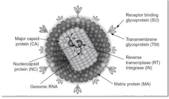
Figure 1: Schematic diagram of a retrovirus virion and its important structures and proteins ... 10
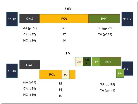
Figure 2: Schematic diagram of genomic structure and major proteins of provirus FeLV and FIV ... 11
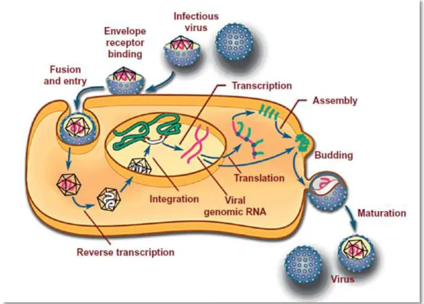
Figure 3: Schematic presentation of replication cycle in retroviruses ... 13
Figure 4: Time course presentation of the three phases of FIV infection ... 31
Figure 5: Severe gingivitis and oral ulcer (tongue) in a FIV-infected cat ... 35
Figure 6: Anisocoria in a FeLV-infected cat ... 41
Figure 7: Different Interferon types and main cellular mechanisms of action ... 62
Figure 8: Clinical parameters observed in some cats evaluated in the study ... 77
Figure 9: Average ± Standard Error of red blood cell count variation in FIV, FeLV and Co-infected cats under treatment with rFeIFNω ... 84
Figure 10: Average ± Standard Error of white blood cell count variation in FIV, FeLV and Co-infected cats under treatment with rFeIFNω ... 84
Figure 11: FCV PCR amplification for FeLV group on D0 ... 85
Figure 12: Real-Time PCR viral load quantification (ng/µl) of FHV-1 excretion in FIV, FeLV and co-infected cats under rFeIFNω therapy ... 86
Figure 13: Real-Time PCR viral load quantification (ng/µl) of FCoV excretion in FIV, FeLV and co-infected cats under rFeIFNω therapy ... 87
Figure 14: Mean ± standard error (SE) of serum concentrations of serum amyloid A (SAA) in naturally retroviral infected cats, before (D0) during (D10, D30) and after (D65) rFeIFNω therapy. ... 100
Figure 15: Mean ± standard error (SE) serum concentrations of alpha-glycoprotein-1 (AGP) in naturally retroviral infected cats, before (D0) during (D10, D30) and after (D65) rFeIFNω therapy. ... 101
Figure 16: Mean ± Standard Error (SE) of serum concentrations of c-reactive protein (CRP) in naturally retroviral infected cats, before (D0) during (D10, D30) and after (D65) rFeIFNω therapy ... 101
Figure 17: Individual clinical scores for each cat of each group, before and after rFeIFN-ω therapy ... 115
Figure 18: Total Proteins, Gammaglobulins and Albumin serum levels of FIV positive Cats treated with two different protocols of rFeIFN-ω ... 117
Figure 19: Individual Mx gene expression of 7 naturally FIV-infected cats treated with oral rFeIFNω protocol 130 Figure 20: Detailed cytokine mRNA variation in naturally FIV-infected cats submitted to two different rFeIFNω protocols... 146
Figure 21: Mean ± SE of plasma IL-12p40, IL-4 and IL-6 concentrations in naturally FIV-infected cats submitted to two different protocols of rFeIFNω ... 147
Figure 22: Mean ± SE of proviral load of FIV in cats submitted to two different rFeIFNω protocols. ... 148
xviii
List of Tables
Table 1: Main clinical and laboratory changes observed in each stage of FeLV-infection. ... 33
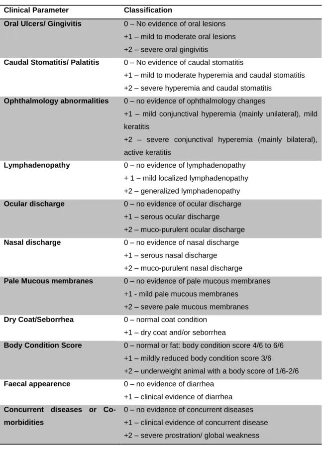
Table 2: Clinical Score - scale used for cats’ clinical evaluation ... 76
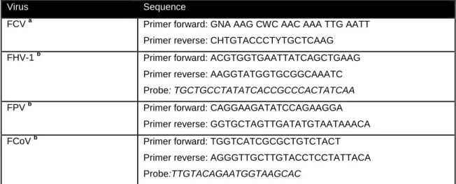
Table 3: Primer nucleotide sequences used for the amplification of FCV, FHV-1, FPV and FCoV... 78
Table 4: PCR and Real Time PCR amplification conditions ... 79
Table 5: Total group and detailed individual clinical score values for each parameter evaluated in FIV cats during rFeIFNω therapy. ... 81
Table 6: Total group and detailed individual clinical score values for each parameter evaluated in FeLV cats during rFeIFNω therapy ... 82
Table 7: Total group and detailed individual clinical score values for each parameter evaluated in Co-Infected cats during rFeIFNω therapy. ... 83
Table 8: Individual variation of clinical scores, concurrent viral excretion and acute phase proteins in FIV, FeLV and FIV/FeLV cats treated with rFeIFN-ω. ... 103
Table 9: Overall Clinical Improvement of FIV positive cats treated with the rFeIFN-ω licensed protocol (SC Group) and rFeIFN-ω PO protocol. ... 115
Table 10: Mean values ± standard error of Serum Amyloid A (SAA), Alpha-1-Glycoprotein (AGP) and C-Reactive Protein (CRP) serum levels in FIV positive cats before and after therapy with licensed rFeIFN-ω (SC group) and oral (PO group) protocols. ... 118
Table 11: Primers used to evaluate cytokine expression by Real-time qPCR in naturally FIV-infected cats treated with rFeIFN protocols. ... 142
Table 12: Real-time qPCR system to assess FIV provirus and viremia changes in naturally FIV-infected cats after rFEIFN therapy ... 143
xix
List of Abreviations
2-5-OAS - 2´-5´ Oligoadenylate synthaseABCD – (European) Advisory Board on Cat Diseases AGP – Alpha-1-glycoprotein
AIDS - Acquired immunodeficiency syndrome APPs – Acute Phase Proteins
APR – Acute Phase Response AZT - zidovudine
BID – twice daily
CBC – complete blood count CNS – central nervous system CRP – C-reactive protein CSF – cerebrospinal fluid CTLs – Cytotoxic T cells
CXCR4 – Chemokine receptor type 4 DM – Diabetes mellitus
DNA- Desoxiribonucleic Acid
ELISA- Enzyme- Linked Immunosorbent Assay ENV- Envelope
EPO - erythropoietin
FA – Fluorescent Antibodies
FCGS - Feline Chronic Gingivostomatits Syndrome FCV – Feline Calicivirus
xx
FeLV-IMHA – FeLV-induced immune-mediated hemolytic anemia FeSV – Feline Sarcoma Virus
feTHTR1 – feline thiamine transport protein FISS – Feline Injection Site Sarcoma FIV - Feline Immunodeficiency Vírus FIV – Feline Infectious Peritonitis
FPLS – Feline panleukopenia-like syndrome FPV – Feline parvovirus
Gag – Group specific antigen
G-CSF – Granulocyte Colony-stimulating factor HIV- Human Immunodeficiency Virus
HuIFN-α- Human Interferon alfa IFNAR – Interferon Receptor Complex IFNs – Interferons
IGF-1 – Insulin Growth Factor 1 IL – Interleukin
IN – Integrase
LTR - long terminal repeats MDS – myelodysplastic syndrome mRNA- Messenger Ribonucleic Acid
NSAID – Non steroidal anti-inflammatory drug NK-cells – natural-killer cells
ORF - open reading frames
PAMPs – Pathogen-associated molecular patterns PBMCs - peripheral blood mononuclear cells
xxi PCR – Polymerase chain reaction
PO group – cats treated with oral rFeIFN-ω Pol- Polymerase
rFeIFN-ω- Recombinant feline interferon omega RI – reference interval
RNA- Ribonucleic Acid RT- Reverse Transcriptase
RT-qPCR – Real-time quantitative PCR SAA – Serum Amyloid A
SC group – cats treated with the subcutaneous rFeIFN-ω licensed protocol SE – standard error
SID – once a day
SPE – Serum Protein Electrophoresis SPF - specific pathogen free
SU- Surface Protein Th1 – T-helper 1 cells Th2 – T-helper 2 cells TLR – toll like receptors TNF – Tumor necrosis factor Tregs – T-regulatory cells VIF- Viral Infectivity Factor
1
2
Theme presentation, justification, objectives
Interferons (IFNs) are key components of the host immune system, being particularly relevant in viral infections (Sadler & Williams, 2008). The large family of IFNs can be divided into different types such as type I-IFNs, commonly used for therapeutic purposes. Among their major functions, type I-IFNs increase and sensitize the immunitary system towards the microbial recognition (Siren, Pirhonen, Julkunen, & Matikainen, 2005), establishing an important link between innate and acquired immunity (Colonna, Trinchieri, & Liu, 2004). Furthermore, they are believed to have some anti-viral properties, blocking viral replication and inducing apoptosis of infected cells (Goodbourn, Didcock, & Randall, 2000; Bracklein, Theise, Metzler, Spiess, & Richter, 2006).
Not only in humans but also in feline medicine, the use of type I-IFNs as immune modulation therapy is common, notably in retroviral infections (Tompkins, 1999; de Mari, Maynard, Sanquer, Lebreux, & Eun, 2004; Domenech, et al., 2011).
Still used in several countries, Human Interferon Alpha (HuIFN-α) was the first interferon used in cats, despite the fact that it is only licensed for humans. In spite of its short term effects, particularly on clinical improvement and increase of the survival time, the development of neutralizing antibodies several weeks after therapy makes HuIFN-α ineffective for long-term immune modulation therapy in cats (Tompkins, 1999; Pedretti, et al., 2006; Hartmann, 2012a). This problem was bypassed by the more recent release of recombinant feline interferon omega (rFeIFN-ω).
RFeIFN-ω is the first interferon compound licensed for use in veterinary medicine. According to the manufacturer’s instructions and license, it should be used in three cycles of five daily subcutaneous injections of 1MU/kg, beginning respectively on days 0, 14 and 60. Despite the fact that it was licensed a few years ago, there are not so many studies that support its clinical benefits, particularly in retroviral infections. The first paper described its clinical application dates from 2004 and reported that treated Feline Leukemia Virus (FeLV) and Feline Immunodeficiency Virus (FIV)/FeLV co-infected cats showed a significant improvement and an increased survival time (de Mari, et al., 2004). More recently, another research group showed that rFeIFN-ω did not induce significant changes on parameters such as hypergammaglobulinemia, proviral load and viremia, suggesting an overall effect mainly on the innate immune reaction rather than on the acquired immunity (Domenech, et al., 2011). Further studies are therefore required in order to clarify the mechanisms of action of rFeIFN-ω.
3
In this sense, the main objective of this work is to explore the main properties of rFeIFN-ω in naturally retroviral infected cats, with special relevance to FIV-infected animals. More than the extension of the current knowledge about the licensed rFeIFN-ω protocol, this work also aims to develop and present a new oral therapeutic protocol, which if successful, can be considered as an alternative immune modulation therapy for FIV-infected cats.
Excluding the literature review (part I), this thesis structurally comprises two parts (part II and III). Part II is based on clinical trials and reports the experimental work which was developed using two specific rFeIFN-ω protocols. The main objectives of the referred experimental work are:
a) To investigate the effect of the licensed rFeIFN-ω protocol on clinical improvement, hematology, biochemistry profile and concurrent viral excretion in naturally retroviral-infected cats living in an animal shelter.
b) To monitor the effect of the licensed rFeIFN-ω protocol on acute phase protein (APP) profile, assessing the role of APPs as potential biomarkers of the innate immune activation in treated animals.
c) To develop and validate a new oral rFeIFN-ω protocol to be used in FIV-infected cats. Recognizing that many FIV-infected cats have a nonspecific clinical presentation and usually does not require a strong immune modulation therapy, this protocol is based on a 10 fold lower dose than the current licensed protocol, to be administered for 3 months (90 continuous days) in these animals. The development of this protocol involves the monitoring of its action on the clinical improvement, hematology, biochemistry profile, concurrent viral excretion and APP profile in treated cats.
d) To assess the effect of the experimental oral rFeIFN-ω protocol on other innate immune parameters such as Mx-protein, a specific biomarker of type-I IFN action.
e) To evaluate proviral load, viremia and cytokine profile [messenger ribonucleic acid (mRNA) expression and concurrent plasma variations] in FIV-infected cats treated with rFeIFN-ω protocols, comparing the main similarities and differences between them. This comparison will allow determining the main mechanisms of action of each rFeIFN-ω protocol, contributing for a better use in clinical practice.
The studies that support this experimental work were converted in five chapters presented on part II. Four of them were submitted/published in international refereed and indexed journals, namely:
Gil, S., Leal, R.O., Duarte, A., McGahie, D., Sepúlveda, N., Siborro, I., Cravo, J., Cartaxeiro, C., Tavares, L., 2013. Relevance of Feline Interferon Omega for Clinical Improvement and
4
Reduction of Concurrent VIral Excretion in Retrovirus Infected Cats from a Rescue Shelter. Research in Veterinary Science. 2013 Jun;94(3):753-63. doi: 10.1016/j.rvsc.2012.09.025. Epub 2012 Oct 31
*These authors contributed equally to the work
Leal RO*, Gil S*, Sepúlveda N, McGahie D, Duarte A, Niza MMRE, Tavares L. 2014 “Monitoring acute phase proteins in retrovirus infected cats undergoing feline interferon omega therapy”Journal of Small Animal Practice 2014 Jan;55(1):39-45. doi: 10.1111/jsap.12160. Epub 2013 Nov 27.
*These authors contributed equally to the work.
Gil. S*, Leal RO*, McGahie D, Sepúlveda N., Duarte A, Niza MMRE, Tavares L 2014 “Oral Recombinant Feline Interferon-Omega as an alternative immune modulation therapy in FIV positive cats: Clinical and laboratory evaluation” Research in Veterinary Science 2014 Feb;96(1):79-85. doi: 10.1016/j.rvsc.2013.11.007. Epub 2013 Nov 25
*These authors contributed equally to the work.
Leal RO, Gil S, Duarte A, McGahie D, Sepulveda N, Niza MMRE, Tavares L 2014 “Evaluation of Viremia, proviral load and Cytokine profile in naturally FIV-infected cats treated with two different protocols of recombinant feline interferon Omega” (Submitted)
Although it is out of the scope of this thesis, part III extrapolates the use of oral protocol as an alternative to steroid therapy in type II feline diabetes mellitus. It is a report of two clinical cases which illustrates the therapeutic potential of this compound in diseases other than retroviral infections. Despite the fact it is only based on two clinical cases, this study was also published as a case report in an international peer-reviewed journal:
Leal RO, Gil S, Brito MTV, McGahie D, Niza MMRE, Tavares L 2013 “The use of oral Recombinant Feline Interferon Omega in two cats with type II diabetes mellitus and concurrent Feline Chronic Gingivostomatitis Complex”. Irish Veterinary Journal 2013 Oct 23;66(1):19. Epub 2013 Oct 23
5
Part I
7
Part I - Chapter I:
Molecular background of retroviral
infections
9
1.1 Retroviruses and taxonomy
Retroviruses are well described in various species, being part of the family retroviridae. The prefix “Retro” refers to reverse and is due to the reverse transcriptase, a particular enzyme which characterizes the virions of this family (MacLachland & Dubovi, 2011). The most widely known virus of this family is the human immunodeficiency virus (HIV), which nowadays has a strong healthy impact, being one of the main subjects of scientific research (Murphy, Gibbs, Horzineck, & Studdert, 1999b). In veterinary medicine, animal retroviruses have also been studied mainly because they are excellent comparative models for human acquired immunodeficiency syndrome (AIDS) research, contributing for the advance of the medical science (Murphy, et al., 1999b; Elder, Lin, Fink, & Grant, 2010; Yamamoto, Sanou, Abbott, & Coleman, 2010).
According to the International Committee of Taxonomy of Viruses, retroviruses are classified into two subfamilies: Orthoretrovirinae and Spumaretrovirinae. This last one includes only one genus, the spumavirus which refers to foamy viruses. In the subfamily Orthoretrovirinae, 6 genera are distinguished: Alpharetrovirinae, Betaretrovirinae and Gammaretrovirinae, which have simple structure and are commonly considered simple retroviruses, in opposition to Deltaretrovirinae, Epsilonretrovirinae and Lentivirinae which are complex retroviruses (MacLachland & Dubovi, 2011).
Despite the phylogenetic classification, Lentivirus and Gammaretrovirus are the most important retroviruses in Veterinary Medicine. Lentiviruses include not only the human immunodeficiency viruses (HIV-1 and HIV-2) but also other ones such as FIV. Then, lentiviruses have a strong impact on the immune system of humans and cats, being widely studied in the last decades. Gammaretrovirus, with more simple structure, includes FeLV, which similarly to FIV, have an important clinical impact in Companion Animal Practice namely in feline medicine (Jarrett, 1999; Dunham & Graham, 2008).
Particularly in cats, retroviral infections seem to be ancestral since the feline genome has always had different genetic elements derived from elderly retroviral infections, also called “endogenous retroviruses” which are vertically transmitted by germ line (Roy-Burman, 1995; Dunham & Graham, 2008).
To a better understanding of this work, it is essential to identify the main genetic and molecular basis of retroviruses, giving special relevance to FIV and FeLV, the most important retroviruses in feline practice.
10
1.2. Genome and molecular basis of retroviruses
1.2.1. The retrovirus virions and their genetic properties
Virions of the family retroviridae are enveloped, having a three-layered structure of 80-100nm of diameter. The inner-layer is the genome of the virion, which is diploid [consisting of a homodimer of two single-stranded ribonucleic acid (RNA)] and includes 30 molecules of reverse transcriptase (RT) in a helical symmetry. It is surrounded by an icosahedral capsid (60nm in diameter) which is involved by an envelope that derived from a host cell membrane (Murphy, et al., 1999b; Goff, 2007; MacLachland & Dubovi, 2011). Schematic diagram of retrovirus virion basic structure is presented on figure 1.
Figure 1: Schematic diagram of a retrovirus virion and its important structures and proteins (MacLachland & Dubovi, 2011).
Retroviruses have different particular findings namely being the only diploid genome (two molecules of single-stranded RNA) and the only viral RNA that requires the host cell enzymes to be synthesized and processed (Murphy, et al., 1999b; Goff, 2007).
The genome of retroviruses contains 3 major genes which encode several proteins:
Gag (group specific antigen): which encodes the virion core (capsid) proteins (Murphy, et al., 1999b).
Pol (polymerase): which encodes the protease, the RT enzyme and integrase (IN). The most important of these enzymes is RT, which contains different domains namely a DNA
11
polymerase (that can have a RNA or DNA template) and a RNase, both particularly relevant for the virus life cycle (Goff, 2007).
Env (envelope): that encodes surface/peripheral proteins, determining cell tropism and contributing for pathogenicity (Pancino, Castelot, & Sonigo, 1995; Roy-Burman, 1995; Verschoor, et al., 1995; Johnston, Silva, & Power, 2002).
Despite the described common basal structure, FIV and FeLV are genomically different (figure 2). FeLV virion is a simple genome (with only these three basal genes) while FIV is a complex one as its genome also encodes some accessory genes which strictly regulate the viral cycle and contribute to a productive infection of different cell types (Murphy, et al., 1999b; Dunham & Graham, 2008; Duarte, Gil, Leal, & Tavares, 2012).
Figure 2: Schematic diagram of genomic structure and major proteins of provirus FeLV and FIV. Proviruses are flanked by long terminal repeat regions (LTR) which regulate gene expression. GAG gene encodes for Matrix proteins (MA), capside (CA) and nucleocapsid (NC); POL encodes for Reverse Transcriptase (RT), Protease (PT) and Integrase (IN), ENV encodes for specific Surface protein (SU) and Transmembrane Protein (TM). Being a complex retrovirus, FIV still have accessory genes namely REV, VIF, ORF-A (open reading frame A) and DUTPase (DP). Adapted from (Dunham & Graham, 2008).
12
In FeLV, the gag gene codes for different proteins namely p10 and p27. This later one is routinely used for rapid diagnostic kits [enzyme-linked immunosorbent assay (ELISA) and immunochromatographic tests] due to the fact that it exists in high amounts in the blood stream being also excreted in tears and saliva. Pol codifies the viral RT and env codes for protein gp70 which define the virus subgroup and is crucial to induce immune response. Antibodies against gp70 can neutralize virus, being a relevant protein as a target for vaccine production. P15e interferes with host cell-immune responses and make viral persistence easier (Murphy, et al., 1999b; Dunham & Graham, 2008; Hartmann, 2012b).
In FIV, Env gene codes for two important envelope proteins namely gpSU (gp95) and gpTM (gp41), which are both mediators of virus interaction to the host (Elder, et al., 2010). In detail, gpSU binds to CD134 and gpTM binds to CxCR4 (further detailed) (Shimojima, et al., 2004). Similarly to other lentiviruses such as HIV, different accessory genes are described. Specifically in FIV, the further ones are known (Troyer, Thompson, Elder, & VandeWoude, 2013):
Rev (Regulator of Expression of Virion Proteins): that encodes a protein which is associated to the splicing of viral RNA transcripts and their export to the cytoplasm, increasing the efficiency of mRNA translation; its cytoplasmic concentration determines the production of virions (Goff, 2007; MacLachland & Dubovi, 2011).
VIF (Viral infectivity factor): that encodes proteins which determines infectivity and is required on the earlier phases pos-infection (Goff, 2007; MacLachland & Dubovi, 2011).
dUTP (dUTPase protein gene): present in nonprimate lentiviruses, this enzyme is encoded in the pol gene. dUTP major function is to reduce levels of dUTP that are incorporated into viral DNA and consequently reduce eventual substitution mutations (Goff, 2007).
OrfA (Open reading frame A): that modulates viral transcription and encodes accessory proteins with similar functions to HIV vpu (that maturates the viral glycoprotein and is associated with virions release, only present in HIV-1), vpr (a transcriptional enhancer), Tat (increase the efficiency of transcription around 1000-fold and preventing premature end of transcription), and nef [crucial for viral replication in macrophages, encoding a protein which down-regulates the expression of CD4 lymphocytes and Interleukin-2 (IL-2)] (Goff, 2007; MacLachland & Dubovi, 2011; Troyer, et al., 2013).
13
1.2.2. Life cycle and basic principles
The life cycle of retroviruses is quite simple and is shown on figure 3.
Figure 3: Schematic presentation of replication cycle in retroviruses (MacLachland & Dubovi, 2011).
After binding of envelope surface proteins to specific cellular receptors, the virion enters the cell (by receptor mediated endocytosis), releasing its RNA genome. In the cytoplasm, still inside the capside and due to the action of the viral enzyme “reverse transcriptase”, RNA is copied into cDNA, which is duplicated to produce a double-stranded DNA (Murphy et al., 1999b). During this phase, 300 to 1300 bps are added in each end of the RNA molecule, constituting long terminal repeats (LTRs) which due to their formed secondary structure are important in the replication of retroviruses (Goff, 2007).
Thereafter, DNA enters the nucleus and by non covalent binding of LTRs and it is embedded in the host genome. The integrated DNA is called provirus (Murphy et al., 1999b). In FIV, this process is potentiated by the integrase, an enzyme which determine the site of binding and integration of FIV provirus into the host DNA, influencing the host function (Shibagaki & Chow, 1997; Shibagaki, Holmes, Appa, & Chow, 1997). In FeLV, the integration occurs randomly, also with the help of the integrase (Hartmann, 2012b).
The DNA remains spliced into the host cell for life as provirus (Dunham & Graham, 2008). This nucleic acid is then used for transcription (Murphy, et al., 1999b). Transcription of the
14
viral genome is done by cellular RNA polymerase which, initiating in the 5´-LTR and finishing in the 3´-LTR produces a new virion RNA. LTRs are important in the initiation of transcription, having promoter regions (such as U3, which encode for positive regulatory elements that enhances viral transcription) (Goff, 2007). Particularly in FeLV, some of these regions are directly involved in viral oncogenesis (Y. Matsumoto, et al., 1992; Nishigaki, et al., 1997). For instance, in FeLV cats U3-LTR upregulates cellular genes (envolved on NFKB pathways) and encharged of the integration of virus, making a specific RNA transcript (Hartmann, 2012b).
All retroviral genomes contain open reading frames (ORF) which are expressed to form precursor proteins that, after translation and viral assembly, form infectious virions (Goff, 2007). The transcription of the retroviral genome is directed into different pathways: a portion of the transcript (corresponding to the truly viral genome) is exported directly to the cytoplasm where it can be packed into new virions; another portion (with identical structure) is exported and submitted to translation, forming Gag and pol; a third part is spliced to form a subgenomic mRNA encoding for env proteins and in some complex retroviruses such as FIV, other multiple auxiliary proteins. The gag-pol and env genes are, subsequently, translated separately and, thereafter, their large precursor proteins are cleaved post-translation.
Env protein, is then translated from a distinct mRNA. After transcription, it is processed firstly in the rough endoplasmic reticulum and after it moves to Golgi complex where it suffers glycosylation. Afterwards, it reaches the plasma membrane by unknown mechanisms (Dunham & Graham, 2008; Goff, 2007; MacLachland & Dubovi, 2011; Murphy, et al., 1999b). Gag-pol poliprotein is transported to the golgi complex where it is processed into several fragments. Once the pol gene encodes for some proteins that are needed at lower levels for viral replication (such as RT and IN), it is not translated in separate but it is expressed as a part of a Gag-pol precursor poliprotein which are thereafter cleaved. Gag poliproteins starts to assembly nucleocapsids on the inner part of the cellular membrane while, by the action of viral proteases, it is cleaved and processed. This process is followed by a binding of nucleocapsids to env proteins which are already fixed in the cellular membrane. Some domains of gag protein also interacts with RNA genome, being responsible for packaging viral RNA (Goff, 2007; MacLachland & Dubovi, 2011).
Finally, budding is complete and virion is released from the host cell. These processes are not strict in time meaning that virion continues its maturation during and after the release from the host’s cell (Goff, 2007).
15
1.2.3. The genetic variation in retroviruses
Due to several mutations and recombination processes, retroviruses have an important genetic variation (Dunham & Graham, 2008). Mutations are mainly due to the lack of a 3´-5´exonuclease proof reading activity by RT enzyme. Gag and Pol genes are usually conserved while certain regions of env, particularly those regions which encode proteins that are antibody targets, are highly variable (MacLachland & Dubovi, 2011). This is more evident in FIV rather than FeLV (Dunham & Graham, 2008). Recombination is also frequent (ranging from 1-20% of genome per replication cycle), mainly if host is infected with more than one virus. These mechanisms tend to occur during reverse transcription when RT jump templates and produce duplications, deletions and inversions (MacLachland & Dubovi, 2011). Consequently, mutations, recombination processes and co-infections lead to the emergency of new subtypes, interfering with the phenotype of the virus and its virulence (Carpenter, Brown, MacDonald, & O'Brien S, 1998; Kann, Seddon, Kyaw-Tanner, & Meers, 2007; Shalev, et al., 2009).
FIV
Based on the hypervariable region of the env sequence, they are at least five FIV subtypes (Pancino, et al., 1993): A, B, C, D and E (Duarte & Tavares, 2006; Sellon & Hartmann, 2012a). Due to the constant new arising of different sequences even within the same subtype, this division is not clear and similarly to HIV, sequences are estimated to diverge up to 30% between subtypes and 2.5 to 15% within the same subtype (Sodora, et al., 1994). Furthermore, co-infection with different subtypes and intersubtype recombination is also possible, contributing for FIV variability (Kann, et al., 2007). Therefore, new sequences have been documented worldwide particularly in Texas, Argentina, Portugal and New Zeland (Pecoraro, et al., 1996; Nishimura, et al., 1998; Weaver, Collisson, Slater, & Zhu, 2004; Duarte & Tavares, 2006; Hayward, Taylor, & Rodrigo, 2007).
In general, the most relevant subtypes are FIV-A and FIV-B which were found to be significantly distant between them (Sodora, et al., 1994; Sellon & Hartmann, 2012a). FIV-B is believed to have a low pathogenicity than FIV-A, also revealing a more advanced state of adaptation to the host (Sodora, et al., 1994).
The prevalence of each subtype is different, according to the country and region of the world. In USA and Canada, FIV-A and FIV-B are predominant, despite the fact that others such as FIV-C and FIV-F, a new suggested subtype, are also present (Bachmann, et al., 1997; Reggeti & Bienzle, 2004; Weaver, 2010). In Africa, FIV-A leads the ranking (Kann, et al., 2006) while in South America, namely in Brasil, subtype B is dominant (Caxito, Coelho, Oliveira, & Resende, 2006; Martins, et al., 2008). In Australia the predominant subtype is FIV-A, although FIV-B is also present (Kann, et al., 2006). In the New Zeland FIV-A is more
16
frequent with documented FIV-C and intersubtype recombination (Hayward, et al., 2007; Hayward & Rodrigo, 2010). In Asia, to be precise in Japan, four FIV subtypes have been also studied (FIV-A, FIV-B, FIV-C and FIV-D) (Nakamura, et al., 2010).
In Europe, subtypes prevalence is also different. In north Europe (namely in Germany), FIV-A is more predominant while in south Europe (Italy, Spain and Portugal), FIV-B leads the subtypes’ prevalence (Pistello, et al., 1997; Duarte, Marques, Tavares, & Fevereiro, 2002; Steinrigl & Klein, 2003). Furthermore, some subtypes can even be divided in subgroups which reflects the genetic variation of FIV (Steinrigl & Klein, 2003). This fact occurs particularly in Portugal where previous epidemiological studies revealed an increased viral diversity among FIV infected cats (Duarte, et al., 2002; Duarte & Tavares, 2006). It was described that FIV-B was predominant in the Portuguese feline population (Duarte, et al., 2002). According to the authors, isolated samples appeared to be a subcluster within B subtype, reinforcing the FIV genetic complexity even within subtypes (Duarte, et al., 2002; Duarte & Tavares, 2006).
FeLV
Also based on the env sequence, FeLV can be divided in 3 subtypes (A, B and C) (Dunham & Graham, 2008; Hartmann, 2012b). FeLV-A is the most common in clinical practice and it is transmitted exogenously (horizontally) among the cat population (Dunham & Graham, 2008; Hartmann, 2012b). FeLV-B occurs in about 50% of infected cats and is believed to be due to a recombination between FeLV-A and an endogenous FeLV-related sequence from the feline genome (Shalev, et al., 2009). It is associated with malignancies namely thymic lymphoma (Dunham & Graham, 2008). FeLV C is characterized by point mutations in env, being associated to fatal non regenerative anemia (Dunham & Graham, 2008; Hartmann, 2011). Although not frequently transmissible among cats, it is belived that B and FeLV-C may have arisen as a chance in FeLV-A infected cats (Dunham & Graham, 2008).
More recently, a new variant, FeLV-T, has been associated with severe immunodeficiency (Anderson, Lauring, Burns, & Overbaugh, 2000). It is believed to come from multiple mutations in FeLV-A and its nomenclature came from the marked tropism and cytotoxicity for T lymphocytes, causing a severe Immunosupression (Lauring, Anderson, & Overbaugh, 2001; Lauring, Cheng, Eiden, & Overbaugh, 2002; Barnett, Wensel, Li, Fass, & Cunningham, 2003).
17
1.3. Retroviruses and evolution – a problem in the future?
The particular life cycle and the subsequent genetic variation potentiate the hypothesis that new subtypes may arise in the future. This is particularly true for FIV and may create real problems on diagnosis, therapeutic and prophylactic approaches (Dunham & Graham, 2008). Therefore, the molecular background in retroviruses is continuously under research, in order to maintain effective prophylactic strategies, avoiding the arising of new subtypes with increased pathogenicity. Only by understanding and following molecular biology of retroviruses, is the scientific committee ready to deal with their clinical properties.
19
Part I - Chapter II:
Retroviruses – from the immunitary to
the clinical perspective
21
2.1. Epidemiology of Retroviruses
Retroviruses are among the most common infectious diseases in feline practice (Hartmann, 2011). Despite their different physiopathology, FIV and FeLV cause a wide range of clinical signs which can easily overlap making an accurate diagnosis difficult. Although there are different ways to prevent retroviral infections, their identification, isolation and treatment of infected cats are the most effective ones (Levy, et al., 2008). In order to recognize the truly epidemiology of retroviruses it is therefore important to improve all of these strategies. FIV
In spite of the retrospective studies which suggest its presence in feline population in 1966, FIV was firstly identified twenty years later, when it was isolated in a cattery from California (Pedersen, Ho, Brown, & Yamamoto, 1987; Shelton, et al., 1990). Regarding FIV hosts, domestic cats are the most prone to be persistent infected although cross-infection and concurrent reactive immune responses to lentiviruses from other species such as lions or pumas have been described (VandeWoude, Hageman, O'Brien, & Hoover, 2002; VandeWoude, Hageman, & Hoover, 2003). Transmission from domestic cats to exotic ones was also documented (Nishimura, et al., 1999). Considering that FIV infection of species other than feline is out of the scope of this work, the further detailed clinical and immunitary features relies only on FIV infection in domestic cats.
Since 1986, FIV has been described worldwide with an estimated prevalence up to 29% in some countries such as Japan, relying on an important disease in clinical practice (Ishida, et al., 1989). In North America, its prevalence is around 2.5%, ranging up to 24%, in healthy cats (Levy, Scott, Lachtara, & Crawford, 2006) while in Canada it is described as 4-5%, ranging up to 23% depending on regional location (S. E. Little, 2005; S. Little, Sears, Lachtara, & Bienzle, 2009; Ravi, Wobeser, Taylor, & Jackson, 2010). In Europe, FIV prevalence is variable, even within each country. In general, FIV is more common and well detailed in southern countries, where free-roaming cats are more frequent (Bandecchi, et al., 1992; Peri, et al., 1994; Arjona, et al., 2000; Dorny, et al., 2002; Muirden, 2002). It is more prevalent in sick cats than in healthy cats (Bandecchi, et al., 1992; Sellon & Hartmann, 2012a).
Risk factors such as age, sex, health status and cat life style have been identified being described that intact young-adult male cats with outdoor access are more prone to FIV infection (Ishida, et al., 1989; Levy, et al., 2006; Gleich, Krieger & Hartmann, 2009).
22 FeLV
Firstly described in 1964 (Jarrett, Crawford, Martin, & Davie, 1964), FeLV is still a feline problematic disease nowadays (Dunham & Graham, 2008). Being one of the most-disease-related deaths reported in cats, his name came from the “contagious tumor” that was firstly associated to it (Addie, et al., 2000; Hartmann, 2011).
FeLV prevalence ranges from 1 to 16% among healthy cats around the world (Arjona, et al., 2000; Bandecchi, Dell'Omodarme, Magi, Palamidessi, & Prati, 2006; Levy, et al., 2006; Solano-Gallego, Hegarty, Espada, Llull, & Breitschwerdt, 2006; Gleich & Hartmann, 2009; Gleich, et al., 2009; Little, et al., 2009). In sick cats, as expected, its prevalence is higher being described as 38% in one study including cats with haemobartonellosis (Harrus, et al., 2002). Concerning its straight relation to neoplasia namely lymphoma, its prevalence in cats with lymphomas is up to 75% (Hartmann, 2012b). With the increase of vaccination and prevention (particulary by removal policy), FeLV prevalence has been decreasing (Lubkin, Romatowski, Zhu, Kulesa, & White, 1996; Hartmann, 2012b). It should not be forgotten that FeLV prevalence is usually based on FeLV p27 antigen detection in blood either by ELISA or immunochromatography techniques. In fact, one study described that 10% of cats were positive for provirus and negative for p27 viremia (Hofmann-Lehmann, et al., 2001). Considering that free antigen can only be detected when animal has productive viremia, prevalence values can be underestimated (Rojko, Hoover, Quackenbush, & Olsen, 1982). In multi-cat environment, the death rate is around 50% in the first two years after infection and 80% in three years (Hartmann, 2009). In single-cat houses, this rate is lower, although the overall median survival time is estimated on 2.4– 3 years (de Mari, et al., 2004; Levy, et al., 2006; Gleich, et al., 2009; Hartmann, 2011, 2012b).
Concerning risk factors, they are similar to FIV, being documented that free-roaming cats with outside lifestyle have an increased risk (Levy, et al., 2006; Gleich, et al., 2009; Hartmann, 2012b). Despite the fact that FeLV is easier spreaded through social contacts and is a “social friendly disease”, aggressive behavior and a common “male attitude” have an important role as risk factors (Gleich, et al., 2009). Therefore, that previous idea of “social disease” should be reconsidered due to the fact that aggressive cats have showed higher risk of FeLV infection (Gleich, et al., 2009; Goldkamp, Levy, Edinboro, & Lachtara, 2008; Hartmann, 2012b). Gender prevalence is controversial; while some authors defend it tend to be the same in male and females (Lee, Levy, Gorman, Crawford, & Slater, 2002), others refer that male cats are more prone to the disease (Gleich, et al., 2009). Pure breeds have a lesser risk for FeLV infection but it is mainly due to the fact that these animals are usually indoor cats (Hartmann, 2012b). Furthermore, breeders are usually sensitive to retroviral infections and tend to regularly test animals. Regarding age of infection, some authors refer
23
that adult animals are more prone to FeLV (Levy, et al., 2006), others defend that younger animals are more likely to be infected (Hosie, Robertson, & Jarrett, 1989), and, more recently, it is believed that FeLV infection is age-independent (Gleich, et al., 2009).
2.2 Transmission routes of feline retroviruses
FIV
It is believed that in a natural field, transmission may occur via blood by parenteral inoculation, namely by bite and direct fight wounds, reason why it is more prevalent in adult male cats, commonly involved in “street fights” (Gleich, et al., 2009; Sellon & Hartmann, 2012a). Experimentally, it is described to be transmitted by bite wounds from infected to healthy cats by intravenous, subcutaneous, intraperitoneal and intramuscular routes (Sellon & Hartmann, 2012a).
Its transmission via saliva is discussable. In fact, FIV can be isolated not only in blood lymphocytes, plasma and serum but also in the saliva and salivary epithelium (Matteucci, et al., 1993; Park, Kyaw-Tanner, Thomas, & Robinson, 1995). However, despite the experimental evidence of transmucosal FIV transmission (Moench, et al., 1993), due to the low amount of infectious virus present in naturally FIV-infected cat’s saliva, this route is doubtful and considered irrelevant (Matteucci, et al., 1993).
In multi-house cat environments and catteries, horizontal transmission divides scientific community once some authors defend that it depends on the behavioral changes and whether a hierarchy among cats is previously established or not (Dandekar, et al., 1992; Addie, et al., 2000; Hosie, et al., 2009; Sellon & Hartmann, 2012a).
About vertical FIV transmission, it was described not only after experimental inoculation but also in natural infection, being a reliable model of fetal/neonatal HIV infection (O'Neil, Burkhard, & Hoover, 1996; Kolenda-Roberts, et al., 2007; Medeiros, Martins, Dias, Tanuri, & Brindeiro, 2012). Although not fully understood, transmission can occur in the prepartum, intra-partum and post-partum via uterus or placenta route and/or by milk ingestion (Wasmoen, et al., 1992; Sellon, Jordan, Kennedy-Stoskopf, Tompkins, & Tompkins, 1994; O'Neil, Burkhard, Diehl, & Hoover, 1995; O'Neil, et al., 1996; Rogers & Hoover, 1998). Although rare in the natural field, transmission via utero can occur inconsistently meaning that some kittens can become infected while others not (Rogers & Hoover, 1998). Detailing FIV transmission by milk, one study reinforced that virus is concentrated mainly in milk once animals showed higher viral loads in milk than in milk secreting cells or blood cells (Allison & Hoover, 2003). Venereal transmission by seminal route is documented experimentally and
24
FIV was also isolated from the semen of naturally FIV-infected male cats (Jordan, et al., 1996; Jordan, et al., 1998). Even though, its transmission in the natural field seems to be reduced (Dunham & Graham, 2008).
More than the described transmission routes, other (more uncommon) ones have been documented such as suture materials or cloned FIV provirus. (Rigby, et al., 1997; Sparger, Louie, Ziomeck, & Luciw, 1997; R. Sellon & Hartmann, 2012a).
FeLV
FeLV horizontal transmission is the most common and occurs not only by bite wounds but also by oronasal route and/or direct contact with infected cat’s saliva or nasal secretions (Jarrett, et al., 1964; Hardy, et al., 1975; Dunham & Graham, 2008; Cattori, et al., 2009; Hartmann, 2012b). In opposition to FIV, this route is particularly relevant in multi-cat environment where mutual grooming, using common litter areas, and the share of water and food dishes are frequent. Actually, several studies have shown that FeLV RNA can be detected in the saliva, being directly correlated to the viremia and clinical signs (Gomes-Keller, Gonczi, et al., 2006; Gomes-(Gomes-Keller, Tandon, et al., 2006). Furthermore, in early stages of progressive infection, saliva shedding may occur earlier than FeLV antigen p27 detection in blood. However, in established infections, animals with low proviral load may not shed FeLV RNA in the saliva meaning that blood screening is still preferable for the diagnosis and outcome prediction of FeLV-infected cats (Gomes-Keller, Tandon, et al., 2006; Cattori, et al., 2009). Although FeLV can infect various tissues, transmission via urine or feces is discussable. In fact, virus can be isolated from urine and feces of cats with progressive infection (Cattori, et al., 2009). It was even documented that cats which contacted with infected feces developed anti-FeLV antibodies despite having remained negative for provirus and viremia (Gomes-Keller, et al., 2009). Then although less relevant, litter sharing in FeLV-infected cats should be avoided (Gomes-Keller, et al., 2009).
Similarly to FIV, vertical transmission can also occur in FeLV. Kittens can be infected via placenta or when queens licks and nurses them. In fact, it can even occur in queens that are regressively infected (false-negative results on routine tests) due to latent infection which can be reactivated during pregnancy (Hartmann, 2012b).
More than the referred infection routes, others have been described namely iatrogenic transmission by blood transfusions, instruments and contaminated needles (Lutz, et al., 2009). Curiously, one study even reported that cat fleas (Ctenocephalides felis) can also be potential vectors of infection (Vobis, D'Haese, Mehlhorn, & Mencke, 2003).