FOOD SAFETY IN THE DOMESTIC ENVIRONMENT
Thesis presented to Escola Superior de Biotecnologia of the Universidade Católica Portuguesa to fulfil the requirements of Master of Science degree in Food Innovation
By Inês Gonçalves de Azevedo Moreira
FOOD SAFETY IN THE DOMESTIC ENVIRONMENT
Thesis presented to Escola Superior de Biotecnologia of the Universidade Católica Portuguesa to fulfil the requirements of Master of Science degree in Food Innovation
By Inês Gonçalves de Azevedo Moreira
Under the supervision of Prof. Doutora Paula Cristina Maia Teixeira and
Doutora Joana Gabriela Laranjeira da Silva
i Resumo
O principal objetivo deste estudo foi avaliar a importância da segurança alimentar em ambientes domésticos.
A prevalência e identificação de bactérias de origem alimentar foi levada a cabo através da recolha de amostras em várias localizações de 15 casas, tais como maçanetas de portas, puxadores do frigorífico e máquina de lavar louça, botões de fogão, superfícies de preparação de alimentos, torneiras e toalhas de cozinha, bem como das patas de animais domésticos que usualmente têm acesso à área da cozinha, e ainda puxadores e torneiras de WC.
Um questionário foi também preparado e efetuado ao responsável pelas tarefas domésticas de modo a avaliar a experiência em práticas de higiene alimentar.
A deteção e quantificação de microrganismos de origem alimentar foram realizadas de acordo com os métodos descritos na International Standards Organization (ISO), resultando num total de 125 isolados de Enterobacteriaceae spp. (19 isolados de Salmonella spp., 46 de Escherichia coli e 60 de outras Enterobacteriaceae), 86 de Staphylococcus coagulase-positive, 5 de Listeria spp. e 13 de Escherichia coli. No entanto, nas 175 amostras analisadas não foi detetado Campylobacter spp..
A resistência aos antibióticos ampicilina, cloranfenicol, ciprofloxacina, gentamicina, ácido nalidíxico, tetraciclina, trimetropin e nitrofurantoína foi avaliada nos 3 grandes grupos dos 125 isolados de Enterobacteriaceae spp. (19 isolados de Salmonella spp., 46 de Escherichia coli e 60 de outras Enterobacteriaceae).
Escherichia coli e Salmonella spp. demonstraram resistência à ampicilina, cloranfenicol, tetraciclina, ácido nalidíxico e nitrofurantoína, enquanto outras Enterobacteriaceae apresentaram resistência apenas à ampicilina, trimetropin e nitrofurantoína. Resistência múltipla aos antibióticos descritos ocorreu maioritariamente nos isolados de Escherichia coli mas também em isolados de Salmonella spp. e de outras Enterobacteriaceae; no entanto, todos os isolados mostraram sensibilidade a antibióticos de grande importância clínica, como as fluoroquinolonas e os aminoglicosídeos.
ii Abstract
The main purpose of the work was to evaluate the significance of food safety in domestic environments. The prevalence and identification of food-borne pathogens were assessed by taking swabs from several points in 15 houses, such as knobs of doors, refrigerators and dishwashers, stove buttons, surfaces of preparation of foods, taps and kitchen towels, as well as from domestic animals’ feet that usually have access to the kitchen area, and WC knobs and taps.
A questionnaire was also prepared and administered to the person responsible for domestic tasks in order to evaluate their experience of hygienic practices.
Detection and quantification of food-borne microorganisms was made according to the methods described in the International Standards Organization (ISO), resulting in a total of 125 Enterobacteriaceae spp. isolates (19 Salmonella spp. isolates, 46 of Escherichia coli and 60 of other Enterobacteriaceae), 86 Staphylococcus coagulase-positive isolates, 5 Listeria spp. isolates and 13 Escherichia coli isolates. No Campylobacter spp. was found in the 175 analyzed samples.
Antibiotic resistance to ampicillin, chloramphenicol, ciprofloxacin, gentamicin, tetracycline, nalidixic acid and trimethoprim was evaluated in the 3 major groups of the 125 isolates of Enterobacteriaceae spp. (19 Salmonella spp. isolates, 46 of Escherichia coli and 60 of other Enterobacteriaceae). Escherichia coli and Salmonella spp. showed resistance to ampicillin, chloramphenicol, tetracycline, nalidixic acid and nitrofurantoin, while other Enterobacteriaceae presented resistance only to ampicillin, trimethoprim and nitrofurantoin. Multiple antibiotic resistance occurred mainly in Escherichia coli isolates but also in Salmonella spp. and other Enterobacteriaceae; nevertheless all the isolates showed sensitivity to antibiotics of clinical importance, such as fluoroquinolones and aminoglycosides.
iii Acknowledgements
To Escola Superior de Biotecnologia de Universidade Católica Portuguesa for accepting me as M.Sc. student.
To my supervisor Prof. Doutora Paula Teixeira I want to present my special thanks. Thank you for this opportunity, for always being available and present in all those critical moments.
To Prof. Doutor Paul Gibbs, thank you for your help with this thesis correction and for all your wise comments about work and life.
To Doutora Joana Silva thank you so much for all the support and presence whenever I felt a little lost. My true gratitude to all who worked and work in Laboratório de Bactérias Lácticas e Pescado, for all the fun and good mood that allowed me a much more healthy and cheerful work, and also for all experiences and advices transmitted, especially to Vânia Ferreira, Joana Barbosa, Helena Albano, Sandra Borges e Ana Carvalheira. I know that my work would not have been possible if you didn't light up my day and I felt blessed to work with so dear and good friends.
To Patrícia Carvalho, I am immeasurably grateful for her invaluable friendship, for all our long conversations and her wise advices, help in all developed work and in life.
To everyone who allowed me into their house I want to express my gratitude. Without you I would never be able to find all my precious "little bugs" for this work.
To all my good friends who were always "annoying" me to finish my thesis a big hug and a unique thank you for all the persistence and determination.
To my great friend Patrícia Pereira, I want to thank for believe in me and, above all, for given me strength all the time even when she is in weakness moments. I am thankful for you being by my side in so many moments, for your friendship, your advices, your care and precious friendship.
My grateful to António Cordeiro, for all that he had to pass through, for many hours listening to me complaining about everything and everyone... Thank you for being part of my life, for being you and for your everlasting love... thank you for all.
To my parents and brothers for everything they've always done and still do for me. I am grateful for all they have taught me, the good values and astute advices, and for always being "next door" in good and bad times... Thank you for making me a more rational and sensible human!
Table of contents Page Resumo i Abstract ii Acknowledgements iii 1. Introduction 1
1.1. Food Safety in the domestic environment 1
1.2. Antibiotic resistance in Enterobacteriaceae species 3
1.3. Food Safety survey: knowledge levels of consumers 4
1.4. Aims of the study 4
2. Material and Methods 5
2.1. Sampling 5
2.2. Campylobacter spp. detection 5
2.3. Coagulase-positive Staphylococcus enumeration 5
2.4. Listeria monocytogenes detection 6
2.5. Escherichia coli enumeration 6
2.6. Enterobacteriaceae spp. 6
2.6.1. Enumeration and detection 6
2.6.2. Identification Tests 7
2.6.2.1. Growth on MacConkey agar plates 7
2.6.2.2. Triple Sugar Iron test 7
2.6.2.3. Indole production from tryptophan 8
2.7. Antibiotic susceptibility of Enterobacteriaceae 8
2.7.1. Minimal Inhibitory Concentration (MIC) estimation 8
2.8. Domestic survey 9
3. Results and Discussion 10
3.1. Campylobacter spp. detection 10
3.2. Coagulase-positive Staphylococcus count 10
3.3. Listeria monocytogenes detection 11
3.4. Escherichia coli count 12
3.5. Enterobacteriaceae spp. count and detection 13
3.6. Enterobacteriaceae antibiotic resistance 19
3.7. Domestic survey 32
4. Conclusion 43
5. Future work 45
6. References 46
Annex
- 1 -
1. Introduction
Every year, millions of people worldwide experience foodborne diseases and illnesses resulting from the consumption of contaminated food, which has become one of the most common public health problems in the contemporary world (Notermans et al., 1995; WHO, 2004).
Foodborne diseases impose a big burden on health and millions of people fall ill and many die as a result of eating unsafe food, so a resolution was adopted by WHO and its Member States to recognize food safety as an essential public health function, and to develop a Global Strategy for reducing the weight of foodborne diseases (WHO, 2002). In May 2010 the World Health Assembly approved a new resolution on food safety - Advancing Food Safety Initiatives (WHA, 2010) – of which the main goal is to update the current WHO Global Strategy for Food Safety (WHO, 2002).
The main purpose of this Master's thesis was to evaluate the significance of food safety in the domestic environment.
1.1. Food Safety in the domestic environment
Food safety is an important issue for consumers; they need to know how to safely prepare and handle food. Knowledge on safe food practices reduces consumer health risks from foodborne diseases that commonly result from poor food-handling and hygiene practices. These are thought to be the cause of a significant amount of foodborne illness, in the domestic environment (Scott, 1996; Fischer et al., 2006).
As consumers, we expect food to be harmless, tasty and nutritious. Yet, every year millions of people become ill as a result of eating contaminated food. In fact, the World Health Organization (WHO) estimates that approximately 10 to 30% of the population in developed countries experience food poisoning annually (WHO, 2007). From farm to fork, microorganisms are transferred to our food through contact with contaminated water, insects, animals, humans, other contaminated foods and air. Microorganisms are able to multiply in our food and sometimes to produce toxins during processing and storage. When foods are eaten, e.g. raw, undercooked or simply cross-contaminated after cooking, we can consume these bacteria and/or their toxins. They can progress into our intestines and invade the cells lining the gut and/or the blood stream and potentially every organ in our bodies (Bolton and Maunsell, 2006).
The expression "diseases of alimentary origin" is vulgar and traditionally used to designate a group of symptoms which include gastric disturbances, usually involving vomiting, diarrhoea, fevers and abdominal pains, that can occur individually or in combination (Pinto, 2007).
Many indicators show that foodborne diseases are increasing in the domestic environment, mostly due to inappropriate food handling preparation and storage by consumers in their own kitchens.The main problem is that home-based outbreaks are not often identified nor reported which understates the real situation (Scott, 1996; Fisher et al., 2006).
- 2 -
It is difficult to estimate the global incidence of foodborne disease, but according to WHO (2007) around 1.8 million people died from diarrheal diseases, mostly due to contamination of food and drinking water. In the United States up to 76 million cases of foodborne diseases, resulting in 325,000 hospitalizations and 5,000 deaths, are estimated to occur every year. In developing countries, a wide range of foodborne illnesses is usually the biggest problem and the high prevalence of diarrheal diseases suggests major primary food safety problems. Although most foodborne diseases are sporadic and often not reported, foodborne disease outbreaks may take on massive proportions. Foodborne diseases are commonly considered as one of the biggest problems of public health in most countries and the reduction of these diseases is one of the main goals in national and international food safety programmes. Poor food handling and hygiene practices in our homes seem to be a key element in the prevention of foodborne diseases (Noronha et al., 2006).
Because of its own nature, the domestic environment is a multifunctional place and this has a direct impact on the need for food safety improvement. First of all, the domestic environment contains occupants of assorted ages and diverse health status. Particularly, the emergent elderly and immunocompromised populations living at home are often at a higher risk for the acquisition of foodborne diseases as well as for a more severe disease outcome (Scott, 2003).
Consumers must know and be aware of the need for good hygiene practices at home to prevent the occurrence of infectious diseases. The biggest problems in achieving these improvements are educating the public and promoting behavioural changes. Inappropriate hand washing, food handling and preparation, short cooking times and long storage in non-appropriate conditions at home, can all permit proliferation of microorganisms. Pathogenic microorganisms are being carried to our homes through people, food, domestic animals, contaminated water and by air. These microorganisms are being disseminated to various surfaces throughout the home by cross-contamination, indicating the need for behavioural changes in our daily life (Gorman et al., 2002). Many consumers don’t know that raw food is one of the sources of bacterial contamination in our kitchen. Even more, consumers are not aware that the human body carries lots of pathogenic microorganisms being the main source of cross-contamination during food handling and preparation (Scott, 1996). Additionally, to its human occupants, the home is often a shelter for pets. Domestic cats and dogs frequently serve as reservoirs for microorganisms and, thus, are potential sources of infection. These animals can transfer their intrinsic microflora to the kitchen food handling surfaces, increasing the risk of cross-contamination to food (Scott, 2003) .
Foods and microorganisms have long and healthy associations such as the nutritional significance and as an ideal culture media for microbial development. Microbial growth in foods can result in preservation or spoilage, depending on the microorganisms involved and food storage conditions. Microorganisms can be used to convert raw foods into gastronomic delights, including cheeses, pickles, sausages, wines, beers and other alcoholic beverages. On the other hand, foods also can act as a vehicle for disease transmission. During the entire sequence of food handling, from the producer to final consumer, microorganisms can affect food quality and human health. Contamination
- 3 -
by disease causing microorganisms can occur at any point in the food handling sequence (Prescott et al., 1999).
Around the world in the near future, foodborne diseases will continue to be an issue of major concern. Public instruction can be seen as a key factor in the improvement of food safety practices at home and the benefits of food hygiene education would include a decrease in the occurrence of foodborne illness as well as a population better prepared to meet the needs for safer food (Scott, 2003).
1.2. Antibiotic resistance in Enterobacteriaceae species
The Enterobacteriaceae family is frequently used as an indicator of faecal contamination during food microbiological analyses, and contains important zoonotic bacteria such as Salmonella spp. and Escherichia coli. Enterobacteriaceae may originate severe infections, and unfortunately several of the most important members of this family are becoming progressively more resistant to currently available antimicrobials such as tetracyclines and fluoroquinolones (Fritsche et al., 2005; Paterson, 2006; Denton, 2007).
Nowadays, the antimicrobial agents used to treat or prevent bacterial infections in animals are basically the same classes of compounds that are used in human medicine. In both cases the use of antibiotics not only causes an increase of resistance in pathogenic bacteria, but also in the endogenous flora of these animals. These animals’ resistant bacteria can infect or reach the human population not only by direct contact, but also via food products of animal origin (van den Bogaard and Stobberingh, 2000).
The choice of antibiotics becomes more limited, since the bacteria are also resistant to other drugs. For example, when established more than two decades ago, the fluoroquinolones, particularly ciprofloxacin, were considered the "new penicillins" because they were secure, bactericidal and exhibited a relatively broad spectrum of activity. Even though resistance to fluoroquinolones was not observed in this present study, over the past decade the emergence of high-level, fluoroquinolone resistance among Escherichia coli and other clinically important pathogens such as Staphylococcus aureus and Pseudomonas aeruginosa, has been witnessed (Piddock, 1999). Nevertheless, E. coli, the leading cause of urinary tract infection and Gram-negative bacteraemia, which was naturally susceptible to ampicillin, nowadays 50-60% of isolates present resistance worldwide (Wu et al., 1992).
This resistance phenomenon requires continual vigilance and measures have to be found in order to control the further spread of resistance by pathogens included in the Enterobacteriaceae family. Another aspect of concern is related to the increase in multi-resistance now common in both community and hospital isolates (Shannon and French, 2004).
- 4 -
However, little information relative to enteric bacteria isolated from domestic settings is currently available. Consequently, a second main goal of the present study was to investigate the prevalence of antimicrobial susceptibility found in Enterobacteriaceae isolates found in the domestic environment and try to make a comparison with some other studies. The potential repercussion of these results in microbiological safety terms, especially concerning the development and spread of antimicrobial resistance to the food chain, will be discussed.
1.3. Food Safety survey: knowledge levels of consumers
Increasingly, food safety awareness levels are essential for food poisoning prevention. The main sources of infection in the domestic environment are people, pests, pets and contaminated food and water. Therefore home hygiene isn’t just daily cleaning the house but also knowing how to prevent contamination. Microbes are constantly transmitted by direct contact with people or animals, through contaminated food, water, surfaces and air. When preparing contaminated food, pathogens easily spread onto cooking utensils, such as cutting boards and knives, or onto surfaces when using kitchen cloths (Beumer and Kusumaningrum, 2003).
Consumers need to know which behaviours are more likely to result in illness in order to make decisions about food handling and consumption behaviours, making education the main focus to reduce foodborne diseases (Jevsnik et al., 2008).
In this study it seemed important to design a questionnaire with some questions related with food safety and cleaning habits which was administered to the responsible persons in each house.
1.4. Aims of the study
In order to evaluate the significance of food safety in domestic environments, the prevalence and identification of food-borne pathogens were assessed by analysing several points in 15 houses and then several objectives were established:
Estimate potential risks of cross contamination in the domestic environment;
Evaluate the prevalence of some foodborne pathogens, namely Enterobacteriaceae spp., E. coli, S. aureus, L. monocytogenes and Campylobacter spp., at various defined points in different houses;
Characterization of the presumptive Enterobacteriaceae spp. isolates in order to obtain representative groups according to their metabolic characteristics;
Determine antibiotic susceptibility of Enterobacteriaceae isolates;
Correlate the microbiological results obtained for each domestic environment with the results of the questionnaire applied at each house.
- 5 -
2. Material and Methods
2.1. Sampling
During the period January 2008 to July 2008, the detection and/or enumeration of Enterobacteriaceae, coagulase-positive Staphylococcus, E. coli, L. monocytogenes and Campylobacter spp. in 15 different private homes was assessed by taking several cotton swabs from various defined points i.e. knobs of doors, refrigerators and dishwashers, stove buttons, surfaces used for preparation of foods, taps and kitchen towels, WC knobs and taps and from domestic animals’ feet that usually have access to the kitchen area. Samples were taken after the normal daily cleaning of the house, then collected, stored in thermo bags and further analysed as soon as they arrived in the laboratory.
2.2. Campylobacter spp. detection
The detection of Campylobacter spp. was performed according to International Standard Organization (ISO) 10272-1 methodology. After sampling, cotton swabs were immediately inoculated in Bolton Broth (Biokar) and incubated at 37 ºC for 4 to 6 hours and subsequently at 41.5 ºC for 44 hours in a microaerobic environment. Using the spread plate technique, 0.1 mL samples were inoculated onto modified Cefoperazone Charcoal Deoxycholate Agar (mCCDA, Oxoid) and incubated for 48 hours at 41.5 ºC under microaerobic conditions, using a specific incubator.
Characteristic colonies (gray, flat, with metallic shine and with swarming tendency) were selected and sub-cultured on Columbia agar with 5% sheep blood (BioMérieux) and incubated between 24 to 48 hours at 41.5 ºC under microaerobic conditions. After this period characteristic colonies were confirmed through direct microbiologic examination, oxidase test and growth on blood agar under aerobic and microaerobic conditions during 44 hours at 41.5 ºC. Small curved bacilli, with rapid motility, corkscrew shape, oxidase positive and that do not grow under aerobic conditions at 41.5 ºC were incubated on Tryptic soy agar (TSA, Biokar), for 24 hours at 37 ºC and then stored, in triplicate, at - 80 ºC in Trypticase Soy Broth (TSB, Pronadisa-Conda Lab) containing 30% (v/v) of glycerol (Sigma). 2.3. Coagulase-positive Staphylococcus enumeration
The enumeration of coagulase-positive Staphylococcus was performed according to the ISO 6888-1 methodology.
After sampling, cotton swabs were immediately inoculated in 10 mL of Buffered Peptone Water (BPW, Oxoid). Decimal dilutions were prepared with sterile Ringer’s solution (Lab M) and enumeration was performed by the spread plate technique on Baird Parker Agar (BPA, Biokar Diagnostic) with egg yolk (Bio-Rad) (0.5 mL from the initial suspension, in duplicate, and 0.1 mL of each dilution) and further incubated at 37 ºC for 48 hours.
Characteristic (with an opaque halo surrounded by a zone of clearing) and non-characteristic black colonies were counted and from each plate, five characteristic and five non-characteristic colonies were selected and then sub-cultured in Brain Heart Infusion broth (BHI, Merck), for 24 hours at 37 ºC.
- 6 -
Coagulase test was performed by adding 150 µL of the BHI suspension to 250 µL of rabbit plasma (Biokar Diagnostic) and incubating for approximately 12 hours at 37 ºC. S. aureus and S. epidermidis were used as positive and negative controls, respectively. All coagulase positive colonies (gelling of the plasma) were isolated on TSA, incubated for 24 hours at 37 ºC and then stored, in triplicate, at - 80 ºC in TSB containing 30% (v/v) of glycerol.
2.4. Listeria monocytogenes detection
The detection of L. monocytogenes was performed according to the ISO 11290-1 methodology. After sampling, cotton swabs were transferred to 10 mL of half-Fraser broth (Biokar Diagnostics) and incubated at 30 ºC for 48 h. Aliquots (1 mL) of these primary enrichments were transferred to 10 mL of secondary enrichment Fraser broth (Biokar Diagnostics) and incubated at 30 ºC for 48 h. A loopful of each primary enrichment culture and of the secondary enrichments after 24 and 48 hours of incubation, were streaked separately onto PALCAM (Merck) and ALOA (BioMérieux) agar plates. Characteristic colonies (blue/green with an opaque halo in ALOA and green/gray with black precipitate in PALCAM) were selected after incubation at 37 ºC for 48 hours, five typical colonies per plate (when possible) were transferred onto PALCAM Agar, incubated at 37 ºC for 48 hours.
Pure cultures were tested for sugars fermentation, mannitol (0.5% w/v), rhamnose (1% w/v) and xylose (0.5% w/v) and CAMP with S. aureus NCTC 1621 and Rhodococcus equi NCTC 25923. L. monocytogenes positive colonies were then stored, in triplicate, at - 80 ºC in TSB containing 30% (v/v) of glycerol.
2.5. Escherichia coli enumeration
The enumeration of E. coli was performed according to the ISO 16649-2 methodology. After sampling, cotton swabs were immediately inoculated in 10 mL of BPW. Decimal dilutions were prepared with sterile Ringer’s solution and enumeration was performed by the pour plate technique (1 mL of each dilution) in Tryptone Bile X-glucuronide Agar (TBX, Bio-Rad). The plates were further incubated at 44 ºC for 48 hours.
Characteristic blue/green colonies were counted and from each plate, five different colonies were selected, sub-cultured in TSA, for 24 hours at 37 ºC and then stored, in triplicate, at - 80 ºC in TSB containing 30% (v/v) of glycerol.
2.6. Enterobacteriaceae spp.
2.6.1. Enumeration and detection
The enumeration and detection of Enterobacteriaceae were performed according to the ISO 21528-2 methodology.
After sampling, cotton swabs were immediately inoculated into 10 mL of BPW. Decimal dilutions were prepared with sterile Ringer’s solution and enumeration was performed by the pour plate technique in Violet Red Bile Glucose Agar (VRBGA, Biokar Diagnostic) (1 mL of each dilution plus
- 7 -
overlay). Plates were then incubated at 37 ºC for 24 hours. Simultaneously, the detection of Enterobacteriaceae as described for the enumeration but with the inclusion of an enrichment step in BPW for 24 hours at 37 ºC before the enumeration.
In both cases, characteristic red colonies were counted. From each plate, five individual colonies were randomly selected and then sub-cultured in TSA, for 24 hours at 37 ºC. Confirmation of isolates was performed according to the results obtained for the glucose fermentation and for the oxidase positive test. Presumptive Enterobacteriaceae, glucose fermenting and oxidase negative isolates, were then stored, in triplicate, at - 80 ºC in TSB with 30% (v/v) of glycerol.
2.6.2. Identification Tests
Different tests were performed in order to confirm the identification of the isolates to the family level and to group them on the basis of specific biochemical characteristics. Controls and working cultures were recovered from frozen storage in TSB for 24 hours at 37 ºC and then inoculated onto TSA (incubated at 37 ºC for 24 hours). All controls used in identification tests are part of ESB culture collection.
2.6.2.1. Growth on MacConkey agar plates
MacConkey agar medium (Merck) is selective for Gram negative bacteria and can differentiate those bacteria that are able to ferment lactose. Isolated colonies of presumptive Enterobacteriaceae grown on TSA were streaked on the surface of MacConkey agar plates and further incubated for 18 to 24 hours at 37 ºC. Salmonella spp. were used as a negative control, colonies of non-lactose fermenting organisms are colourless. E. coli was used as a positive control - colonies of lactose fermenting organisms are red and surrounded by a turbid zone due to the precipitation of bile acids as a result of acid pH.
2.6.2.2. Triple Sugar Iron test
A colony of presumptive Enterobacteriaceae grown in TSA was inoculated onto Triple Sugar Iron (TSI) slants and incubated for 24 h at 37 °C. This medium was used to observe the degree of acid produced and to differentiate between non-fermenters, glucose-fermenters (which produce a relatively small amount of acid) and those which ferment lactose and/or sucrose in addition to glucose (producing a relatively large amount of acid which diffuses throughout the medium). Organisms which produce hydrogen sulfide from the reduction of thiosulfate are easily detected because the H2S reacts with the iron in the
medium to produce ferrous sulfide, a black precipitate. Five controls were used, namely Klebsiella spp. Salmonella spp., E. coli, Proteus vulgaris, and a negative without inoculum. In the case of Klebsiella spp. the TSI tube became yellow with some cracks because it ferments all three sugars producing gas. For Salmonella spp. the butt of the tube presented
- 8 -
a black cracked precipitate indicating glucose fermentation with gas and H2S production but
the slant colour is red because only glucose is fermented and the bacterium is capable of utilizing and fermenting glucose, but not lactose or sucrose. E. coli fermented all sugars with gas formation and that’s why the tube presented a yellow colour with big cracks. Proteus vulgaris fermented all three sugars with gas and H2S formation (yellow with a black
precipitate colour with cracks). The non-inoculated tube remained red, the characteristic colour of the original medium.
2.2.2.3. Indole production from tryptophan
The indole test determines the ability of an organism to produce indole from the degradation of the amino acid tryptophan, which is hydrolyzed by tryptophanase to produce three possible end products, one of which is indole. BPW was inoculated with one isolated colony grown in TSA and further incubated at 37 °C for 24 to 28 hours. After this period 0.5 mL of Kovac’s reagent (Merck) was gently added. The presence of a red or red-violet colour in the surface alcohol layer of the broth was considered a positive result. A negative result appeared yellow. E. coli and Salmonella spp were used as positive and negative controls, respectively.
2.7. Antibiotic susceptibility of Enterobacteriaceae spp.
2.7.1. Minimal Inhibitory Concentration (MIC) estimation
For each isolate, the minimum inhibitory concentration MIC (µg/mL) of eight antibiotics was determined by the agar microdilution method, according to the Clinical and Laboratory Standards Institute (CLSI, 2007). Antibiotics were chosen on the basis of their ability to provide a diverse representation of different classes of antimicrobial agents.
Each test was carried out on Muller-Hinton Agar (MHA) (BioMérieux) with cation adjusted for ampicillin (AMP) (Fluka) and on MHA for the seven other tested antibiotics – ciprofloxacin (CIP), chloramphenicol (CHL), gentamicin (GEN), nalidixic acid (NAL), nitrofurantoin (NIT), tetracycline (TET) and trimethoprim (TMP) (kindly supplied by the company Labesfal, Portugal). With the exception of TMP ranging from 0.0156 to 128 µg/mL; all the other antibiotic concentrations ranged from 0.0156 to 512 µg/mL. Inocula were prepared from overnight cultures on TSA plates, by suspension in sterile Ringer’s solution in order to obtain turbidity equivalent to 0.5 McFarland standards. Approximately 1 µL was positioned on each plate containing antibiotic with an automatic plating system (Mast Group, Ltd.). All isolates were grown in plates of MHA and MHA with cation adjusted with no antibiotic. The quality control strains Enterococcus faecalis ATCC 29212 and E. coli ATCC 25922 were used to monitor the accuracy of MICs (CLSI, 2007). Plates were incubated for 24 hours at 37 ºC. Classification of isolates according to their susceptibility (as sensitive, intermediate or resistant) was based on the values recommended by the CLSI (2007; Table 2A – MIC Interpretative
- 9 -
Standards (µg/mL) for Enterobacteriaceae). Isolates exhibiting resistance to at least two of the antimicrobial agents were considered to be multi-resistant strains.
2.8. Domestic survey
A questionnaire was designed and some questions related with food safety and cleaning habits were administered to the responsible persons in each house.
This questionnaire included the following questions: Is there any domestic animal in your house?
Does your pet stay inside, outside your house or both? When it’s inside your house does it stay in the kitchen area? What kind of pet do you possess? A cat, a dog or something else? If you own a cat, what sort of sand do you buy?
When you use WC do you wash your hands always, most of times, rarely or never?
In the kitchen area do you usually wash your hands when you go to WC, handle raw, cooked or ready to eat food?
How often do you normally clean door knobs?
Do you use detergent, disinfectant, water or something else for knobs cleaning? How often do you normally clean kitchen taps?
Do you use detergent, disinfectant, water or something else for taps cleaning? How often do you normally clean kitchen counter?
Do you use detergent, disinfectant, water or something else for kitchen counter cleaning? How often do you normally clean kitchen stove buttons?
Do you use detergent, disinfectant, water or something else for kitchen stove buttons cleaning? How often do you normally clean the dishwasher knob?
Do you use detergent, disinfectant, water or something else for dishwasher knob cleaning? How often do you normally clean the refrigerator knob?
Do you use detergent, disinfectant, water or something else for refrigerator knob cleaning? Do you use kitchen cloths?
Do you use wood or plastic cutting board?
Do you have different cutting boards for vegetables, meat, fish, cooked and raw food? Do you have any kind of doubt about proper food safety behaviours?
All the data was evaluated and combined, using Excel, in order to obtain comparative results between different houses and surfaces.
- 10 -
3. Results and Discussion
Increasingly, food safety awareness levels are essential for preventing food poisoning. Domestic environment is frequently contaminated by people, pests, pets, food and water and daily cleaning the home hygiene may not be enough therefore it is rather important to know how to prevent contamination. Taking this into consideration, in this section, all the results obtained from the domestic setting will be presented and discussed.
3.1. Campylobacter spp. detection
Although Campylobacter is often present in domestic environments when high risk foods, like chicken, are prepared in domestic kitchens, resulting in cross contamination (Humphrey et al., 2001), this bacteria was not detected in any of the 15 houses sampled in this study. This agrees with the results of Speirs et al. (1995), who also failed to identify this pathogen in a large variety of sites examined in 46 domestic kitchens.
These bacteria are generally sensitive to the extra-intestinal environment and conditions common in kitchens, such as high or low temperature and drying environment, that may not only cause a reduction in the viable population, but also injure surviving cells. There is much to be learned about Campylobacter behaviour, and particularly about the best methods for their isolation from non-clinical samples. Usually Campylobacter can take a very long time to repair cellular damage and begin to grow, which can result in some false negative results being obtained (Humphrey et al., 2001).
3.2. Coagulase-positive Staphylococcus count
A total of eighty six samples of coagulase-positive Staphylococcus were collected from all places in all 15 houses analysed. As can be seen in Figure 1, the greatest numbers were detected from the feet of domestic animals, WC tap and knob, kitchen counter, cooking stove buttons, refrigerator and dishwasher handles. According to some authors it is quite common to find S. aureus in domestic environments since it is a common inhabitant of the human nose, throat and skin (Arbuthnott et al., 1990) and therefore more likely to contaminate foods by direct or indirect human contact during domestic food handling (Kusumaningrum et al., 2003). This microorganism can survive for between 2 and 4 days on surfaces, and is easily transferred from such sites to food by a range of mechanisms (Kusumaningrum et al., 2003). S. aureus is also commonly found in a wide range of food products such as meat, cheese and milk and from environmental sources such as soil, air and water (Kloos and Schleifer,1986).
Domestic animals analysed in this study, both canine and feline species, demonstrated high carrier rates of coagulase-positive Staphylococcus on their feet, which is considered normal since they are usually found inside and outside of the house. These animals may possibly serve as a source of
- 11 -
pathogenic staphylococci, since the rates isolated are indicative of a potential reservoir of pathogenic organisms (Morrison et al., 1961).
Up to 39% of domestic food poisoning outbreaks are due to food preparers’ hands (Ryan et al., 1996) and therefore all sites which are directly contacted by fingers, are potentially contaminated. Places like taps, knobs, stove buttons, kitchen counter and handles (refrigerator and dishwasher) were shown to carry high counts since cross-contamination to other surfaces by hands, occurs easily. Many studies have noted the ability of pre-inoculated foods to cause cross-contamination of other surfaces and sites in the domestic kitchen (de Wit et al., 1979; de Boer and Hahné, 1990; Scott and Bloomfield, 1990; Bradford et al., 1997; Zhao et al., 1998), thereby identifying the ability of foodborne disease microorganisms to become disseminated from naturally contaminated foods to various hand and food contact surfaces in the domestic kitchen.
3.3. Listeria monocytogenes detection
Since Listeria spp. are commonly found in the general environment (Beumer et al., 1996; Azevedo et al., 2005), the presence of these organisms in the domestic environment is not surprising. Several food products have been associated with Listeria contamination, such as milk and dairy products, various meats and meat products such as beef, pork, fermented sausages, fresh produce such as radishes, cabbage, seafood and fish products (Gadhi and Chikindas, 2007). Through cross contamination this pathogen can spread, adapt to survive and grow in a wide range of environmental conditions.
In this study Listeria spp. were present in low numbers and only on five of the 11 sites analysed (Figure 2). L. monocytogenes was not isolated from any of the samples, however L. seeligeri, L. innocua and L. grayi were found in the WC (tap and knob), kitchen tap and kitchen counter, respectively (Table 1).
Azevedo et al. (2005) found that L. monocytogenes was present in three domestic refrigerators out of the 86 investigated and L. grayi and L. innocua were also isolated from four and one refrigerators, respectively. This may indicate that these pathogens may normally exist in our kitchen, although apparently at low numbers and frequency.
Table 1 - Occurrence of Listeria spp. in the domestic environment.
Type of sample (Number) Listeria number present (%) Isolated Listeria spp.
Domestic Animal (30) 1 (3.3) 1 (Listeria spp.)
WC Tap (15) 1 (6.7) 1 (Listeria seeligeri)
WC Knob (15) 1 (6.7) 1 (Listeria seeligeri)
Kitchen Tap (15) 1 (6.7) 1 (Listeria innocua)
Kitchen Counter (15) 1 (6.7) 1 (Listeria grayi)
A study carried out by Beumer et al. (1996) demonstrated that L. monocytogenes and L. innocua are the usual Listeria spp. found in domestic environments. According to Beumer’s (1996) study, Listeria spp. is frequently found in wet places and up to 37% was found in dishcloths. Taps are by their nature
- 12 -
places were water is normally present and that can explain Listeria presence in our samples. Generally dishcloths are used to clean other surfaces in the kitchen and in the course of cleaning cross contamination can occur which may explain Listeria presence on the kitchen counter. Other workers have recognized the potential for spread of microbial contamination via cleaning utensils and the potential for some microorganisms to persist in the environment (Davis et al., 1968; Westwood and Mitchell, 1971).
Humans are exposed to this pathogen on a regular basis, because of its ubiquity in food products and the wider environment (Farber and Losos, 1988; Farber and Peterkin, 1991), therefore it is likely that domestic animals’ paws will also be contaminated with this kind of pathogen.
L. monocytogenes has also been shown to adhere to various surface materials normally in contact with foods, such as stainless steel, rubber, glass and polypropylene (Blackman and Frank, 1996; Mafu et al., 1990) which can explain the WC knob contamination.
3.4. Escherichia coli count
Beumer and Kusumaningrum (2003) stated that places or objects with high numbers of microorganisms, which can simply be transmitted to other surfaces, are considered as reservoirs/disseminators and that even though raw material is most likely the major cause of contamination in the kitchen, the adjacent areas could also act as sources of free-living bacterial populations.
In a study carried out by Gorman et al. (2002), E. coli was isolated from chicken samples which cross-contaminated one or more surfaces in the domestic kitchen, namely dishcloths, person's hands, refrigerator handles, oven door handles and counter-tops. This was not surprising since E. coli is a normal inhabitant of the chicken intestine and contamination may occur during evisceration. E. coli are frequently isolated from human or animal faeces or from food products, like poultry (Saénz et al., 2001).
In this study several surfaces and utensils were contaminated with E. coli (Figure 3), resulting in collection of 13 samples, which is in agreement with several other studies where domestic kitchens were investigated for the presence of food pathogens (de Wit et al., 1979; de Boer and Hahné, 1990; Beumer and Giffel, 1999). High numbers of E. coli ( > 103 CFU/swab) were detected on domestic animals, WC and kitchen taps, kitchen knob and counter, stove buttons, cutting board, refrigerator and dishwasher handle. Surprisingly the kitchen cloth, which is generally recognized as a potential source for spreading microorganisms, since bacteria tend to persist in these vehicles (Josephson et al., 1997; Rusin et al., 1998), presented low contamination level (< 102 CFU/swab).
According to Adiga et al. (2012) among the different places in the kitchen, water taps were found to be most contaminated followed by stove knob, towel and refrigerator handle. The high incidence of pathogens on water taps and stove knobs/buttons, which are usually touched with unwashed hands
- 13 -
during cleaning of raw food, was not a surprise since the moisture creates an ideal environment for bacterial growth.
Previous studies acknowledged that the kitchen generally shows more bacterial contamination than the bathroom (Finch et al., 1978; Scott et al., 1982; Speirs et al., 1995; Rusin et al., 1998) and there is evidence that the survival and transfer of potentially pathogenic bacteria via environmental surfaces is important (Sanborn, 1963; Humphrey et al., 1994). de Wit et al. (1979) showed that following the domestic preparation of chickens contaminated with E. coli, the bacteria were isolated from the cutting board, door handles and faucet handles where hand transfer must have occurred.
The presence of enteric bacteria such as E. coli, a widely accepted indicator of faecal contamination, might be introduced into the kitchen through raw foods, mainly of animal origin, people, pets and insects and may be a sign of a low level of hygiene among the kitchen users (Scott et al., 1982; William E. Oswald et al., 2007), i.e. poor hand washing.
3.5. Enterobacteriaceae spp. count and detection
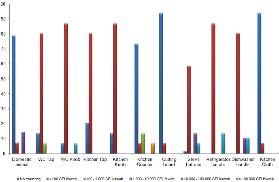
Enterobacteriaceae isolates were collected and Figure 4 summarizes Enterobacteriaceae distribution in the domestic setting. As can be seen these kinds of microorganisms are distributed all around the house with high isolation rates (> 102 CFU/swab). Localities as domestic animal paws, kitchen tap, kitchen counter, stove buttons, refrigerator and dishwasher handle or kitchen cloth show levels of contamination higher than 105 CFU/swab.
In a study carried out by Scott et al. (1982), more than 80% of the 201 homes examined contained one or more species of enterobacteria and wet places like taps and dishcloths were highly contaminated. The normal contamination of dishcloths and other wet items with large numbers of organisms including enterobacteria, suggests that these objects may act not only as reservoirs but also as disseminators of contamination in the kitchen and although enteropathogenic organisms probably originate from the toilet and toilet usage, hands and cleaning cloths harbour and may disseminate these organisms. Experimental studies with Salmonella spp. and E. coli show the likelihood for spread from toilets to bathroom surfaces and hands (Gerba et al., 1975; Barker and Bloomfield, 2000) and from hands to other surfaces (Rheinbaben et al., 2000).
In a study by Curtis et al. (2003), microbiological samples proved that faecal contamination of the domestic environment does occur, since faecal coliforms were found at a number of sites, not only in toilets and bathrooms but also in kitchens and on a variety of objects. The fact that a number of bathroom and toilet sites including door handles, were found to confirm signs of faecal contamination, suggests that hand-washing after using the toilet is not always regularly practised.
Several studies show that the intestinal tracts of animals generally harbour Enterobacteriaceae (Beutin, 1999; Guardabassi et al., 2004; Cobeljic et al., 2005; Jimenez et al., 2011); therefore it is normal that domestic animals may introduce these types of pathogens into the domestic setting.
- 14 -
Adiga et al. (2012) studies demonstrated that bacterial contamination in the kitchen is common and among 10 kitchens analysed several sites, like refrigerator handle, kitchen stove and water taps, were infected with faecal microorganisms.
Enterobacteriaceae presence in all 11 places of the house, analysed in this study, is in accordance with several studies which showed that various species of bacteria can live on kitchen surfaces and cross-contamination can easily occur contaminating the food preparation counter, cloths, utensils and hands (de Wit et al., 1979; Ak et al., 1994; Scott, 1996; Gorman et al., 2002; Beumer and Kusumaningrum, 2003).
- 15 -
- 16 - Figure 2 - Listeria spp. detection (%) by locality in the domestic environment.
- 17 - Figure 3 - Escherichia coli counts (%) by locality in the domestic environment.
- 18 - Figure 4 - Enterobacteriaceae counts (%) by locality in the domestic environment.
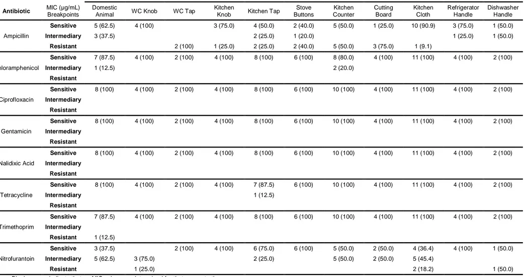
- 19 - 3.6. Enterobacteriaceae antibiotic resistance
For antibiotic resistance, all 351 isolates suspected to be Enterobacteriaceae were Gram-stained and tested for oxidase, catalase activity and fermentation of glucose. A total of 125 isolates glucose-fermenting, Gram-negative, oxidase-negative, catalase-positive were considered to belong to the family Enterobacteriaceae, and only these were included in further testing. After primary identification, E. coli and Salmonella spp. were differentiated from all the other Enterobacteriaceae as shown in Table 2.
Table 2 - Escherichia coli and Salmonella spp. differentiation by biochemical tests Microorganism
Biochemical Tests
Indole Triple Sugar Iron Agar (TSI)
Glucose Lactose Sucrose H2S Gas
Escherichia coli + + + + - +
Salmonella spp. - + - - + +
From this differentiation all 125 samples were separated in 3 major groups, for antibiotic resistance tests, namely 46 of E. coli, 19 of Salmonella spp. and all the remaining 60 isolates of other Enterobacteriaceae (Table 3 - see pag. 25).
High frequencies of antimicrobial resistance have been previously found in Enterobacteriaceae, in faecal flora as well as in clinical isolates (Kelch and Lee, 1978; Levy et al., 1988; Lester et al., 1990; Bonten et al., 1992; Leistevuo et al., 1996). Yet little or nothing is reported about antibiotic resistance in Enterobacteriaceae isolates found in the domestic setting, but there is plenty of evidence for enteric organisms found in this study with their origins in animals, humans and/or food.
Both animals and humans can introduce enteric pathogens into the dosmestic environment by cross-contamination when hands are poorly washed and paws have a direct contact to domestic surfaces. Food related problems arise when antimicrobials are used to treat infections and resistance is developed. In animals as in humans misuse of antibiotics may not only cause an increase of resistance in pathogenic bateria, but also in the endogenous flora of these animals. Resistant bacteria from these animals may be transferred to the human population, not only by direct contact, but also through food products of animal origin. These resistant bacteria may then either colonise humans and/or transfer their resistance genes to other bacteria in the human intestinal flora (van den Bogaard and Stobberingh, 2000).
At this time, it is well acknowledged that several antimicrobial resistant bacteria isolated from humans originated mainly from animals raised for human consumption (Aarestrups, 2000) and that such resistant bacteria may contaminate the meat derived from those animals (Sáenz et al., 2001). As a result, development of antimicrobial resistance amongst bacterial isolates from animal supplies can represent potential hazards to consumers through foodborne infections caused by these bacteria. In the past years, several studies have reported the antimicrobial resistance of some Enterobacteriaceae genera isolated from poultry, such as E. coli and Salmonella spp. (Antunes et al., 2003; Cormican et
- 20 -
al., 2001; Guerra et al., 2003; Kijima-Tanaka et al., 2003; Sáenz et al., 2001; van den Bogaard et al., 2001).
Enterobacteriaceae family, a group containing some highly pathogenic Gram negative organisms, is universally used as an indicator of faecal contamination during food microbiology analyses and it includes zoonotic bacteria like Salmonella and E. coli. These microorganisms may cause severe infections and are becoming gradually more resistant to the generally used treatment antibiotics like tetracyclines, aminoglycosides, trimethoprim, fluoroquinolones and chloramphenicol (Paterson 2006), demonstrating multiple resistance and declining activity of several antibiotic groups such as the fluoroquinolones (Rhomberg et al., 2006).
According to Table 3, in this study Enterobacteriaceae strains were found to be resistant to ampicillin (28.3%), trimethoprim (1.7%) and nitrofurantoin (5.0%). However, all strains were also sensitive to ciprofloxacin, gentamicin and nalidixic acid although resistance to Chloramphenicol and Tetracycline was practically non-existent (95 and 98.3% of sensitive strains, respectively).
Resistance of E. coli was found to ampicillin (41.3%), chloramphenicol (4.3%), tetracycline (6.5%), nalidixic acid (6.5%) and nitrofurantoin (4.3%). Nevertheless all strains showed sensitivity to ciprofloxacin, gentamicin and trimethoprim.
Among Salmonella spp. isolates, 26.3% were resistant to ampicillin, 5.3% to chloramphenicol, 10.5% to tetracycline, 5.3% to nalidixic acid and 15.8% to nitrofurantoin. Ciprofloxacin, gentamicin and trimethoprim were shown to be very effective with 100% of sensitivity detected.
For all eight antimicrobials tested, overall no resistance was found to ciprofloxacin and gentamicin and for all 3 major groups resistance to ampicillin was common with a top score distinguished for E. coli with 41.3% of strains resistant, followed by Enterobacteriaceae with 28.3% and Salmonella spp. with 26.3%. For nitrofurantoin 15.8% of Salmonella spp. strains were resistant, followed by Enterobacteriaceae spp. with 5.0% and E. coli with 4.3%. This is in accordance with a study carried out by Osterblad et al. (1999) where antimicrobial sensitivity was shown in Enterobacteriaceae isolated from vegetables.
For example, urinary tract infections is a common illness that afects both community and hospital patients and is often caused by E. coli which is naturally susceptible to ampicillin even though about 50 - 60% of isolates are now resistant worlwide (Wu et al., 1992; Chomarat, 2000; Sefton, 2000; Gupta, 2001).
For more than 50 years nitrofurantoin has been an option for the management of urinary tract infection but its use declined with the introduction of alternative antimicrobials, like trimethoprim, although there has been a slow reappearance in its use because of continued low rates of resistance among common urologic pathogens (Hooton and Stamm, 1997). Nowadays, the only indication for nitrofurantoin is the management of bladder infection resulting from susceptible strains of E. coli, once it appears to be associated with lower cure rates (approximately 85%) than other first line agents (90% to 95%), like trimethoprim (Warren et al., 1999). In the present study, isolates showed low levels of resistance to nitrofurantoin (15.8%, 5.0% and 4.3% for Salmonella spp., Enterobacteriaceae and E. coli samples,
- 21 -
respectively). A study conducted by Rampling et al., (1990) stated that Salmonella Enteritidis, isolated from poultry and from human enteric infection in the United Kingdom, showed high resistance rates which can be explained by the use of nitrofurans in the poultry industry.
Trimethoprim has been the core of therapy for urinary tract infection for the past many years with a 90% success rate as first-line agent indicated for the supervision of acute urinary tract infection and pyelonephritis (Hooton et al., 1995). It is very effective against most Enterobacteriaceae like E. coli, Klebsiella species, Enterobacter species, Morganella morganii, Proteus mirabilis and Proteus vulgaris although there has been a significant increase in the prevalence of resistance of E coli.
Enterobacteriaceae family has shown worldwide trimethoprim resistance in chicken, pork, fish and even water (Tao et al., 2010; Su et al., 2011; Schwaiger et al., 2012). Generally E. coli and Salmonella spp. present higher resistance rates although this is not what was found in this study. Domestic environment Enterobacteriaceae showed a very low rate of resistance (1.7%) while E. coli and Salmonella spp. strains were all sensitive to this kind of antimicrobial.
In some countries Salmonella and E. coli antibiotic resistance rates are reported to be high (Oppegaard et al., 2001; Ronald, 2002; Threlfall, 2002). In a study with 752 E. coli isolates from human and animal agriculture sources in several countries, tetracycline high frequency resistance rates were found in isolates from human and turkey samples (56% and 71%, respectively). The resistance profiles for cattle, chicken, and swine were similar with approximately 47% of cattle isolates resistant to tetracycline (Schroeder et al., 2002). This can be explained by the fact that tetracycline is the drug most often used in animal husbandry and is only a drug of second choice in human medicine (Mayrhofer et al., 2004). In another study performed by Schroeder et al. (2003) samples taken from retail beef, chicken, pork, and turkey resulted in 472 E. coli isolates, 59% of which were resistant to tetracycline, nalidixic acid (8%), and chloramphenicol (6%).
In contrast with these results, domestic isolates showed comparatively low rates of resistance to tetracycline with 6.5% and 10.5% for E. coli and Salmonella spp., respectively. All other Enterobacteriaceae were sensitive to this antimicrobial (98.3%). Although resistance rates related to nalidixic acid (6.5% for E. coli and 5.3% for Salmonella spp.) and chloramphenicol (4.3% for E. coli and 5.3% for Salmonella spp.) were much lower, this is in conformity with Schroeder et al. (2003) study. In our study no resistance was determined for gentamicin or ciprofloxacin. This is in agreement with several studies but antimicrobial resistance can be modified depending on the nature of the food production system considered (Osterblad et al., 1999; van den Bogaard et al., 2000; Bywater et al., 2004; Fluckey et al., 2007; Miranda et al., 2008; Knezeviz and Petrovic, 2008).
In an antimicrobial susceptibility study of Enterobacteriaceae isolated from vegetables, no resistance was found to nalidixic acid but tetracycline and chloramphenicol showed low resistance rates (5.5 and 12%, respectively) (Osterblad et al., 1999). This is consistent with some other antibiotic resistance studies where Enterobacteriaceae were isolated from milk, cheese and other dairy products. Isolates from milk products presented no resistance to nalidixic acid but some resistance was detected for tetracycline (14.28%) and chloramphenicol (9.52%). Among samples of cheese, 24% of isolates were
- 22 -
resistant to tetracycline but no resistance was found to chloramphenicol and nalidixic acid. Dairy products showed high rates of resistance to tetracycline (52.38%), but also no resistance was found to chloramphenicol and nalidixic acid (Hleba et al., 2011).
In our study nalidixic acid, chloramphenicol and tetracycline were shown to be effective against Enterobacteriaceae since no resistance was found.
Several studies reported Salmonella and E. coli food-related isolates showed resistance to trimethoprim and some declare that this resistance profile is due to treatments used in animal medicine (van den Bogaard et al., 2000; Cormican et al., 2001; Sáenz et al., 2001; van den Bogaard et al., 2001; Kijima-Tanaka et al., 2003; Bywater et al., 2004; Fluckey et al., 2007). Although some studies also stated food-related isolates showed sensitivity to trimethoprim (Lundin et al., 2008; Erdington et al., 2009) which is in accordance with this study where all Salmonella and E. coli strains were sensitive to this antibiotic.
Another interesting way of analysing our results was to organize antibiotic resistance rates by house (Table 4 to 6 - see pag. 26 to 28) and surface (Table 7 to 9 - see pag. 29 to 31).
From data in Table 4 it can be concluded that although other Enterobacteriaceae are not present in one house they are widely spread in all the others. Several antibiotic resistant strains were detected, mainly to ampicillin (House 1, 6, 9, 10, 11, 12 and 15) and ranging from 25.0% of isolates in House 9 to 66.7% in House 12, and nitrofurantoin (House 3, 4, 8, 12 and 15), ranging from 16.7% in House 12 to 50.0% in House 3. Only one strain showed trimethoprim resistance in House 1 (9.1%). All isolates were sensitive to ciprofloxacin, gentamicin and nalidixic acid although chloramphenicol and tetracycline present some intermediary isolates.
In conclusion, resistance to more than one antibiotic is verified in Enterobacteriaceae isolates from House 1 (ampicillin and trimethoprim), House 12 and House 15 (ampicillin and nitrofurantoin, in both cases). However, isolates from House 5 and House 13 showed no resistance to all 8 antibiotics tested. In Table 5 it can be seen that not all houses are contaminated with E. coli which is good news since some strains of these bacteria are considered a severe faecal pathogen. Once more strains showed resistance to ampicillin (House 3, 7, 9, 10, 13, 14 and 15) and nitrofurantoin (House 4 and 15) with rates ranging from 20.0% in House 10 to 100% in House 3 and 8.3% in House 15 to 50.0% in House 4, respectively. Moreover in House 13, 50.0% strains were found to be resistant to chloramphenicol, 75.0% to nalidixic acid and another 50.0% to tetracycline. Isolates from House 15 also showed some extra resistance to tetracycline although in low proportion (1 in 11 isolates). All E. coli domestic isolates showed no resistance to ciprofloxacin, gentamicin and trimethoprim.
In E. coli isolates resistance to more than one antibiotic was confirmed in House 13 (ampicillin, chloramphenicol, nalidixic acid and tetracycline) and House 15 (ampicillin, tetracycline and nitrofurantoin). Nevertheless, strains from House 5, House 8 and House 11 showed no resistance against all 8 antibiotic tested.
Table 6 presents Salmonella spp. resistance rates by house and it can can be seen that contamination is lower than for other bacteria presented above, since only 6 in 15 houses show contamination. In this
- 23 -
case isolates exhibit high resistance rates against ampicillin in House 2, 3, 4 and 11 (varying from 25.0% in House 3 to 100% in House 4) while strains were resistant to nitrofurantoin in House 1 (50.0%) and House 3 (25.0%). Some strains were resistant to chloramphenicol (12.5% in House 3), nalidixic acid (50.0% in House 11) and tetracycline (25.0% in House 3). Ciprofloxacin, gentamicin and trimethoprim are once more the most effective antibiotics with 100% sensitive strains.
In this case, Salmonella spp. isolates from two houses proved to be resistant to more than one antibiotic namely House 3 (ampicillin, chloramphenicol, tetracycline and nitrofurantoin) and House 11 (ampicillin and nalidixic acid). On the other hand, only House 7 showed no resistance for all 8 antibiotic tested.
Related to Table 7 it can be seen that Enterobacteriaceae show no resistance to chloramphenicol, ciprofloxacin, gentamicin, nalidixic acid and tetracycline even though some strains show intermediary breakpoints for chloramphenicol (domestic animal and kitchen counter) and tetracycline (kitchen tap). Resistance rates were detected against ampicillin (ranging from 9.1% in kitchen cloth to 100% in WC tap) and nitrofurantoin (ranging from 18.2% in kitchen cloth to 50% in dishwasher handle).
In summary, only those Enterobacteriaceae isolates found in the kitchen cloth showed resistance to more than one antibiotic (ampicillin and nitrofurantoin) while the refrigerator handle isolates presented no resistance to all 8 antibiotics tested.
From the data in Table 8, no E. coli was detected in WC knob but in all 9 other surfaces it is widely distributed. High resistance to ampicillin was found 9 localities, not including domestic animals’ feet. The kitchen cloth and dishwasher handle isolates presented resistance to chloramphenicol, nalidixic acid and tetracycline while nitrofurantoin resistance was found in the strains from the kitchen counter and dishwasher handle. No resistance was found to ciprofloxacin, gentamicin and trimethoprim.
In conclusion, E. coli isolates found in the kitchen cloth and dishwasher handle showed resistance to more than one antibiotic. Resistance to ampicillin (75.0%) and nitrofurantoin (50.0%) was detected in kitchen counter isolates, while those from the kitchen cloth demonstrated resistance to ampicillin (54.5%), chloramphenicol (9.1%), nalidixic acid (18.2%) and tetracycline (9.1%). The dishwasher handle isolates showed low resistance (16.7%) to 5 antibiotics namely ampicillin, chloramphenicol, nalidixic acid, tetracycline and nitrofurantoin. Only strains from domestic animals presented no resistance to all 8 antibiotics tested.
Analysing Table 9 Salmonella spp. shows no isolates from the WC tap, kitchen knob and stove buttons. Resistance was detected for 5 of the 8 antibiotics detected with ampicillin taking the lead with high rates in isolates from kitchen tap (50.0%), cutting board (100%) and kitchen cloth (50.0%). Cutting board also showed evidence for resistance to chloramphenicol (50.0%), tetracycline (100%) and nitrofurantoin (100%) and only one strain (33.3) showed resistance to nalidixic acid (from the kitchen counter). No isolates were resistant to ciprofloxacin, gentamicin or trimethoprim.
Therefore, of the Salmonella spp. isolates, only those from the cutting board presented resistance to more than one antibiotic namely, ampicillin, chloramphenicol, tetracycline and nitrofurantoin. In contrast, the isolates from the WC knob, refrigerator and dishwasher handles revealed no resistance.
- 24 -
Overall, multi-resistance, to more than one antibiotic, was found in this study. Analysing Annex 1, where all the different antibiotic profiles are shown, in all 15 Houses, we can assume that diverse sources of enteric bacteria were found probably from different origins as animals, humans and/or foodstuff.
- 25 -
Table 3 - In vitro susceptibility of Enterobacteriaceae isolates to several antibiotics and minimum inhibitory concentration (MIC) breakpoints
MIC (µg/mL) Breakpoints Other Enterobacteriaceae isolates Escherichia coli isolates Salmonella spp. isolates
Class Antibiotic Sensitive (S) Intermediary (I) Resistant (R)
Number of sensitive isolates (%) Number of intermediary isolates (%) Number of resistant isolates (%) Number of sensitive isolates (%) Number of intermediary isolates (%) Number of resistant isolates (%) Number of sensitive isolates (%) Number of intermediary isolates (%) Number of resistant isolates (%) Penicillins Ampicillin 8 16 32 37 (61.7) 6 (10.0) 17 (28.3) 21 (45.6) 6 (13.0) 19 (41.3) 10 (52.6) 4 (21.1) 5 (26.3) Phenicols Chloramphenicol 8 16 32 57 (95.0) 3 (5.0) 44 (95.7) 2 (4.3) 16 (84.2) 2 (10.5) 1 (5.3) Fluoroquinolones Ciprofloxacin 1 2 4 60 (100) 46 (100) 19 (100) Aminoglycosides Gentamicin 4 8 16 60 (100) 46 (100) 19 (100) Tetracyclines Tetracycline 4 8 16 59 (98.3) 1 (1.7) 42 (91.3) 1 (2.2) 3 (6.5) 13 (68.4) 4 (21.1) 2 (10.5) Quinolones Nalidixic Acid 16 --- 32 60 (100) 43 (93.5) 3 (6.5) 18 (94.7) 1 (5.3) Folate pathway
inhibitor Trimethoprim 8 --- 16 59 (98.3) 1 (1.7) 46 (100) 19 (100)
Nitrofurantoins Nitrofurantoin 32 64 128 34 (56.7) 23 (38.3) 3 (5.0) 39 (84.8) 5 (10.9) 2 (4.3) 13 (68.4) 3 (15.8) 3 (15.8) Blank spaces indicate that no MIC value was determined for that concentration.