Bacterial, fungal and yeast contamination
in six brands of irreversible hydrocolloid
impression materials
Contaminação por bactérias, fungos e
leveduras em seis marcas comerciais de
materiais de moldagem à base de
hidrocolóide irreversível
Abstract: This study assessed the level of contamination of six commercially available ir-reversible hydrocolloids (two containing chlorhexidine) and identiied the contamination present in the materials. Petri dishes containing selective and enriched culture media were inoculated with alginate powder (0.06 g), in triplicate. After incubation (37°C/7 days), the colony-forming units (CFU) were counted and Gram stained. Biochemical identiication of the different morphotypes was also performed. The contamination levels for the mate-rials were: Jeltrate - 389 CFU/g; Jeltrate Plus - 516 CFU/g; Jeltrate Chromatic - 135 CFU/ g; Hydrogum - 1,455 CFU/g; Kromopan - 840 CFU/g; and Greengel - 59 CFU/g. Gram staining revealed the presence of Gram-positive bacillus and Gram-positive cocci. The bacteria Staphylococcus epidermidis, Bacillus subtilis, Bacillus sp., Bacillus coagulans,
Bacillus licheniformis, Bacillus cereus, Micrococcus luteus, and Nocardia sp.; the ila-mentous fungi Aspergillus niger, Aspergillus lavus, Rhizopus sp., Neurospora sp.; and the yeast Candida sp. were isolated. The contamination detected in the impression materi-als points out the need for adopting measures to improve the microbiological quality of these materials. The use of contaminated materials in the oral cavity goes against the ba-sic principles for controlling cross-contamination and may represent a risk for debilitated or immunocompromised patients.
Descriptors: Dental materials; Microbiology; Infection control.
Resumo: Este estudo avaliou o nível de contaminação de seis marcas comerciais de algi-nato (duas contendo clorexidina) e identiicou a contaminação presente nesses materiais. Alíquotas de alginato (0,06 g) foram semeadas em meios de cultura seletivos e enrique-cidos, em triplicata. Após incubação (37°C/7 dias), as unidades formadoras de colônia (UFC) foram contadas e foram realizadas as identiicações morfotintorial (Gram) e bio-química. Os níveis de contaminação dos materiais foram: Jeltrate - 389 UFC/g; Jeltrate Plus - 516 UFC/g; Jeltrate Chromatic - 135 UFC/g; Hydrogum - 1.455 UFC/g; Kromopan - 840 UFC/g; e Greengel - 59 UFC/g. A coloração de Gram revelou a presença de bacilos Gram-positivos e cocos Gram-positivos. As bactérias Staphylococcus epidermidis, Ba-cillus subtilis, Bacillus sp., Bacillus coagulans, Bacillus licheniformis, Bacillus cereus,
Micrococcus luteus e Nocardia sp.; os fungos ilamentosos Aspergillus niger, Aspergillus lavus, Rhizopus sp., Neurospora sp.; e a levedura Candida sp. foram isolados. A conta-minação detectada nos materiais aponta a necessidade de adoção de medidas para melho-rar seu controle de qualidade microbiológica. O uso de materiais contaminados na boca contradiz os princípios básicos de controle de infecção-cruzada e pode representar um risco para pacientes debilitados ou imunocomprometidos.
Descritores: Materiais dentários; Microbiologia; Controle de infecções. Luciana Assirati Casemiro(a)
Carlos Henrique Gomes Martins(b)
Fernanda de Carvalho Panzeri Pires de Souza(c)
Heitor Panzeri(c)
Isabel Yoko Ito(d)
(a) PhD, Professor, School of Dentistry of
Franca; (b)PhD, Professor, Laboratory of
Microbiology – University of Franca.
(c) PhDs, Professors; (d)Full Professor – School
of Dentistry of Ribeirão Preto, University of São Paulo.
Corresponding author: Luciana Assirati Casemiro Avenida Caramuru, 2100 ap. 901 Ribeirão Preto - SP - Brazil CEP: 14030-000
E-mail: lucianacasemiro@hotmail.com
Introduction
For years, researchers and manufacturers have put much effort in developing and enhancing dental materials in the search for excellence in their physi-cal, mechaniphysi-cal, and biological properties. None-theless, certain materials, such as irreversible hydro-colloids, still show deiciencies, and it is possible to isolate and identify viable microorganisms and fungi in the powder of commercialized containers.16-19
Rice et al.19 (1990) found viable Gram-negative
cocci and Gram-negative rods in 25% of the as-sessed alginate samples. According to those authors, the scarcity of data concerning alginate contamina-tion in the literature may be the reason for the little questioning about the possibility of contaminating immunocompromised patients with these materials. After that, other studies were performed,16-18
point-ing out the contamination of various brands, includ-ing those containinclud-ing antimicrobial agents.16
As to the addition of antimicrobial agents to dental materials,7 this has been a current tendency,
with the goal of inhibiting or avoiding the adhesion and growth of microorganisms. In irreversible hy-drocolloids,11 besides promoting disinfection of the
impressions, the added antimicrobial agents may act as preservatives, reducing the presence of viable mi-croorganisms in the powder. These materials show a better microbiologic quality when compared to al-ginates without antimicrobial agents; however, add-ing antimicrobial agents to these materials does not mean they will be free from microorganisms.16
Be-cause alginates have polysaccharide structures simi-lar to those of agar (excellent substrate for micro-organisms), it seems unlikely that alginate powders would be free from microorganisms.
The possibility of contaminating immunocom-promised patients in dental procedures of minor complexity, since they are susceptible to infections by microorganisms of low virulence, has been point-ed out.12 Given that it is impossible to determine the
patients’ immunological condition during each den-tal treatment, there is a need for adopting the con-cept of universal precaution.2
Impression procedures frequently cause bleeding of the mouth’s soft tissues. Considering that blood is a rich culture and microbial transportation medium,
and that any rupture of skin integrity offers an open-ing for the entrance of potentially pathogenic micro-organisms,5 there is a risk of accidental
transmis-sion of this infectious substrate to undesired places. Hence, the use of contaminated impression materi-als may represent an additional risk of inoculation of microorganisms and, consequently, of occurrence of diseases in immunocompromised patients.
It has long been known that irreversible hydro-colloids may contain viable microorganisms;16-19
however, it seems that, until today, no measures have been taken by manufacturers to avoid such contamination. Due to the risk posed by contami-nated irreversible hydrocolloids, there is a need for improvement from a microbiological point of view. Sterilization methods should be incorporated, such as irradiation with gamma rays at the end of produc-tion. Nonetheless, such method requires a previous knowledge concerning microbial load, both qualita-tively and quantitaqualita-tively, in order to determine the doses to be applied. This fact was the motivation for the present study.
This study’s goal was to assess the level of tamination of six irreversible hydrocolloids (two con-taining antimicrobial agents), and isolate and iden-tify the contamination present in these materials.
Material and Methods
The morphology of the colonies was observed in a stereoscopic microscope (SMZ645, Nikon, Tokyo, Japan), under relected light. The biochemical iden-tiication14 of the different morphotypes was
per-formed in the following manner.
Bacteria Identiication: Fermentation of glucose, lactose, sucrose, maltose, mannitol and sorbitol. Use of citrate and malonate as the only carbon source. Hydrolyses of urea, gelatin, starch, and lecithin. Decarboxylation of lysine, ornithine and arginine. Production of nitrite, acetoin, H2S, indole, and phenylpyruvic acid.
Fungi Identiication: The identiication of fungi was based on their macroscopic colonial mor-phology8,10 and on their microscopic aspect,
us-ing the microculture technique.10
Results
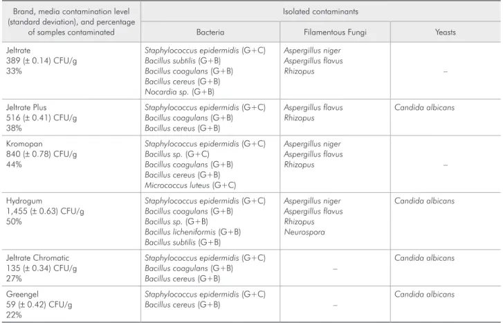
Table 2 shows the levels of contamination detect-ed in the powders and the isolatdetect-ed contaminants. The samples inoculated in Blood Agar, Mueller-Hinton Agar, and Sabouraud Dextrose Agar pre-sented microbial and fungal growth.
Discussion
The overall level of contamination was high for the materials studied, ranging from 1,455 CFU/g to 59 CFU/g; these means were signiicantly different (p < 0.05). In a previous study,16 the alginates
with-out antimicrobial agents showed a contamination of 9-161.1 CFU/g, which was smaller than that
ob-•
•
served for most of the alginates assessed in this study. However, alginates containing antimicrobial agents showed a better microbiologic quality, 135 CFU/ g (Jeltrate Chromatic) and 59 CFU/g (Greengel). Comparing this best result with those obtained in another study16 for alginates with chlorhexidine
(2.8 CFU/g) and cetylpyridinium chloride (2.8 CFU/ g), it was observed that the latter were smaller than those observed in the present study. The best results obtained in this study may be due to the presence of chlorhexidine diacetate 0.05%, which may have acted as a preservative. The quality of the raw mate-rial and the manufacturing, packaging and storing procedures may also have inluenced this result.3
The materials containing chlorhexidine – Green-gel and Jeltrate Chromatic – presented the lowest contamination levels, respectively 59 CFU/g and 135 CFU/g. Chlorhexidine is a cationic agent with a broad-spectrum antibacterial and antifungal ac-tivity. It is also biocompatible with mouth tissues. It has substantivity, which is the ability to remain on a particular surface and be gradually released.15
Its excellent properties have motivated its increasing use in dentistry.
Besides chlorhexidine, other components in the formulation of alginates may have inluenced the results. Fluoride, for instance, is acknowledged as being antimicrobial.1 Its action mechanisms against
microorganisms include inhibiting sugar transporta-tion to the interior of the bacteria, and affecting the energetic mechanisms, the glycolytic pathway, and
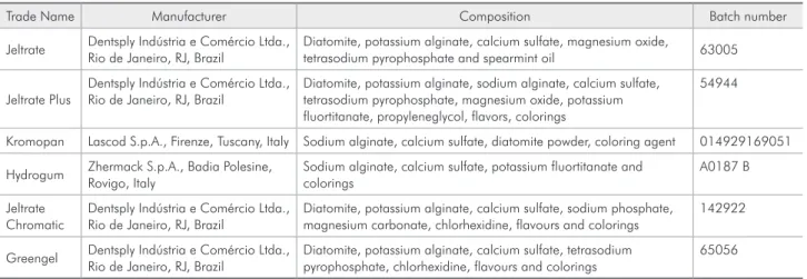
Table 1 - Irreversible hydrocolloids, their manufacturers, compositions, and batches.
Trade Name Manufacturer Composition Batch number
Jeltrate Dentsply Indústria e Comércio Ltda., Rio de Janeiro, RJ, Brazil
Diatomite, potassium alginate, calcium sulfate, magnesium oxide,
tetrasodium pyrophosphate and spearmint oil 63005
Jeltrate Plus
Dentsply Indústria e Comércio Ltda., Rio de Janeiro, RJ, Brazil
Diatomite, potassium alginate, sodium alginate, calcium sulfate, tetrasodium pyrophosphate, magnesium oxide, potassium fluortitanate, propyleneglycol, flavors, colorings
54944
Kromopan Lascod S.p.A., Firenze, Tuscany, Italy Sodium alginate, calcium sulfate, diatomite powder, coloring agent 014929169051
Hydrogum Zhermack S.p.A., Badia Polesine, Rovigo, Italy
Sodium alginate, calcium sulfate, potassium fluortitanate and colorings
A0187 B
Jeltrate Chromatic
Dentsply Indústria e Comércio Ltda., Rio de Janeiro, RJ, Brazil
Diatomite, potassium alginate, calcium sulfate, sodium phosphate, magnesium carbonate, chlorhexidine, flavours and colorings
142922
Greengel Dentsply Indústria e Comércio Ltda., Rio de Janeiro, RJ, Brazil
Diatomite, potassium alginate, calcium sulfate, tetrasodium pyrophosphate, chlorhexidine, flavours and colorings
the synthesis of glycogen and metalloenzymes.6 Two
of the materials evaluated – Greengel and Jeltrate Chromatic – contain chlorhexidine in their compo-sitions, but only the former contains Fluoride, which may explain the different levels of contamination (59 ± 0.42 for Greengel and 135 ± 0.34 for Jeltrate Chromatic).
Another component with antimicrobial activity is magnesium oxide.20,21 When its powder becomes
in contact with vegetative cells of Escherichia coli,
Bacillus cereus, or Bacillus globigii, it kills over 90% of them in a few minutes.9 It is also active
against fungi and yeasts, such as Candida albicans
NBRC1060, Saccharomyces cerevisiae NBRC1950,
Aspergillus niger NBRC4067, and Rhizopus stoloni-fer NBRC4781.21 Essential oils such as spearmint oil
also have antibacterial and antifungal activities.13,22
These components may also have helped to reduce the contamination levels presented by the evaluated impression materials; however, lower contamination
levels were observed for the irreversible hydrocol-loids containing chlorhexidine and Fluoride.
The inoculated samples showed variable contam-ination levels with microorganisms and fungi. For Jeltrate (33%), Jetrate Plus (38%), Kromopan (44%), Hydrogum (50%), and Jeltrate Chromatic (27%), the percentages were above the 25% found by Rice et al.17 (1991). The Greengel product, however, showed
a lower percentage of contamination (22%).
In the results, the contamination levels were ex-pressed in CFU/g, but it is important to consider that the mean quantity of material used to make an impression is about 20 g, which results in an inocu-lation of a high number of CFUs for each impres-sion.
Regarding the morphotypes, Gram-positive ba-cilli and Gram-positive cocci were isolated. How-ever, other researchers19 have observed the presence
of Gram-negative cocci and Gram-positive rods in materials without antimicrobial agents.
Table 2 - Microorganisms isolated in the impression materials.
Brand, media contamination level (standard deviation), and percentage
of samples contaminated
Isolated contaminants
Bacteria Filamentous Fungi Yeasts
Jeltrate
389 (± 0.14) CFU/g 33%
Staphylococcus epidermidis (G+C)
Bacillus subtilis (G+B)
Bacillus coagulans (G+B)
Bacillus cereus (G+B)
Nocardia sp. (G+B)
Aspergillus niger Aspergillus flavus
Rhizopus –
Jeltrate Plus 516 (± 0.41) CFU/g 38%
Staphylococcus epidermidis (G+C)
Bacillus coagulans (G+B)
Bacillus cereus (G+B)
Aspergillus flavus Rhizopus
Candida albicans
Kromopan 840 (± 0.78) CFU/g 44%
Staphylococcus epidermidis (G+C)
Bacillus sp. (G+C)
Bacillus coagulans (G+B)
Bacillus cereus (G+B)
Micrococcus luteus (G+C)
Aspergillus niger Aspergillus flavus
Rhizopus –
Hydrogum
1,455 (± 0.63) CFU/g 50%
Staphylococcus epidermidis (G+C)
Bacillus coagulans (G+B)
Bacillus sp. (G+B)
Bacillus licheniformis (G+B)
Bacillus subtilis (G+B)
Aspergillus niger Aspergillus flavus Rhizopus Neurospora
Candida albicans
Jeltrate Chromatic 135 (± 0.34) CFU/g 27%
Staphylococcus epidermidis (G+C)
Bacillus coagulans (G+B)
Bacillus cereus (G+B)
–
Candida albicans
Greengel 59 (± 0.42) CFU/g 22%
Staphylococcus epidermidis (G+C)
Bacillus cereus (G+B) –
Candida albicans
The microorganisms isolated in all alginates were similar to those detected previously,16-19 mostly
com-mensal ones in healthy human beings, and incapable of causing any diseases in a situation of equilibrium.
Micrococcus luteus, for example, is not considered a pathogenic, or “disease-causing”, organism in healthy people. However, when the patient’s immunological conditions are deicient, commensal as well as patho-genic microorganisms may be involved in developing diseases, which is a result of the complex interactions between the infecting organism and the host.2,14
Regarding the origin of the contamination pres-ent, it may be due to the materials used, to the manufacturing process (contaminated equipment, operators, air and packages), to the casing, trans-portation, and even caused by the user.3 As to the
latter, even if the alginates were free of contamina-tion at irst, there would certainly still be a possibil-ity of contamination because the material is kept in a large package, whose content is suficient to per-form many impressions. By opening the container,
microorganisms from the environment could be introduced into it, which would make the material inadequate for further use, from a microbiological point of view.
It would therefore be ideal to improve the microbi-ological quality of commercially available alginates, taking into consideration the Good Manufacturing Codes of Practices,4,23 and to determine, by
perform-ing other studies, if these materials should be sterile or should be allowed a microbiological limit, both qualitatively and quantitatively. It is also recom-mended that the packages be presented with a single-use size (for a single impression) in order to preserve the microbiological quality of the material.24
Conclusion
All the irreversible hydrocolloids studied here presented viable bacteria, fungi, and yeasts. How-ever, the materials containing chlorhexidine showed the lowest contamination levels (Greengel, 59 CFU/ g, and Jeltrate Chromatic, 135 CFU/g).
References
1. Bowden GH. Effects of fluoride on the microbial ecology of dental plaque. J Dent Res. 1990;69 Spec No:653-9. 2. Cottone JA. Practical Infection Control in Dentistry.
Phila-delphia: Lea & Febiger; 1991.
3. Fassihi RA. Preservation of medicines against microbial con-tamination. In: Block SS. Disinfection, sterilization and pres-ervation. 4th ed. Malvern: Lea & Febiger; 1991. p. 871-86.
4. Good manufacturing practices, including quality assurance for dental materials. Report to General Assembly, Federation Dentaire Internationale. Int Dent J. 1990;40(4):253-6. 5. Gurevich I. Infection control: Applying theory to clinical
prac-tice. In: Block SS. Disinfection, sterilization and preservation. 4th ed. Malvern: Lea & Febiger; 1991. p. 655-62.
6. Hamilton IR. Biochemical effects of fluoride on oral bacteria. J Dent Res. 1990;69 Spec No:660-7.
7. Imazato S, Torii M, Tsuchitani Y, McCabe JF, Russell RR. Incorporation of bacterial inhibitor into resin composite. J Dent Res. 1994;73(8):1437-43.
8. Kern ME, Blevins KS. Medical Mycology: A Self-Instructional Text. 2nd ed. Philadelphia: FA Davis; 1997.
9. Koper OB, Klabunde JS, Marchin GL, Klabunde KJ, Stoimenov P, Bohra L. Nanoscale powders and formulations with biocidal activity toward spores and vegetative cells of bacillus species, viruses, and toxins. Curr Microbiol. 2002;44(1):49-55.
10. Lacaz CS, Porto E, Martins JEC. Micologia Médica. 7th ed.
São Paulo: Sarvier; 1984.
11. Lott G, Gribi HK. Aseptic dental alginate impressions. Quin-tessence Int. 1988;19(8):571-4.
12. McGhee JR, Michalek SM, Cassel GH. Dental Microbiology. New York: Harper Row; 1982.
13. Mimica-Dukic N, Bozin B, Sokovic M, Mihajlovic B, Mata-vulj M. Antimicrobial and antioxidant activities of three Men-tha species essential oils. Planta Med. 2003;69(5):413-9. 14. Murray PR. Manual of Clinical Microbiology. 7th ed.
Wash-ington: ASM Press; 1999.
15. Ribeiro J, Ericson D. In vitro bacterial effect of chlorhexi-dine added to glass ionomer cements. Scand J Dent Res. 1991;99(6):533-40.
16. Rice CD, Dykstra MA, Feil PH. Microbial contamination in two antimicrobial and four control brands of alginate impres-sion material. J Prosthet Dent. 1992;67(4):535-40.
17. Rice CD, Dykstra MA, Gier RE. Bacterial contamination in irreversible hydrocolloid impression material and gingival retraction cord. J Prosthet Dent. 1991;65(4):496-9.
19. Rice CD, Moghadam B, Gier RE, Cobb CM. Aerobic bacterial contamination in dental materials. Oral Surg Oral Med Oral Pathol. 1990;70(4):537-9.
20. Sawai J. Quantitative evaluation of antibacterial activities of metallic oxide powders (ZnO, MgO and CaO) by conducti-metric assay. J Microbiol Methods. 2003;54(2):177-82. 21. Sawai J, Yoshikawa T. Quantitative evaluation of antifungal
activity of metallic oxide powders (MgO, CaO and ZnO) by an indirect conductimetric assay. J Appl Microbiol. 2004;96(4):803-9.
22. Soliman KM, Badeaa RI. Effect of oil extracted from some medicinal plants on different mycotoxigenic fungi. Food Chem Toxicol. 2002;40(11):1669-75.
23. Von Bockelmann B. Aseptic Packaging. In: Block SS. Disinfec-tion, sterilization and preservation. 4th ed. Malvern: Lea &
Febiger; 1991. p. 833-45.

