ABSTRACT
A new improved protocol for
in vit r o
intratubular
dentinal bacterial contamination for antimicrobial
endodontic tests: standardization and validation
by confocal laser scanning microscopy
Flaviana Bombarda de ANDRADE1, Marcela Paola Castro ARIAS1, Amanda Garcia Alves MALIZA1, Marco Antonio Hungaro DUARTE1, Márcia Sirlene Zardin GRAEFF2, Pablo Andrés AMOROSO-SILVA1, Raquel Zanin MIDENA1,
Ivaldo Gomes de MORAES1
1- Department of Operative Dentistry, Endodontics and Dental Materials, Bauru School of Dentistry, University of São Paulo, Bauru, SP, Brazil 2- Integrated Research Center, Bauru School of Dentistry, University of São Paulo, Bauru, SP, Brazil.
Corresponding address: Flaviana Bombarda de Andrade - Faculdade de Odontologia de Bauru - Universidade de São Paulo - Al. Dr. Octávio Pinheiro Brisolla 9-75 - Vila Universitária - Bauru - SP - Brazil - 17012-901 - Phone: +55 (14) 3235 8344 / +55 (14) 99750 1313 - Fax: +55 (14) 3235 8323 - e-mail:
ÀDYLDQD#IREXVSEU
6XEPLWWHG-XO\0RGL¿FDWLRQ1RYHPEHU$FFHSWHG1RYHPEHU
O
bjectives: To compare three methods of intratubular contamination that simulate endodontic infections using confocal laser scanning microscopy (CLSM). Material and Methods: Two pre-existing models of dentinal contamination were used to induce intratubular infection (groups A and ). These methods were modi ed in an attempt to improve the model (group C). Among the modi cations the following were included:specimen contamination for ve days ultrasonic bath with broth after specimen
sterilization, use of E. faecalis during the exponential growth phase, greater concentration of inoculum, and two cycles of centrifugation on alternate days with changes of culture media. All specimens were longitudinally sectioned and stained with of LIVE/DEAD® for 20
min. Specimens were assessed using CLSM, which provided images of the depth of viable bacterial proliferation inside the dentinal tubules. Additionally, three examiners used scores to classify the CLSM images according to the following parameters: homogeneity, density, and depth of the bacterial contamination inside the dentinal tubules. Kruskal-Wallis and Dunn’s tests were used to evaluate the live and dead cells rates, and the scores obtained. Results: The contamination scores revealed higher contamination levels in group C when compared with groups A and B (p<0.05). No differences were observed between group A and B (p>0.05). The volume of live cells in group C was higher than in groups A and B (p<0.05). Conclusion: The new protocol for intratubular infection resulted in high and uniform patterns of bacterial contamination and higher cell viability in all specimens when compared with the current methods.
Ke y w or ds: Confocal microscopy. Contamination. Dentin. Ent er ococcus faecalis.
I N TROD UCTI ON
Bacteria and their toxins are the major cause of the development, progression, and persistence of pulp and periapical diseases12,17,25. Appropriate
biomechanical preparations, assisted by irrigants and intracanal medications to promote the elimination of bacteria and their products, are required to obtain success in root canal treatment11,14,22,23,30. A
reason for the persistence of endodontic infections is bacterial penetration inside the dentinal tubules, rendering its elimination dif cult10,27,29.
scanning microscopy
Some authors veri ed this bacterial penetration by means of microbiological culture3,10 and
histological analysis with Brown & Brenn staining15,16,
both of them showing interesting results and images of dentinal contamination; however, they are time-consuming methods. Alternatively, scanning electron microscopy (SEM) was used to verify the disinfection of the root canal walls4,15,22,28. Because
SEM achieves extremely high magni cation, each eld of analysis is therefore very small. Thus, many SEM images are necessary to accurately assess the outcome. Optical microscopy (OM)16 and confocal
laser scanning microscopy (CLSM)18,20,22,28 have
lower magni cation abilities than SEM, therefore OM and CLSM produce larger analysis elds, which means that less images are required.
urthermore, with the help of uorescent stains such as LIVE/DEAD® BacLightTM 20, it is possible
to distinguish between live (stained with green) and dead (stained with red) bacteria by their color and to determine the viability pro le and spatial distribution of bacteria inside the dentinal tubules.
Therefore, the aim of this study was to test and improve the recognized in v it r o dentinal tubule contamination methods currently available in the literature3,10,16,18,20. Particularly, using CLSM and
LIVE/DEAD® BacLightTM as evaluation tools, it aimed
to develop a bovine root canal-based contamination protocol that produced a more homogeneous pattern of live bacterial distribution inside the dentine. It is expected that the use of this model will result in more accurate antimicrobial endodontic testing results.
M ATERI AL AN D M ETH OD S
Fifteen bovine teeth were divided into three equal groups (n=5) in order to test three distinct protocols of dentinal tubule contamination. The contamination was performed using the microorganism Ent er ococcus faecalis. The teeth of group A were contaminated according to the method of Haapasalo and Orstavik10 (1987),
while group B followed the protocol of Ma, et al.18
(2011), and group C consisted of a new method of contamination, described below. Three teeth were used as negative controls. These teeth were processed and evaluated in an identical fashion to the experimental groups, except that they did not undergo any contamination. Eight CLSM images were obtained for each tooth (four images of each half). Each experimental group was therefore comprised of forty images.
All specimens of the three groups were prepared using the following procedure. Freshly extracted bovine teeth were stored for 48 hours in 1% sodium hypochlorite solution for initial decontamination, and were subsequently immersed in distilled water.
The teeth crowns and ve apical mm were removed. These radicular samples were standardized in 12 mm lengths using an Isomet saw (Isomet 1000, Buehler Ltd, Lake Bluff, IL, USA) with a diamond disk at 250 rpm, under irrigation. The root canals were prepared using K les (Dentsply, ork, PA, USA) up to #120. Teeth with a wider root canal were discarded. The inorganic part of the smear layer was removed with 17% ethylenediaminetetraacetic acid (EDTA) (Biodinâmica Química e Farmacêutica, Ibiporã, PR, Brazil) for 5 min, and the canals were washed with deionized water. Two layers of red nail polish (L’Oréal Colorama, Rio de Janeiro, RJ, Brazil) were then used to cover the external surface of the samples. They were dried for 24 h before being autoclaved at 121°C.
E. faecalis (ATCC 29212) was reactivated in Brain Heart Infusion broth (BHI, Difco, Kansas City, MO, USA) and maintained at 37°C for 24 h. The bacterial culture was transferred to another BHI ask and incubated for further 24 h in order to achieve exponential growth. This culture was adjusted to McFarland standard No. 1 (3×10⁸ CFU/mL) using a
spectrophotometer (Bel Photonics do Brasil Ltda, Osasco, SP, Brazil).
Group A contamination followed the protocol established by Haapasalo and Orstavik10 (1987).
Brie y, following the preparation of cylindrical dentin specimens from bovine incisors, the tubules were opened by a four min treatment with 17% EDTA. The infection was then established over 21 days of incubation with E. faecalis, with daily culture media changes.
Group B contamination followed the protocol of Ma, et al.18 (2011). Brie y, following the preparation
of cylindrical dentin specimens from bovine incisors, an E. faecalis suspension was added to a centrifuge tube containing the dentin specimen. The tubes were centrifuged in sequence at 1,400, 2,000, 3,600, and 5,600 g, over two cycles of 5 min for each speed. A fresh solution of bacteria was added between every centrifugation, and the solution that had penetrated through the dentin piece was discarded. Following centrifugation, all tubes were incubated at 37°C in BHI broth under aerobic conditions for 24 h, in order to facilitate bacterial recovery.
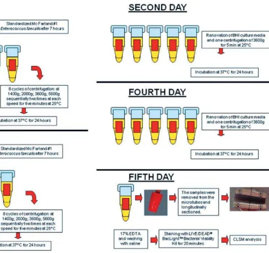
Figure 1- Flowchart of the group C contamination protocol
adjustment of the E. faecalis culture to McFarland standard No. 1, it was incubated again at 37°C for seven hours in order to achieve exponential growth. This amount of time (7 h) was de ned in a pilot study (data not shown). This inoculum (800 μL) was inserted into the microtubes with the samples and centrifuged (Eppendorf 5417R centrifuge, Eppendorf, Hamburg, Germany) as described above for group B. The inoculum was renewed at each cycle of centrifugation. Following the eight centrifugation cycles, sterilized BHI broth was inserted into the microtubes, agitated in a vortex (Vortex-mix VX200, Edison, NJ, USA), and incubated at 37°C for 24 h.
Following incubation (day two), the samples were agitated in a vortex for 10 s, and the inocula from the microtubes were discarded. One milliliter of sterilized BHI broth was inserted, followed by a centrifuge cycle of 3,600 g for 5 min at 25°C, and the microtubes were incubated again at 37°C for 24 h.
On the third day, a new inoculum of E. faecalis at exponential growth phase after seven hours of last subculture was inserted into the sample tubes, and the centrifugation protocol (twice at each speed, at 25°C) was repeated. On the fourth day, the procedures were as described for the second day. On the fth day, the samples were removed from the microtubes and observed with CLSM.
Thus, group A underwent three weeks of contamination, group B 24 h, and group C ve days. All three groups were analyzed by CLSM and uorescent LIVE/DEAD® BacLightTM Bacterial
Viability stain (Molecular Probes, Eugene, OR, USA) to verify the bacterial penetration.
Following the contamination procedures, all specimens were longitudinally sectioned using an Isomet saw with a diamond disc. Constant irrigation with a sterilized saline solution occurred during the sectioning process. The smear layer produced by these cuttings was removed with 17% EDTA for 10 min, and the samples were washed in sterilized saline solution. The halves of the dentinal tubes were stained with 30 μL of LIVE/DEAD® stain for
20 min. This kit contains SYTO 9® dye, which stains
live bacteria with a green pigment, and propidium iodine dye, which stains dead bacteria with a red pigment, thus enabling the easy identi cation of viable bacteria.
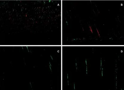
Figure 2- Confocal laser scanning microscopy (CLSM) images following contamination with E. faecalis. A- Longitudinal
view of the medial third of a root canal following group A contamination. Low level penetration of bacteria inside the tubules can be observed. Red color denotes dead bacteria; B- Longitudinal view of the cervical third of a root canal following group A contamination. Compared to Figure 2A, there are slightly more bacteria inside the tubules and some of them are green, denoting live bacteria; C- Longitudinal view of the cervical third of a root canal following group B contamination. A small amount of live bacteria can be observed; D- Longitudinal view of the medial third of the root canal following group B contamination. A small amount of live bacteria can be observed
Figure 3- Confocal laser scanning microscopy (CLSM) images following group C contamination protocol. A, B, and C-
Longitudinal views of the cervical third of a root canal. A greater quantity of bacteria and a predominance of live (green) bacteria can be observed; D, E, and F- Longitudinal views of the medial third of a root canal. Live bacteria can be observed.
7KHPHGLDOWKLUGDVLQDOO¿JXUHVLVOHVVFRQWDPLQDWHGWKDQWKHFHUYLFDOWKLUG scanning microscopy
were acquired and fragmented using the Leica Application Suite-Advanced Fluorescence software (LAS AF, Leica, Mannheim, Baden-Württemberg, Germany).
For an objective analysis, the CLSM images were fragmented into many layers and were converted into “tiff” format by the LAS AF software. These images were exported to the bioImageL TM v21 software7 in order to quantify the amounts of
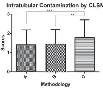
Figure 4- &ODVVL¿FDWLRQ RI VFRUHV RI LQWUDWXEXODU
contamination observed in the confocal laser scanning microscopy (CLSM) images, performed by three examiners (A×C, p=0.015) (B×C, p=0.022). The symbol ** shows the statistical difference between groups B and C (p<0.05), and the symbol *** shows the statistical difference between groups A and C (p<0.01)
Figure 5-4XDQWL¿FDWLRQLQSHUFHQWDJHRIFXELFPLFURPHWHUVRIWKHELRYROXPHRIOLYHDQGGHDGEDFWHULDE\ELR,PDJH/Y
software, from confocal laser scanning microscopy (CLSM) images. The symbol ** shows the statistical difference between groups B and C in the analysis of biovolume of live bacteria (p<0.05), and the symbol *** shows the statistical differences between groups A and C in the analysis of biovolume of live bacteria, and between groups A and C, and B and C, in the analysis of biovolume of dead bacteria (p<0.01)
Dunn’s tests. A p value of <0.05 was considered statistically signi cant.
In addition to the objective CLSM analysis, a subjective evaluation of contamination was performed by three blinded calibrated examiners using the CLSM images. Classi cation was de ned according to the level of homogeneity, density, and depth of the bacterial contamination inside the dentinal tubules. Prior to the evaluation, the examiners were trained by examining other CLSM images of intradentinal contamination, as well as observing the images obtained from our three experimental groups, with no knowledge of which group the images were from.
The images were classi ed according to the following scores (determined in this study): 0, no
contamination; 1, low contamination; 2, moderate contamination; and 3, high contamination. No contamination was de ned as less than one-fourth of the analyzed eld showing live bacteria inside the dentinal tubules. Low contamination was de ned as half of the analyzed eld showing live bacteria inside the tubules. Moderate contamination was de ned as of the analyzed eld showing live bacteria inside the tubules, and high contamination was de ned as all of the analyzed eld showing live bacteria, along with high bacterial proliferation observed within the dentinal tubules. The data were analyzed by the kappa agreement test, and a result of 0.7 (classi ed as good) was obtained. The subjective analysis was done by observation of the composite image from each site, i.e., each CLSM image was composed of 23 images at 1 μm intervals, giving a nal composite image of 23 μm in depth. After the normality test, the Kruskal-Wallis and Dunn’s tests were used to perform the statistical analysis. p values <0.05 were considered statistically signi cant.
RESULTS
The CLSM images obtained from group C demonstrated a greater bacterial penetration from the main root canal. This contamination was more dense and homogeneous, and a greater quantity of live bacteria were present (Figures 2 and 3).
After classifying the CLSM images in scores, the examiners observed greater ap<0.05); however, no differences were noted between groups A and B (p>0.05) (Figure 4).
scanning microscopy
and dead bacteria (p<0.05).
The negative controls did not show any bacteria at the images.
D I SCUSSI ON
This study showed that a novel method for the intratubular contamination of bovine dentine is superior to previously reported contamination protocols10,18. This was demonstrated both by
subjective score analyses and by quantitatively comparing the biovolume of live and dead bacteria inside the dentinal tubules.
Bovine teeth are commonly used as an experimental substitute for human teeth due to their ease of availability26. However, compared to human
dentin, bovine dentin has a higher concentration of dentin tubules per square millimeter, although this difference is small. On average, the diameter of the dentin tubules of bovine teeth is greater than that of human teeth, although this difference is not statistically signi cant. The percentage of intertubular dentin in bovine teeth is the same as in humans24,26. In this study, the bacterial tubule
penetration procedure was standardized, and since bovine tubules are generally larger, this process was easier to achieve than if human teeth had been used. A further advantage of using bovine teeth is that human teeth can show greater variations, due to age, microanatomy, and degree of tooth mineralization.
Haapasalo and Orstavik10 (1987) revolutionized
the study of antimicrobial substances by introducing a new protocol for dentin contamination. This technique involved contamination with a pure colony of E. f aecalis for 21 days, followed by evaluation with SEM and Brown and Brenn staining. For medication tests, specimens infected for three weeks were used, and a layer of nail varnish was used to cover the root surfaces of the dentin blocks. This procedure rapidly became the method of choice for the evaluation of substances and treatment protocols in endodontics8,13,19.
Later, the same authors modi ed their protocol, using 14-day contamination periods and allowed cultures of dentin dust from sequential bur samples to be analyzed21. This modi ed protocol was effective
for all microorganisms tested, with different times of contamination, and E. faecalis rapidly infected the whole length of the tubules. Furthermore, this protocol could be utilized in another study that evaluated the ef ciency of nickel-titanium rotary instrumentation9. Additional improvements to the
protocol by other scientists resulted in an even more rapid and simple methodology, requiring just seven days of contamination1,2.
In 2011, Ma, et al.18 introduced a new method
that could induce biofilm on the dentin block
surface beside the internal dentin contamination. The contamination was achieved in just 24 h and used a sequential centrifugation protocol; between every centrifugation, a fresh solution of bacteria was added. This protocol was developed to allow for the analysis of microorganisms in dentinal depth using uorescence detected by CLSM. However, the experimental model comprised a small 4 mm dentin fragment to promote bio lm formation and contamination of the internal surface of the root canal, which have made the contamination easier.
This study con rmed that the classic protocol of Haapasalo and Orstavik10 (1987) (group A)
produced a satisfactory amount of bacterial contamination, as con rmed by the biovolume and score analyses. However, the biovolume analysis also revealed large numbers of dead bacteria. Our results using the protocol of Ma, et al.18 (2011)
(group B) were deemed unsatisfactory, because the contamination included intermediate amounts of dead bacteria, and some areas revealed no contamination. Teeth from groups B and C received inocula between every centrifugation, but group B had only one centrifugation and two centrifugations were performed in alternate days for group C. No statistical differences between the group A and group B protocols were observed (p>0.05).
The new experimental model attempted to reproduce the oral environment of endodontic infections in bovine dentinal tubes (12 mm length and canal internal diameter of 1.2 mm), with an external surface covered with two layers of nail varnish. The goal was to ensure that the internal contamination occurred only via the root canal. This model, which is based on modi cations to the group B method, began by placing microcentrifuge tubes containing the samples and sterile culture media into an ultrasonic bath. This allowed for a greater penetration of the culture medium into the tubules, thus providing nutrients for the subsequent contamination step, which lasted four days. An exponential phase inoculum of high cell density was used for contamination and the centrifuge protocol of Ma, et al.18 (2011) was followed. In this
CLSM analysis.
All of the modi cations introduced into this protocol were added gradually over many pilot studies, in order to obtain the best contamination. For instance, slicing the dentinal tubes into two halves was found to be preferable to fracturing because some fractured fragments in the pilot studies did not provide satisfactory CLSM images. Furthermore, the use of a diamond disk for slicing was necessary to obtain a regular and plain surface, which improved the focus adjustment and view. After slicing, the smear layer was removed with 17% EDTA for 10 minutes, and the image of microorganisms within the tubules could then be captured.
EDTA has the capacity of killing microorganisms, but is a weak acid. With proper application time, EDTA could promote removal of smear layer without elimination of microorganisms, which is why only EDTA was used. In addition to their cleaning ability, chelators may detach bio lms adhering to the root canal walls21,30, but it has been demonstrated that its
antiseptic capacity is relatively limited6,28,30. In the
present work, EDTA was applied for a short time (10 minutes) for the removal of the smear layer only. Therefore, the contamination of the specimens was higher and deeper than in the tests that con rmed that EDTA presents antimicrobial capacity. A pilot study was performed in order to verify the in uence of this super cial EDTA application and any in uence of this weak acid over the bacterial viability was not observed.
It may be noted that images in the paper of Ma, et al.18 (2011) demonstrate a greater contamination
of tubules than our new protocol. However, this can be explained by differences in dentine fragment preparation. In the previous study, the tooth was cut into four pieces, without a cement layer, thus allowing for easier microbial penetration. In the present work, the dentine fragment was larger, and it contained cement and an external nail polish layer. Therefore, bacteria could access the dentinal mass only by the main root canal. Another factor is that in the Ma, et al.18 (2011) images, it is possible
to observe stained tubules because the dye has an af nity for dentine collagen5.
The adoption of the new endodontic protocol studies of antimicrobial substances should ensure a greater accuracy of results, particularly regarding the avoidance of false negatives due to failed contamination. It is important that this procedure be repeated with human teeth in order to verify that a satisfactory standard of contamination occurs in narrow and curved canals, and that an accurate replication of the clinical conditions of endodontic infections is achieved.
CON CLUSI ON
This research demonstrates a new protocol for the contamination of bovine dentinal tubules. It promotes greater and more homogeneous live bacterial proliferation compared to other methods. These aspects are essential for antimicrobial studies using in vit ro models of endodontic infections, since they produce more accurate results and prevent false negatives.
ACKN OW LED GEM EN TS
This research was supported by the National Council for Scienti c and Technological Development (CNPq) and São Paulo Research Foundation (FAPESP) (process no. 2010/20186-3). The authors would like to thank Dr Luis Chavez de Paz for the facilities for using the bioimageL software.
REFEREN CES
1- Atila-Pekta B, Yurdakul P, Gülmez D, G rduysus O. Antimicrobial effects of root canal medicaments against Ent er ococcus faecalis
and St r ept ococcus m ut ans. Int Endod J. 2013;46(5):413-8. 2- Bago I, Ple ko V, Gabri Panduri D, Schauperl , Baraba A, Ani I. Antimicrobial ef cacy of a high-power diode laser, photo-activated disinfection, conventional and sonic photo-activated irrigation during root canal treatment. Int Endod J. 2013;46(4):339-47. 3- Berber VB, Gomes BP, Sena NT, Vianna ME, Ferraz CC, Zaia AA, et al. Ef cacy of various concentrations of NaOCl and instrumentation techniques in reducing Ent er ococcus faecalis within root canals and dentinal tubules. Int Endod J. 2006;39(1):10-7.
4- Bergmans L, Moisiadis P, Huybrechts B, Van Meerbeek B, Quirynen M, Lambrechts P. Effect of photo-activated disinfection on endodontic pathogens ex vivo. Int Endod J. 2008;41(3):227-39. 5- Bryce G, O’Donnell D, Ready D, Ng Y, Pratten J, Gulabivala K. Contemporary root canal irrigants are able to disrupt and eradicate single- and dual-species bio lms. J Endod. 2009;35(9):1243-8. 6- Bystr m A, Sundqvist G. The antibacterial action of sodium hypochlorite and EDTA in 60 cases of endodontic therapy. Int Endod J. 1985;18(1):35-40.
7- Chávez de Paz LE. Image analysis software based on color segmentation for characterization of viability and physiological activity of bio lms. Appl Environ Microbiol. 2009;75(6):1734-9. 8- Cheng X, Guan S, Lu H, Zhao C, Chen X, Li N, et al. Evaluation of the bactericidal effect of Nd:YAG, Er:YAG, Er,Cr:YSGG laser radiation, and antimicrobial photodynamic therapy (aPDT) in experimentally infected root canals. Lasers Surg Med. 2012;44(10):824-31.
9- Coldero LG, McHugh S, MacKenzie D, Saunders WP. Reduction in intracanal bacteria during root canal preparation with and without apical enlargement. Int Endod J. 2002;35(5):437-46.
10- Haapasalo M, Orstavik D. I n vit r o infection and disinfection of dentinal tubules. J Dent Res. 1987;66(8):1375-9.
11- Haapasalo M, Shen Y, Qian W, Gao Y. Irrigation in endodontics. Dent Clin North Am. 2010;54(2):291-312.
12- Kakehashi S, Stanley HR, Fitzgerald RJ. The effects of surgical exposures of dental pulps in germ-free and conventional laboratory rats. Oral Surg Oral Med Oral Pathol. 1965;20:340-9.
scanning microscopy
14- Kishen A, Sum CP, Mathew S, Lim CT. In uence of irrigation regimens on the adherence of Ent er ococcus faecalis to root canal dentin. J Endod. 2008;34(7):850-4.
15- Kitagawa R, Kitagawa H, Izutani N, Hirose N, Hayashi M, Imazato S. Development of an antibacterial root canal lling system containing MDPB. J Dent Res. 2014;93(12):1277-82. 16- Love RM. Ent er ococcus faecalis - a mechanism for its role in endodontic failure. Int Endod J. 2001;34(5):399-405.
17- Love RM, Chandler NP, Jenkinson HF. Penetration of smeared or nonsmeared dentine by St r ept ococcus gor donii. Int Endod J. 1996;29(1):2-12.
18- Ma J, Wang Z, Shen Y, Haapasalo M. A new noninvasive model to study the effectiveness of dentin disinfection by using confocal laser scanning microscopy. J Endod. 2011;37(10):1380-5. 19- Miranda RG, Santos EB, Souto RM, Gusman H, Colombo AP. Ex v iv o antimicrobial ef cacy of the EndoVac system plus photodynamic therapy associated with calcium hydroxide against intracanal Ent er ococcus faecalis. Int Endod J. 2013;46(6):499-505.
20- Ordinola Zapata R, Bramante CM, Moraes IG, Bernardineli N, Gasparoto TH, Graeff MS, et al. Confocal laser scanning microscopy is appropriate to detect viability of Ent erococcus faecalis in infected dentin. J Endod. 2008;34(10):1198-201.
21- Orstavik D, Haapasalo M. Disinfection by endodontic irrigants and dressings of experimentally infected dentinal tubules. Endod Dent Traumatol. 1990;6(4):142-9.
22- Ozdemir HO, Buzoglu HD, Calt S, Stabholz A, Steinberg D. Effect of ethylenediaminetetraacetic acid and sodium hypochlorite irrigation on Ent er ococcus faecalis bio lm colonization in young and old human root canal dentin: in v it r o study. J Endod. 2010;36(5):842-6.
23- Paquette L, Legner M, Fillery ED, Friedman S. Antibacterial ef cacy of chlorhexidine gluconate intracanal medication in vivo. J Endod. 2007;33(7):788-95.
24- Schilke R, Lisson JA, Bauss O, Geurtsen W. Comparison of the number and diameter of dentinal tubules in human and bovine dentine by scanning electron microscopic investigation. Arch Oral Biol. 2000;45(5):355-61.
25- Sundqvist G. Taxonomy, ecology, and pathogenicity of the root canal ora. Oral Surg Oral Med Oral Pathol. 1994;78(4):522-30. 26- Tanaka JL, Medici Filho E, Salgado JA, Sangado MA, Moraes LC, Moraes ME, et al. Comparative analysis of human and bovine teeth: radiographic density. Braz Oral Res. 2008;22(4):346-51. 27- Vera J, Siqueira JF Jr, Ricucci D, Loghin S, Fernández N, Flores B, et al. One-versus two-visit endodontic treatment of teeth with apical periodontitis: a histobacteriologic study. J Endod. 2012;38(8):1040-52.
28- Wong DT, Cheung GS. Extension of bactericidal effect of sodium hypochlorite into dentinal tubules. J Endod. 2014;40(6):825-9. 29- Wu MK, Dummer PMH, Wesselink PR. Consequences of and strategies to deal with residual post-treatment root canal infection. Int Endod J. 2006;39(5):343-56.