Dissertation presented to obtain the Ph.D degree in Biology | Neuroscience
Instituto de Tecnologia Química e Biológica António Xavier | Universidade Nova de Lisboa
Oeiras,
João Afonso
cognitive and motor features, in the mouse medial
prefrontal cortex, during a memory guided behavior
João Afonso
Dissertation presented to obtain the Ph.D degree in Biology |
Neuroscience
Instituto de Tecnologia Química e Biológica António Xavier | Universidade Nova de Lisboa
Oeiras, February, 2019
Multiplexed simultaneous representations of
cognitive and motor features, in the mouse
medial prefrontal cortex, during a memory
guided behavior
I would like to thank:
My parents and family, for the unconditional support.
Rita,
for the happiness, patience and love.
Alfonso,
for the trust, idealism and guidance.
The Champalimaud Research community, for the nurturing and stimulating environment.
Contents
List of Figures iii
List of Acronyms vii
Summary / Resumo ix
1 General Introduction 1
1.1 The Prefrontal Cortex . . . 2
1.2 The Rodent Prefrontal Cortex . . . 5
1.3 Prefrontal Cortex and Working Memory . . . 7
1.4 Heterogeneous Representations in the Prefrontal Cortex . . . 15
2 Head-Fixed Delayed Response Task on a Treadmill 31 2.1 Abstract . . . 33
2.2 Introduction . . . 34
2.3 Methods and Materials . . . 39
2.4 Results . . . 50
3.2 Introduction . . . 84
3.3 Methods and Materials . . . 89
3.4 Results . . . 98
3.5 Discussion . . . 119
4 Single Trial Decoding of Speed and Memory 133 4.1 Abstract . . . 135
4.2 Introduction . . . 136
4.3 Methods and Materials . . . 140
4.4 Results . . . 145
4.5 Discussion . . . 166
5 General Discussion 177 5.1 Objective . . . 178
5.2 Properties of Recorded Neurons . . . 178
5.3 Multiplexed Representations . . . 180
List of Figures
1.1 Schematic Diagram of Some PFC’s Connections . . . 3
1.2 Schematic Diagram of the Rat Prefrontal Cortex . . . 6
1.3 Delay Period Activity in ODR Task . . . 10
1.4 Neural Mechanisms of WM . . . 14
1.5 Low and High Dimensional Representations and Mixed Selec-tivity . . . 18
2.1 Head-fixed Delayed Response Task on a Treadmill . . . 50
2.2 Mice Performance . . . 51
2.3 Performance Across Sound Start Locations . . . 52
2.4 Mice’s Speed Behavior . . . 54
2.5 Mean Speed Behavior . . . 55
2.6 Mean Speed Behavior All Animals . . . 57
2.7 Mean Speed Behavior in Incorrect Trials . . . 58
2.8 Predicting Stop Behavior . . . 59
2.9 Predicting the Stopping Behavior of One Animal . . . 60
2.10 Predicting the Stopping Behavior of All Animals . . . 61
2.13 Performance in Catch Trials . . . 65
2.14 Mean Speed Behavior Catch Trials . . . 66
2.15 Performance in Session with Stopping Area in Different Location 67 2.16 Mean Speed Behavior in Session with Stopping Area in Dif-ferent Location . . . 68
2.17 Performance in No Area Session . . . 69
2.18 Mean Speed Behavior No Area Session . . . 70
2.19 Mean Speed Behavior First Trials of No Area Session . . . . 71
3.1 Acute Recordings on Awake Behaving Mice . . . 98
3.2 Recording Locations . . . 100
3.3 Trial Space Histograms of Firing Rates . . . 102
3.4 Firing Rate Selectivity in the Memory Period . . . 103
3.5 Peri Event Raster Plots and Time Histograms . . . 105
3.6 Significant FR Modulation by Task Events . . . 106
3.7 Demixed Principal Component Analysis . . . 108
3.8 dPCA Input Trial Space Histograms . . . 110
3.9 dPCA Results for Sound and Decision as Parameters . . . . 111
3.10 Differences Between Correct and Incorrect Trials . . . 113
3.11 PCA on Session Speeds . . . 115
3.12 dPCA Results Stop Trials Speeds . . . 116
3.13 dPCA Results No Stop Trials Speeds . . . 117
4.2 Performance of the Speed Prediction Model . . . 146 4.3 Real and Predicted Mean Speeds . . . 148 4.4 Trial to Trial Vs. Mean Trial type Speed Prediction . . . . 149 4.5 Speed Prediction Kernels Coefficients . . . 151 4.6 Speed-Acceleration and Past-Future Indices . . . 152 4.7 Speed-Acceleration and Past-Future Indices All Sessions . . 153 4.8 Predicting Speed with Past and Future Neural Activity . . . 154 4.9 Predicting Speed in a Condition Specific Manner . . . 156 4.10 Predicting Trial Identity from Single Trial Neural Activity . 158 4.11 Performance of the Trial Identity Prediction Model . . . 159 4.12 Real and Predicted Mean Trial Identity . . . 160 4.13 Trial Identity Prediction Kernels Coefficients . . . 162 4.14 Speed and Trial Identity Prediction Kernels Comparison . . 163 4.15 Speed and Trial Identity Prediction in Different Segments of
List of Acronyms
AP anterior-posterior. 41, 45, 46, 89, 90, 97–99
AUC area under the curve. 48, 59, 60, 62, 156, 157, 162
dlPFC dorsolateral prefrontal cortex. 2, 4, 5, 7, 14
dPCA demixed principal components analysis. iv, ix, x, xii, 83, 87, 88, 93–96, 106–111, 113, 115, 116, 120, 122, 168, 178–182, 185
DV dorsal-ventral. 40, 41, 90, 98
FR firing rate. iv, 91–93, 102, 103, 105, 112, 113, 118, 138–140, 144, 146, 149, 151–153, 177
IL infralimbic. 6, 87
IR infrared. 40, 41
ITI intra trial interval. 39, 41, 49, 105, 119–121, 181
ML medial-lateral. 45, 98
176–179, 182–187
ODR oculomotor delayed response. iii, 10, 11
PBS phosphate-buffered saline solution. 89
PCA principal components analysis. iv, 95, 96, 110, 113–115
PETH Peri-event time histogram. 93, 103–105
PFC prefrontal cortex. iii, ix, 2–7, 9–13, 15–17, 83–85, 120, 121, 133, 134, 177, 182, 184–186
PFI past future index. 139, 151
PRL prelimbic. 6, 87, 97, 98
ROC receiver operating characteristic. 48, 59, 141
SAI speed acceleration index. 140
SD standard deviation. 51, 54, 59, 114, 145, 147, 148
SEM standard error of the mean. 50, 51, 56, 64–69, 102, 147
TSH trial space histogram. 87, 92, 101, 109, 118
WM working memory. ix, x, 4, 8, 9, 11–13, 33, 35, 36, 52, 57, 64, 65, 71, 72, 74, 75, 83–85, 87, 98, 118, 121, 133, 135, 136, 158, 168, 176–179, 181, 185–187
Summary
To develop complex models of the environment, coordinate extended sequences of actions and plane ahead organisms must break free from rigid stimulus response associations and be able to link events and behaviors that are separated in time. Essential to these is the ability of the brain to maintain sustained representations of environmental absent information: a process generally designated as working memory (WM).
Seating at the apex of the cortical hierarchy the prefrontal cortex (PFC) as been strongly implicated, by both lesions and physiology studies, in WM dependent behaviors. Given its integrative nature activity in the area it’s a combination of preprocessed sensory and motor inputs with time varying cognitive internal mechanisms, making a complete understanding of its func-tion only possible in the context of a moment to moment comparison with behavior.
To investigate how the joint activity of neurons, in prefrontal regions, encodes task relevant information, during a WM dependent behavior, we developed a head-fixed delayed response task on a treadmill for mice. Our task allowed us to minutely monitor the animals’ behavior while acutely recording the simultaneous activity of dozens of cells in the mPFC of the mice. Such features enabled us to have a good understanding of how the animals were solving the task, confirming its WM, and to relate representa-tions encoded in the joint neural activity with the mice’s behavior in each trial.
neu-of both cue and decision. Also demixable was a trial length stable signal seemingly related to the animals’ engagement in the task. dPCA further revealed that the mice’s movement strategies on the treadmill had a strong influence in the recorded activity.
Taking advantage of our simultaneously recorded neurons we also dis-covered that in each trial the mouse mPFC encodes both WM sustained information, during the cue free memory period, and a faithful representa-tion of the mice speed strategy on the treadmill.
Together these results show that the joint activity of neurons in the mPFC of the mice encodes, in a multiplexed way, multimodal representations of informative sensory features, future goals or decisions, speed strategies and the animals’ internal state - engagement. All the aforementioned variables are relevant when considering a putative function of the area in organizing context adapted, WM dependent, goal directed behavior.
Resumo
Para conseguirem formar modelos complexos do ambiente, coordenar sequências de acções que se desenrolam no tempo e fazer planos, os organ-ismos não podem depender apenas de associações rígidas entre estímulos e repostas, tendo que ser capazes de ligar eventos e comportamentos que acon-tecem separados no tempo. Essencial para tal é a possibilidade do cérebro manter e suster representações de informação que já não se encontra pre-sente no exterior, um processo genericamente denominado por memória de trabalho.
Posicionado no cume da hierarquia cortical o córtex pré-frontal tem sido implicado, por estudos baseados em lesões e fisiologia, em comportamentos dependentes da memória de trabalho. Dado a sua natureza integradora a actividade nesta área é uma combinação de estímulos sensoriais e motores com processos cognitivos que variam no tempo, tornando um entendimento completo da sua função apenas possível no contexto de uma comparação, momento a momento, com o comportamento.
De modo a investigar como a actividade conjunta dos neurónios, nas regiões pré-frontais, codifica informação relevante, durante um comporta-mento dependente da memória de trabalho, desenvolvemos uma tarefa de resposta tardia, numa passadeira rolante, para ratinhos com cabeça imobi-lizada por um implante. A tarefa permitiu-nos monitorizar com precisão o comportamento dos animais enquanto gravávamos a actividade simultânea de dezenas de células no seu córtex pré-frontal medial. Estas características permitiram-nos perceber bem como é que os animais resolviam a tarefa,
con-ronal com o comportamento dos ratinhos em cada ensaio.
Usando uma técnica de redução dimensional, dPCA, descobrimos que era possível separar da actividade conjunta dos neurónios representações de memória de trabalho, retrospectivas e prospectivas, das pistas e decisões im-plicadas na tarefa. Também separável foi um sinal estável, presente durante todo o ensaio, aparentemente relacionado com o envolvimento dos animais na tarefa. Para além destes o dPCA também revelou que a estratégia de movimento dos ratinhos na passadeira tinha uma grande influência na ac-tividade que gravámos.
Tirando partido das nossas gravações simultâneas da actividade neuronal também percebemos que, em cada ensaio, o córtex pré-frontal medial do ratinho codifica informação em memória de trabalho, durante um período de memória livre de pistas, juntamente com uma representação fiel da estratégia de velocidade dos animais na passadeira.
Juntos estes resultados demonstram que a actividade conjunta dos neurónios no córtex pré-frontal dos ratinhos codifica, de uma maneira multiplexada, representações multi-modais de variáveis sensoriais informativas, objectivos futuros ou decisões, estratégias de velocidade e estados internos dos animais. Todas estas variáveis são relevantes para uma possível função da área em organizar comportamento deliberado, e adaptado ao contexto, dependente de memória de trabalho.
Chapter 1
1.1
The Prefrontal Cortex
Measured by our success in adapting, prospering and becoming dominant in almost all environments there seems to be something strikingly different between humans and the other species. We are not the most adapted of animals, when considering the relation with a specific habitat, but we seem to be the more flexible in creating new maps between our possible repertoire of behaviors and the specific challenges imposed by different or evolving contexts. When looking into the brain in search for what endows us with this particular ability the spotlight has been historically centered on the PFC. A set of features seems to point to the distinctive character of the area: the PFC attained maximum relative growth in the human brain ( 29% of the total cortical surface against 17% in the chimpanzee and 3.5% in the cat, for instance) also, in the timeline of brain evolution, it was one of the last cortical regions to develop [37], a pattern mimicked by ontogeny, with it maturing late, in humans [36] and monkeys [29], and not attaining full maturity until adolescence [70].
The PFC is located at the anterior pole of the mammalian brain and is generally considered to be formed by three main regions: dorsolateral, medial and orbital prefrontal cortices. The boundaries between these (and respective subdivisions not mentioned here) have been traced in different ways, dependent on the applied methodologies and followed criteria, and despite the existence of common motifs (the dorsolateral prefrontal cortex (dlPFC) is usually associated with cognitive functions supporting behavior and the medial and orbital with regulating emotional behavior and basic
Chapter 1. General Introduction
drives) there are no strong evidences to consider the whole area, or its sub-divisions, as structural entities with unitary functions [25].
Figure 1.1: Schematic Diagram of Some PFC’s Connections: Inputs from several brain systems converge in the interconnected PFC. Most connections are reciprocal with the exceptions indicated by arrows. Figure from Miller and Cohen, 2001 [53].
Determinant for the understanding and description of PFC function and organization are its profuse and reciprocal connection with virtually all sen-sory and motor systems and many subcortical structures [53] (Figure 1.1 ). The diversity of information streams that converge in it imply that complex
sensory and motor landscapes, as well as the cognitive mechanisms to op-erate on them, can be found in the PFC. The neural networks underlying these, however, are not confined to topographically demarcated areas, and the computations they perform are many times concomitant in the context of the same behavior and common across several behaviors. Such is one of the main reasons why it as been historically difficult to reconcile a wide collection of, many times, seemingly unrelated facts into a coherent whole.
General theories put forward to bound prefrontal function (particularly the dlPFC) have tried to frame its involvement in disparate cognitive mech-anisms (e.g attention [10], WM [21], planning [84], decision making [43] and inhibitory control [2]) in the broader context of executive function [22] and cognitive control [52] responsible for context adapted organization of goal-directed behavior [19]. Such idea is supported by its aforementioned connections as well as hierarchical position on top of the motor oriented frontal half of the brain, which highlights his role as, in essence, an action and execution cortex [26].
The PFC is not critical for performing simple, automatic forms of behav-ior [52][53], it may be involved in learning them, but, with sufficient training, they are automatized and completely implemented by lower hierarchical ar-eas. The PFC is, however, particularly necessary in situations and contexts that because of their novelty, ambiguity, complexity or extension in time demand for top-down control of behavior. The area, in a position where its representations encapsulate goals and schemes of actions containing in themselves subordinate actions and subgoals, can then be said to have a cen-tral part in integrating cross-temporal contingencies, percepts and actions,
Chapter 1. General Introduction
by maintaining time sustained representations and monitoring their progress against a general plan.
1.2
The Rodent Prefrontal Cortex
Even if the PFC, and PFC related behaviors, attain their maximal ex-pression in primates, and in particular in humans, such doesn’t mean that other species don’t have a PFC or exhibit prefrontal mediated behaviors. There has been then, since long, a strong practical interest in being able to study the area in more cost and manipulation amenable model animals, with rodents being strong candidates.
Establishing parallels between the prefrontal function in primates and rodents is not easy [78]. First, the area general role and regional special-izations are not well understood in both model animals, creating a base problem about what exactly is being compared; second, there are enor-mous cross-species variations in the cortical cytoarchitectonics and connec-tions[57]; third, a wide array of criteria and nomenclatures has been used, seemingly ad-hoc, in both species [46].
Combining anatomical with functional information to address the issue different authors proposed that, even if not possessing a granular structure directly equivalent to the human and monkey dlPFC [61] [79], rodents have behaviors that engage functional mechanisms akin to the ones normally as-cribed to that area, mechanisms that are shared among several regions of their own PFC [8][40]. The rodent prefrontal is, for sure, not as differenti-ated as the primates’ one, with later specializations likely to have occurred, but dorsolateral-like features, both anatomical and functional, are present
and can be revealed by the use of assays probing species-relevant executive functions [79] [8].
Figure 1.2: Schematic Diagram of the Rat Prefrontal Cortex: (a) Lateral view, 0.9 mm from the midline. (b) Unilateral coronal section, AP location depicted by the arrow above. The different shadings represent the three major sub- divisions of the prefrontal cortex (medial, ventral and lateral). Abbreviations: ACg, anterior cingulate cortex; AID, dorsal agranular insular cortex; AIV, ventral agranular insular cortex; AOM, medial anterior olfactory nucleus; AOV, ventral anterior olfactory nucleus; cc, corpus callosum; Cg2, cingulate cortex area 2; gcc, genu of corpus callosum; IL, infralimbic cortex; LO, lateral orbital cortex; M1, primary motor area; MO, medial orbital cortex; OB, olfactory bulb; PrL, prelimbic cortex; PrC, precentral cortex; VLO, ventrolateral orbital cortex; VO, ventral orbital cortex. Figure from Dalley et.al, 2004 [15].
The rodent PFC can be organized in medial, lateral and ventral areas (see [79] and [40] for variations on this arrangement). The medial can be subdivided in a dorsal region, with precentral and anterior cingulate cortices, and a ventral with the prelimbic (PRL), infralimbic (IL) and medial orbital (MO) cortices. The lateral includes the ventral agranular, insular and lateral
Chapter 1. General Introduction
orbital cortices. The ventral region encompasses the ventral orbital and ventral lateral orbital cortices (Figure 2 ) [15].
Based on its anatomical connections, in particular the reciprocal ones with the mediodorsal thalamus [68], and involvement in specific behaviors, the rodent mPFC as long been considered to take part in cognitive pro-cesses analogous to some of the ones ascribed to the primate dlPFC [40]. Some authors have investigated and pointed to the existence of functional heterogeneity within the area, particularly between its dorsal and ventral parts [15][40][19][69], but such differences, and its reasons, are many times difficult to interpret out of the context of specific tasks and in the broader spectrum of prefrontal function.
Hence, as with the PFC (especially dorsolateral) of humans and non-human primates, and even if in the scope of less differentiated forms of be-havior, the rodent mPFC can be said to be implicated in a set of cognitive control processes needed for the optimal scheduling of complex sequences of behavior including decision making [76], attentional selection [39], monitor-ing [30], behavioral inhibition [44] and task switchmonitor-ing[60]. Crucially, it has also been systematically considered critical for the online maintenance of memory representations necessary for the organization of actions over time [35][33][63].
1.3
Prefrontal Cortex and Working Memory
To go from simple, but inflexible, bottom up determined behaviors, in-cluding reflexes and habits, to complex and versatile streams of actions, animals need to be able to integrate context relevant information and
fu-ture plans, over variable periods of time, and use them to generate adapted conducts.
The recognition of such need, different from the long term storage of information in reference memory, led researchers to come up with the concept of WM, a mechanism by which the brain would be able to sustain and manipulate, for a short period of time, representations that could provide the backbone to high level cognitive operations such as thinking, planning, reasoning and decision-making [21]. Given this central role in goal-directed behavior, establishing the neural basis of WM has been a primary focus of neuroscience research.
The terminologies working and short-term memory have been used largely interchangeably when scientists want to refer, at the behavioral or mecha-nistic level, to something that needs to be remembered for a short period of time. There is no general consensus about the terms [1] and whether they refer to distinct mechanisms or qualitative levels within the same general mechanism. The more accepted distinction, though, implies a complexity difference, with short-term memory referring to the passive maintenance of information and WM also to the processes through which that information is manipulated [22]. In what relates to our work here, even taking in consid-eration the complexity arguments and the heavy assumptions loaded on the term by human and primate research, we’ll use the designation WM. Our decision is mainly based on the fact that it is the more generally used by the field and that to go back and forth between terminologies, when referring to our or others’ work, would result confusing. It should be clear, though, that, despite using the term WM, we interpret it as the basic mechanism
Chapter 1. General Introduction
through which information is maintained active in the brain and imply no distinction with short-term memory.
Based on psychological evidence several conceptual WM models were de-veloped [54], but the more well-known and influential is the one proposed by Baddeley and Hitch [3]. Badelley’s model includes one master compo-nent (the central executive) and three slave compocompo-nents, responsible for processing and maintaining information from several modalities: the visu-ospatial sketchpad (visuvisu-ospatial information), the phonological loop (speech perception and language comprehension) and the episodic buffer (integrating chunks of information from a different variety of sources). The central exec-utive supervises the performance of the slave components and it’s considered to be an attentional system with a limited memory capability. Baddeley’s proposal is an abstract, not mechanistic, model and establishing a compari-son between its components and particular brain structures it’s difficult and of limited usefulness. Despite, and in a loose sense, it’s central executive component has been related to the function played in the brain by the PFC [16].
The PFC involvement in WM supported behaviors has been consistently established by lesion studies, both in primates [25][59] and other mammalian species [35], and non-invasive brain activation experiments in humans [75], but it was the finding of neurons with elevated activity throughout a short WM dependent period that, by providing a simple explanation for the mech-anism of information storage, contributed the most for the association of the area with the process.
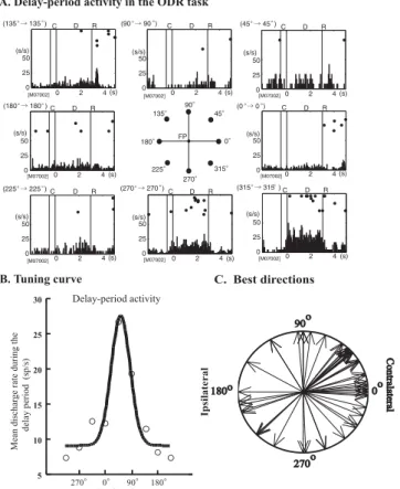
Figure 1.3: Delay Period Activity in ODR Task: (A) An example of directional delay-period activity. (B) An example of a tuning curve of directional delay-delay-period activity. (C) Polar distribution of the best directions of delay-period activity. A majority of the best directions were directed toward the contralateral visual field. Figure from Funahashi, 2006 [15].
Neurons in the PFC with firing rate higher than the baseline, between the presentation of a cue and the response of the subject, were first found in delayed response tasks [45] [27] [56], where the former and the later were separated by a time interval (delay). Their hypothetical role as the mech-anism for temporal active maintenance of information was, however, deci-sively established and investigated in the context of the oculomotor delayed
Chapter 1. General Introduction
response (ODR) task[23]. In it monkeys had to fixate a central point on a screen. After fixation an image was shown for some time and then disap-peared, marking the beginning of a delay period. During the delay animals had to keep fixating the central point, controlling for movement related neu-ral activity. With the subsequent disappearance of the centneu-ral point the monkey had to make a saccade to the location where the image had been initially displayed.
Neural activity, during the delay period of the ODR task, showed charac-teristics that made it an ideal candidate mechanism for the maintenance of information in WM: it was prolonged or shortened depending on the duration of the delay period [42], was only present when the animals performed cor-rect responses, being truncated or absent in error trials [27] and, importantly, exhibited directional preference, with specific neurons firing only when the visual cue was presented at one or a few adjacent positions [64] (Figure 1.3 ). Also, and curiously, the neurons seemed to encode preferentially the loca-tion of the cue and not the direcloca-tion of the saccade [77]. The clarity of the results and the seeming simplicity of the mechanism, PFC neurons, or neu-ronal populations, selectively tuned to the to-be-remembered information, hold it in an active state through persistent activation [32], made this the predominant model in the WM field, inspiring several proposed theoretical mechanisms [17] [11].
Recently, however, several lines of evidence have questioned the role of the PFC, and stable persistent neural activity, in WM related behaviors [71]. Following the ODR task results, subsequent studies, and tasks, revealed PFC neurons with activity that showed selectivity regarding a varied panoply of
information (e.g. tactile [67], auditory [41], task rules [83] and temporal order of stimuli [24]). This led to a progressively compartmentalized view of the area in which it apparently contained specialized neurons for every type of potentially useful information. Such idea seems rather implausible, and not parsimonious, if one thinks that the same information is already being represented, and processed, in lower hierarchical areas and that these also possess the hypothetical requisites to maintain it over short periods of time, with cells with sustained activity having been observed throughout the brain (e.g. parietal cortex [31], primary visual cortex [28], superior colliculus [6], thalamus [82] and even the spinal cord[62]).
Adding to such, and more revealing, temporal and occipital areas were shown to also encode, during the entire memory period, task relevant rep-resentations[80] that were tied to WM precision and the behavior of the subjects [18]. Moreover, information in sensory areas seems to better repre-sent the characteristics of the remembered item during the delay period than activity in the PFC [47], where representations were shown to be more cat-egorical in nature [50]. Together, these led to an alternative view according to which the PFC’s primary function, in WM, is not to store but rather to influence representations stored in hierarchically lower areas[51] [20] through top-down signals [12][53].
The relevance of the PFC’s fixed selectivity persistent neural activity has also been questioned. This type of activity in the PFC as practically become a symbol for WM, but, as before mentioned, sustained activity can be found nearly everywhere in the brain, questioning any privileged status of the area. Also, despite its recognized importance in forming temporal links [26] [14],
Chapter 1. General Introduction
short-term lived representations can be stored in different ways and are not dependent on selective sustained activity, as proved by works in which infor-mation could still be decoded, during the delay period, despite the removal of cells with selective elevated activity [4] [66] [72], or the observation of relatively silent periods, between encoding and response preparation, where, despite the inexistence of overt sustained activity, memory is still maintained [5].
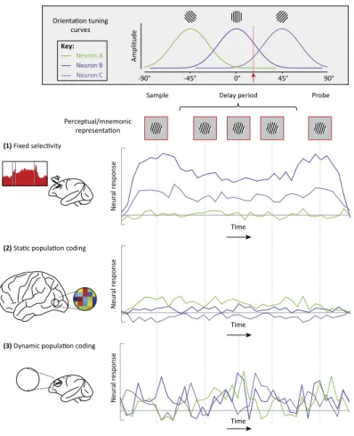
Because of the before mentioned arguments, and following developments on brain monitoring technologies and data analysis capabilities, the focus of research has been shifting from information encoded by neurons, or groups of neurons, with delay period selective elevated activity, to WM encoding based on the joint activity of neuron ensembles (Figure 4 ). Indeed, an increasing number of studies reports that, rather than utilizing distinct populations to encode each task variable, activity in the PFC encodes multiple task param-eters within a single neuronal population [38] [74]. Further, researchers have also discovered that the encoding is often done through dynamic spatiotem-poral patterns (Figure 1.4 3 ) [50][74][4], which evolve through state space trajectories and in which the encoding might be entirely different in distinct time points. Although the precise mechanisms underlying dynamic popula-tion coding are still not well understood, recent works have highlighted its possible coupling with functional and short term synaptic plasticity [55][73]. Dynamic coding is certainly not limited to the PFC, but the area’s hi-erarchical location and temporal integration role, allow it to fully explore a general principle of WM dynamic coding in organizing flexible cognition and behavior.
0° 45° 90° -45° -90° Key: A m p lit u d e
Sample Delay period Probe
Neuron A Neuron B Neuron C N eu ra lr es p o n se N eu ra lr es p o n se N eu ra lr es p o n se Perceptual/mnemonic representa�on Time Time Time
(2) Sta�c popula�on coding
(3) Dynamic popula�on coding (1) Fixed selec�vity
Orienta�on tuning curves
Figure 1.4: Neural Mechanisms of WM: A simplified schematic comparing and contrasting the fixed-selectivity model with population coding models involving static and dynamic temporal codes. Orientation tuning curves for three hypothetical neurons, A, B, and C, are shown in the top inset. Below this are schematics for three different potential neural models of WM. Top row: The fixed-selectivity model, primarily derived from single-unit recordings in monkey dlPFC. Middle row: Evidence for static population coding comes primarily from fMRI decoding and forward encoding studies of visual cortex. Here the pattern of activity across neurons can encode stimulus orientation in the absence of highly selective neural responses. This pattern is sustained throughout maintenance. Bottom row: Dynamic population coding has been demonstrated largely in monkey lPFC. Despite time-varying activity in all three neurons, the representation of orientation remains stable. The relevant orientation is encoded by a different combination of neural responses at each point in time. Figure from Sreenivasan et.al, 2014 [71].
Chapter 1. General Introduction
1.4
Heterogeneous Representations in the
Prefrontal Cortex
An interpretation of the neural activity mediating the perception-action cycle solely based on a function that takes us from stimuli to behavior is made difficult by the fact that the brain doesn’t simple represent the world in a different way. Personal significant preexistant attributions modulate the primary representations of sensory stimuli and motor implementations through a collection of cognitive mechanisms. At any given moment, thus, what is being encoded by the neurons are representations of environmental and behavioral features coloured and contextualized according to an organ-ism’s interpretation, based on its world model and future goals [9]. Such fact is more and more evident when moving away from the periphery, with the activity of each neuron being less and less determined by external drivers and more a reflection of activity in lower information processing levels. In its position at the apex of a hierarchy through which feedforward informa-tion ascends, being processed in the way by multiple intermediate loops [26], the PFC deals essentially with representations that are already highly abstracted, filtered and integrated for task relevance[58].
A direct consequence of what as been described is that neurons in the PFC often have complex responses that are not organized anatomically and may reflect multiple parameters such as stimuli, rules, responses or combi-nations of these [48] [42] [7]. Traditionally this heterogeneity was neglected and considered a difficulty in understanding the mechanistic roles of certain neurons and brain regions. Consequently, in an effort to identify the
com-ponents involved in the processes they were interested in, scientists tended to handpick cells that could be directly related with features of interest, or average the activity of a given neuron over repetitions of the same feature, blurring all other information its response might also contain.
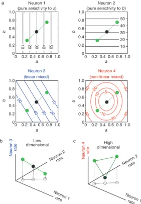
Recently though, this mixed selectivity, common characteristic of neu-rons in high order areas, as been associated to high-dimensional neural rep-resentations and the way PFC encodes information. Rigotti et.al, 2013 [66] showed that activity in a population of neurons, in the PFC of monkeys, simultaneously encoded all task relevant variables, in an object sequence memory task, even when classic neural selectivity was artificially abolished. The authors propose nonlinear mixed selectivity as the crucial character-istic that allows for high-dimensional neural representations (Figure 1.5 ) and show that artificially abolishing it reduces the representations dimen-sionality. High-dimensionality seemingly allows task relevant aspects to be accessible to linear classifiers, such as simple neuron models, that can only separate representations through planes, which would be impossible, for in-stance, in the case of the pure and linear mixed selective neurons in Figure 1.5 b.
The characteristic response profile of neurons found in the PFC, with its heterogeneity and mixed selectivity, might thus be at the core of the mechanisms responsible for the adaptability of the area and its seemingly limitless capacity to represent a multiplicity of information. Riding the wave of technological and computational power development, both at the level of brain activity recording and data analysis, scientists have been discovering that the joint activity of neural ensembles in high order areas encode,
simul-Chapter 1. General Introduction
taneously and without the need for feature selectivity, multiple task relevant variables. Findings, both in the primate [65] and rodent PFC [49], revealed evidence for multiplexed encoding, in diverse task moments, of contextually relevant information concerning stimuli, goals, rules and strategies [81] [48] [4] [34], the basic necessary ingredients to orchestrate behavior.
Recording population activity allows a shift of focus from cells with easily interpretable response tuning to the information contained in mixed selec-tivity neurons. Leveraging statistical power from simultaneously recorded activity [13] researchers can move away from trial averaging and extract meaningful representations from the activity in individual trials, something particularly important if the questions being addressed involves brain areas where neurons are not directly, and reliable, driven by external stimuli or actions. As said before, in the PFC the observed activity results from a combination of external influences with the brain appraisal of these and en-sembles in the area, working at multiple temporal and spatial scales, reflect a panoply of internal processing mechanisms taking place in networks at several hierarchical levels. Hence, even if the contingencies of a given trial type are kept constant, the time course of relevant internal mechanisms may differ substantially, making them only fully intelligible when analyzed trial by trial, the importance of such increasing with the cognitive complexity of task or problem.
It seems so that to fully understand the mechanisms behind the func-tional role of an highly integrative area, like the PFC, one needs to combine, and contrast, an understanding of the moment to moment neural activity, with a thorough description, and comprehension, of the synchronous
behav-ior. 1 0 20 30 40 50 a b 0 0.2 0.4 0.6 0.8 1.0 0 0.2 0.4 0.6 0.8 1.0 10 20 30 40 50 a b 0 0.2 0.4 0.6 0.8 1.0 10 20 30 40 50 a b 0 0.2 0.4 0.6 0.8 1.0 Neuron 1 (pure selectivity to a) Neuron 2 (pure selectivity to b) Neuron 3 (linear mixed) Neuron 4 (non-linear mixed) 5 10 10 20 20 40 40 40 40 a b 0 0.2 0.4 0.6 0.8 1.0 a Neuron 1 rate Neuro n 2 rate N e u ro n 3 ra te Low dimensional N e u ro n 4 ra te High dimensional b c Neuro n 2 rate 0 0.2 0.4 0.6 0.8 1.0 0 0.2 0.4 0.6 0.8 1.0 0 0.2 0.4 0.6 0.8 1.0 Neuron 1 rate
Figure 1.5: Low and High Dimensional Representations and Mixed Selectivity: (a), Con-tour plots of the responses (spikes per s) of four hypothetical neurons to two continuous parameters that characterize two task-relevant aspects (a,b, varying between 0 and 1) corresponding to relevant stimulus features. Neurons 1,2 are pure selectivity neurons, selective to individual parameters (a and b, respectively). Neuron 3 is a linear mixed selectivity neuron: its response is a linear combination of the responses to parameters a and b. Neuron 4 is a nonlinear mixed selectivity neuron: its response cannot be explained by a linear superposition of responses to the individual parameters. The green circles indicate the responses to three sensory stimuli parameterized by three a,b combinations. (b), The responses of the pure and linear mixed selectivity neurons from a in the space of activity patterns elicited by the three stimuli indicated by the green circles in a lie on a line, therefore spanning a low-dimensional space. (c), As in (b), with the third neuron being the nonlinear mixed selectivity Neuron 4 in a.The representations of the stimuli lie on a plane, no longer being confined on a line . Figure from Rigotti et.al, 2013 [66].
REFERENCES
References
[1] B. Aben, S. Stapert, and A. Blokland. “About the Distinction between Working Memory and Short-Term Memory.” In: Frontiers in psychol-ogy 3 (2012), p. 301 (cit. on p. 8).
[2] A. R. Aron, Trevor W. Robbins, and R. A. Poldrack. “Inhibition and the right inferior frontal cortex.” In: Trends in cognitive sciences 8 (4 Apr. 2004), pp. 170–177 (cit. on p. 4).
[3] A. Baddeley. “Working memory: theories, models, and controversies.” In: Annual review of psychology 63 (2012), pp. 1–29 (cit. on p. 9). [4] E. H. Baeg et al. “Dynamics of population code for working memory
in the prefrontal cortex.” In: Neuron 40 (1 Sept. 2003), pp. 177–188 (cit. on pp. 13, 17).
[5] O. Barak, M. Tsodyks, and R. Romo. “Neuronal Population Coding of Parametric Working Memory”. In: Journal of Neuroscience 30.28 (July 2010), pp. 9424–9430 (cit. on p. 13).
[6] M. A. Basso and R. H. Wurtz. “Modulation of neuronal activity in superior colliculus by changes in target probability.” In: The Journal of neuroscience 18 (18 Sept. 1998), pp. 7519–7534 (cit. on p. 12). [7] C. D. Brody et al. “Timing and neural encoding of somatosensory
parametric working memory in macaque prefrontal cortex.” In: Cere-bral cortex (New York, N.Y. : 1991) 13 (11 Nov. 2003), pp. 1196–1207 (cit. on p. 15).
[8] V. J. Brown and E. M. Bowman. “Rodent models of prefrontal cortical function.” In: Trends in neurosciences 25 (7 July 2002), pp. 340–343 (cit. on pp. 5, 6).
[9] G. Buzsáki. “Large-scale recording of neuronal ensembles.” In: Nature neuroscience 7 (5 May 2004), pp. 446–451 (cit. on p. 15).
[10] L. L. Chao and R. T. Knight. “Human prefrontal lesions increase dis-tractibility to irrelevant sensory inputs.” In: Neuroreport 6 (12 Aug. 1995), pp. 1605–1610 (cit. on p. 4).
[11] A. Compte et al. “Synaptic mechanisms and network dynamics un-derlying spatial working memory in a cortical network model.” In: Cerebral cortex (New York, N.Y. : 1991) 10 (9 Sept. 2000), pp. 910– 923 (cit. on p. 11).
[12] D. A. Crowe et al. “Prefrontal neurons transmit signals to parietal neurons that reflect executive control of cognition.” In: Nature neuro-science 16 (10 Oct. 2013), pp. 1484–1491 (cit. on p. 12).
[13] J. P. Cunningham and B. M. Yu. “Dimensionality reduction for large-scale neural recordings.” In: Nature neuroscience 17 (11 Nov. 2014), pp. 1500–1509 (cit. on p. 17).
[14] C. E. Curtis and D. Lee. “Beyond working memory: the role of persis-tent activity in decision making.” In: Trends in cognitive sciences 14 (5 May 2010), pp. 216–222 (cit. on p. 12).
[15] J. W. Dalley, R. N. Cardinal, and T. W. Robbins. “Prefrontal execu-tive and cogniexecu-tive functions in rodents: neural and neurochemical
sub-REFERENCES
strates.” In: Neuroscience and biobehavioral reviews 28 (7 Nov. 2004), pp. 771–784 (cit. on pp. 6, 7, 10).
[16] M. D’Esposito et al. “The neural basis of the central executive system of working memory.” In: Nature 378 (6554 Nov. 1995), pp. 279–281 (cit. on p. 9).
[17] D. Durstewitz, J. K. Seamans, and T. J. Sejnowski. “Neurocomputa-tional models of working memory.” In: Nature neuroscience 3 Suppl (Nov. 2000), pp. 1184–1191 (cit. on p. 11).
[18] E. F. Ester et al. “A Neural Measure of Precision in Visual Work-ing Memory”. In: Journal of Cognitive Neuroscience 25.5 (May 2013), pp. 754–761 (cit. on p. 12).
[19] D. R. Euston, A. J. Gruber, and B. L. McNaughton. “The role of medial prefrontal cortex in memory and decision making.” In: Neuron 76 (6 Dec. 2012), pp. 1057–1070 (cit. on pp. 4, 7).
[20] E. Feredoes et al. “Causal evidence for frontal involvement in memory target maintenance by posterior brain areas during distracter inter-ference of visual working memory.” In: Proceedings of the National Academy of Sciences of the United States of America 108 (42 Oct. 2011), pp. 17510–17515 (cit. on p. 12).
[21] S. Funahashi. “Prefrontal cortex and working memory processes.” In: Neuroscience 139 (1 Apr. 2006), pp. 251–261 (cit. on pp. 4, 8).
[22] S. Funahashi and J. M. Andreau. “Prefrontal cortex and neural mech-anisms of executive function.” In: Journal of physiology, Paris 107 (6 Dec. 2013), pp. 471–482 (cit. on pp. 4, 8).
[23] S. Funahashi, C. J. Bruce, and P. S. Goldman-Rakic. “Mnemonic cod-ing of visual space in the monkey’s dorsolateral prefrontal cortex.” In: Journal of neurophysiology 61 (2 Feb. 1989), pp. 331–349 (cit. on p. 11).
[24] S. Funahashi, M. Inoue, and K. Kubota. “Delay-period activity in the primate prefrontal cortex encoding multiple spatial positions and their order of presentation.” In: Behavioural brain research 84 (1-2 Mar. 1997), pp. 203–223 (cit. on p. 12).
[25] J. M. Fuster. The Prefrontal Cortex. Academic Press, 2008 (cit. on pp. 3, 9).
[26] J. M. Fuster. “The Prefrontal Cortex—An Update”. In: Neuron 30.2 (May 2001), pp. 319–333 (cit. on pp. 4, 12, 15).
[27] J. M. Fuster. “Unit activity in prefrontal cortex during delayed-response performance: neuronal correlates of transient memory.” In: Journal of neurophysiology 36 (1 Jan. 1973), pp. 61–78 (cit. on pp. 10, 11). [28] J. R. Gibson and J. H. Maunsell. “Sensory modality specificity of
neu-ral activity related to memory in visual cortex.” In: Journal of neuro-physiology 78 (3 Sept. 1997), pp. 1263–1275 (cit. on p. 12).
[29] K. R. Gibson. “Myelination and behavioral development: a compara-tive perspeccompara-tive on questions of neoteny, altriciality and intelligence.” In: Brain Maturation and Cognitive Development. 1991 (cit. on p. 2). [30] P. Gisquet-Verrier and B. Delatour. “The role of the rat prelimbic/infralimbic
REFERENCES
but in monitoring and processing functions.” In: Neuroscience 141 (2 Aug. 2006), pp. 585–596 (cit. on p. 7).
[31] J. W. Gnadt and R. A. Andersen. “Memory related motor planning ac-tivity in posterior parietal cortex of macaque.” In: Experimental brain research 70 (1 1988), pp. 216–220 (cit. on p. 12).
[32] P. S. Goldman-Rakic. “Cellular basis of working memory.” In: Neuron 14 (3 Mar. 1995), pp. 477–485 (cit. on p. 11).
[33] S. Granon et al. “Working memory, response selection, and effortful processing in rats with medial prefrontal lesions.” In: Behavioral neu-roscience 108 (5 Oct. 1994), pp. 883–891 (cit. on p. 7).
[34] V. Hok et al. “Coding for spatial goals in the prelimbic/infralimbic area of the rat frontal cortex.” In: Proceedings of the National Academy of Sciences of the United States of America 102 (12 Mar. 2005), pp. 4602– 4607 (cit. on p. 17).
[35] N. K. Horst and M. Laubach. “The role of rat dorsomedial prefrontal cortex in spatial working memory.” In: Neuroscience 164 (2 Dec. 2009), pp. 444–456 (cit. on pp. 7, 9).
[36] P. R. Huttenlocher. “Morphometric study of human cerebral cortex development.” In: Neuropsychologia 28 (6 1990), pp. 517–527 (cit. on p. 2).
[37] H. J. Jerison. “Evolution of the brain”. In: Neuropsychology. 1994 (cit. on p. 2).
[38] J. K. Jun et al. “Heterogenous population coding of a short-term mem-ory and decision task.” In: The Journal of neuroscience : the official journal of the Society for Neuroscience 30 (3 Jan. 2010), pp. 916–929 (cit. on p. 13).
[39] J. B. Kahn et al. “Medial prefrontal lesions in mice impair sustained attention but spare maintenance of information in working memory.” In: Learning & memory (Cold Spring Harbor, N.Y.) 19 (11 Oct. 2012), pp. 513–517 (cit. on p. 7).
[40] R. P. Kesner and J. C. Churchwell. “An analysis of rat prefrontal cortex in mediating executive function.” In: Neurobiology of learning and memory 96 (3 Oct. 2011), pp. 417–431 (cit. on pp. 5–7).
[41] Y. Kikuchi-Yorioka and T. Sawaguchi. “Parallel visuospatial and au-diospatial working memory processes in the monkey dorsolateral pre-frontal cortex.” In: Nature neuroscience 3 (11 Nov. 2000), pp. 1075– 1076 (cit. on p. 12).
[42] S. Kojima and P. S. Goldman-Rakic. “Delay-related activity of pre-frontal neurons in rhesus monkeys performing delayed response.” In: Brain research 248 (1 Sept. 1982), pp. 43–49 (cit. on pp. 11, 15). [43] D. C. Krawczyk. “Contributions of the prefrontal cortex to the neural
basis of human decision making.” In: Neuroscience and biobehavioral reviews 26 (6 Oct. 2002), pp. 631–664 (cit. on p. 4).
[44] U. M. Krfffdfffdmer et al. “The role of the lateral prefrontal cortex in inhibitory motor control.” In: Cortex; a journal devoted to the study of
REFERENCES
the nervous system and behavior 49 (3 Mar. 2013), pp. 837–849 (cit. on p. 7).
[45] K. Kubota and H. Niki. “Prefrontal cortical unit activity and delayed alternation performance in monkeys.” In: Journal of Neurophysiology 34.3 (May 1971), pp. 337–347 (cit. on p. 10).
[46] M. Laubach et al. “What, If Anything, Is Rodent Prefrontal Cortex?” In: eNeuro 5 (5 2018) (cit. on p. 5).
[47] S. Lee, D. J. Kravitz, and C. I. Baker. “Goal-dependent dissociation of visual and prefrontal cortices during working memory.” In: Nature neuroscience 16 (8 Aug. 2013), pp. 997–999 (cit. on p. 12).
[48] C. K. Machens, R. Romo, and C. D. Brody. “Functional, But Not Anatomical, Separation of "What" and "When" in Prefrontal Cortex”. In: Journal of Neuroscience 30.1 (Jan. 2010), pp. 350–360 (cit. on pp. 15, 17).
[49] S. Maggi, A. Peyrache, and M. D. Humphries. “An ensemble code in medial prefrontal cortex links prior events to outcomes during learn-ing”. In: Nature Communications 9.1 (June 2018) (cit. on p. 17). [50] E. M. Meyers et al. “Dynamic Population Coding of Category
Infor-mation in Inferior Temporal and Prefrontal Cortex”. In: Journal of Neurophysiology 100.3 (Sept. 2008), pp. 1407–1419 (cit. on pp. 12, 13).
[51] B. T. Miller et al. “The prefrontal cortex modulates category selectiv-ity in human extrastriate cortex.” In: Journal of cognitive neuroscience 23 (1 Jan. 2011), pp. 1–10 (cit. on p. 12).
[52] E. K. Miller. “The prefrontal cortex and cognitive control.” In: Nature reviews. Neuroscience 1 (1 Oct. 2000), pp. 59–65 (cit. on p. 4). [53] E. K. Miller and J. D. Cohen. “An integrative theory of prefrontal
cortex function.” In: Annual review of neuroscience 24 (2001), pp. 167– 202 (cit. on pp. 3, 4, 12).
[54] A. Miyake and P. Shah. Models of Working Memory. Ed. by Akira Miyake and Priti Shah. Cambridge University Press, 1999 (cit. on p. 9). [55] G. Mongillo, O. Barak, and M. Tsodyks. “Synaptic Theory of Working Memory”. In: Science 319.5869 (Mar. 2008), pp. 1543–1546 (cit. on p. 13).
[56] H. Niki. “Differential activity of prefrontal units during right and left delayed response trials”. In: Brain Research 70.2 (Apr. 1974), pp. 346– 349 (cit. on p. 10).
[57] D. Ongfffdfffdr and J. L. Price. “The organization of networks within the orbital and medial prefrontal cortex of rats, monkeys and humans.” In: Cerebral cortex (New York, N.Y. : 1991) 10 (3 Mar. 2000), pp. 206– 219 (cit. on p. 5).
[58] D. Passingham and K. Sakai. “The prefrontal cortex and working memory: physiology and brain imaging”. In: Current Opinion in Neu-robiology 14.2 (Apr. 2004), pp. 163–168 (cit. on p. 15).
[59] M. Petrides. “Frontal lobes and working memory: evidence from in-vestigations of the effects of cortical excisions in nonhuman primates”. In: Handbook of neuropsychology, Vol. 9 (Boller. 1994 (cit. on p. 9).
REFERENCES
[60] N. J. Powell and A. D. Redish. “Complex neural codes in rat prelimbic cortex are stable across days on a spatial decision task.” In: Frontiers in behavioral neuroscience 8 (2014), p. 120 (cit. on p. 7).
[61] T. M. Preuss. “Do rats have prefrontal cortex? The rose-woolsey-akert program reconsidered.” In: Journal of cognitive neuroscience 7 (1 1995), pp. 1–24 (cit. on p. 5).
[62] Y. Prut and E. E. Fetz. “Primate spinal interneurons show pre-movement instructed delay activity.” In: Nature 401 (6753 Oct. 1999), pp. 590– 594 (cit. on p. 12).
[63] M. E. Ragozzino, S. Adams, and R. P. Kesner. “Differential involve-ment of the dorsal anterior cingulate and prelimbic-infralimbic areas of the rodent prefrontal cortex in spatial working memory.” In: Behav-ioral neuroscience 112 (2 Apr. 1998), pp. 293–303 (cit. on p. 7). [64] G. Rainer, S. C. Rao, and E. K. Miller. “Prospective coding for
ob-jects in primate prefrontal cortex.” In: The Journal of neuroscience : the official journal of the Society for Neuroscience 19 (13 July 1999), pp. 5493–5505 (cit. on p. 11).
[65] M. Rigotti et al. “Internal representation of task rules by recurrent dynamics: the importance of the diversity of neural responses.” In: Frontiers in computational neuroscience 4 (2010), p. 24 (cit. on p. 17). [66] M. Rigotti et al. “The importance of mixed selectivity in complex cognitive tasks”. In: Nature 497.7451 (May 2013), pp. 585–590 (cit. on pp. 13, 16, 18).
[67] R. Romo et al. “Neuronal correlates of parametric working memory in the prefrontal cortex.” In: Nature 399 (6735 June 1999), pp. 470–473 (cit. on p. 12).
[68] J. E. Rose and C. N. Woolsey. “The orbitofrontal cortex and its con-nections with the mediodorsal nucleus in rabbit, sheep and cat.” In: Research publications - Association for Research in Nervous and Men-tal Disease 27 (1 vol.) (1948), pp. 210–232 (cit. on p. 7).
[69] J. K. Seamans, S. B. Floresco, and A. G. Phillips. “Functional dif-ferences between the prelimbic and anterior cingulate regions of the rat prefrontal cortex.” In: Behavioral neuroscience 109 (6 Dec. 1995), pp. 1063–1073 (cit. on p. 7).
[70] E. R. Sowel et al. “In vivo evidence for post-adolescent brain matura-tion in frontal and striatal regions”. In: Nature neuroscience 2.10 (Oct. 1999), pp. 859–861 (cit. on p. 2).
[71] K. K. Sreenivasan, E. K. Curtis, and M. D’Esposito. “Revisiting the role of persistent neural activity during working memory”. In: Trends in Cognitive Sciences 18.2 (Feb. 2014), pp. 82–89 (cit. on pp. 11, 14). [72] K. K. Sreenivasan, J. Vytlacil, and M. D’Esposito. “Distributed and dynamic storage of working memory stimulus information in extras-triate cortex.” In: Journal of cognitive neuroscience 26 (5 May 2014), pp. 1141–1153 (cit. on p. 13).
[73] M. G. Stokes. “’Activity-silent’ working memory in prefrontal cortex: a dynamic coding framework.” In: Trends in cognitive sciences 19 (7 July 2015), pp. 394–405 (cit. on p. 13).
REFERENCES
[74] M. G. Stokes et al. “Dynamic coding for cognitive control in prefrontal cortex.” In: Neuron 78 (2 Apr. 2013), pp. 364–375 (cit. on p. 13). [75] D. T. Stuss and R. T. Knight. Principles of Frontal Lobe Function.
Oxford University Press, 2002 (cit. on p. 9).
[76] J. H. Sul et al. “Distinct roles of rodent orbitofrontal and medial prefrontal cortex in decision making.” In: Neuron 66 (3 May 2010), pp. 449–460 (cit. on p. 7).
[77] K. Takeda and S. Funahashi. “Prefrontal task-related activity rep-resenting visual cue location or saccade direction in spatial work-ing memory tasks.” In: Journal of neurophysiology 87 (1 Jan. 2002), pp. 567–588 (cit. on p. 11).
[78] H. B. Uylings and C. G. van Eden. “Qualitative and quantitative com-parison of the prefrontal cortex in rat and in primates, including hu-mans.” In: Progress in brain research 85 (1990), pp. 31–62 (cit. on p. 5).
[79] H. B. Uylings, H. J. Groenewegen, and B. Kolb. “Do rats have a pre-frontal cortex?” In: Behavioural brain research 146 (1-2 Nov. 2003), pp. 3–17 (cit. on pp. 5, 6).
[80] E. K. Vogel, A. W. McCollough, and M. G. Machizawa. “Neural mea-sures reveal individual differences in controlling access to working memory.” In: Nature 438 (7067 Nov. 2005), pp. 500–503 (cit. on p. 12). [81] M. R. Warden and E. K. Miller. “The representation of multiple objects in prefrontal neuronal delay activity.” In: Cerebral cortex (New York, N.Y. : 1991) 17 Suppl 1 (Sept. 2007), pp. i41–i50 (cit. on p. 17).
[82] Y. Watanabe and S. Funahashi. “Neuronal activity throughout the pri-mate mediodorsal nucleus of the thalamus during oculomotor delayed-responses. I. Cue-, delay-, and response-period activity.” In: Journal of neurophysiology 92 (3 Sept. 2004), pp. 1738–1755 (cit. on p. 12). [83] K. G. White, A. C. Ruske, and M. Colombo. “Memory procedures,
performance and processes in pigeons.” In: Brain research. Cognitive brain research 3 (3-4 June 1996), pp. 309–317 (cit. on p. 12).
[84] T. Zalla et al. “Action planning in a virtual context after prefrontal cortex damage.” In: Neuropsychologia 39 (8 2001), pp. 759–770 (cit. on p. 4).
Chapter 2
Head-Fixed Delayed Response
Task on a Treadmill
Chapter Authors
João Afonso, André Monteiro, Sebastien Royer and Alfonso Renart
Authors’ Contributions
Built and designed the experimental setups: João Afonso and Sebastien Royer
Conceived and designed the behavior protocol, analysis and controls: João Afonso and Alfonso Renart
Performed the surgical procedures: João Afonso
Trained the animals: João Afonso and André Monteiro Analysed the behavioral data: João Afonso
Acknowledgements
We thank the staff of the Champalimaud Centre for the Unknown Vivarium for assistance and support in mice care related issues. We are also in debt to the Hardware Platform for building dedicated electronics for our setups and assisting us on all matters of electronics related issues.
2.1. Abstract
2.1
Abstract
When using behavioral paradigms to investigate the neural basis of cer-tain behaviors, or cognitive processes, one must first make sure to completely understand how the subjects are solving them. Delayed response tasks have been successfully used in investigating WM at the behavioral and neural level, but, given their design, with the cue immediately giving away the future response, subjects have been found to use behavioral strategies to avoid the need of keeping a memory during their cue absent period. Here we present an head-fixed delayed response task on a treadmill, for mice, that al-lows us to precisely monitor the behavior of the animals while simultaneously performing multi-electrode acute recordings. Mice perform consistently well in the task and, through a combination of analysis and behavior based con-trols, we show that a WM representation is effectively needed for correct performance, paving the way for a meaningful interpretation of the neural activity.
2.2
Introduction
Behavior is one of those pervasive concepts for which seemingly simple and clear intuitions become suddenly muddled when a formal definition is attempted. Nevertheless, from Tinbergen’s "The total movements made by the intact animal" [26], to a more recent definition that emerged from a survey performed in the behavioral biologists’ community: "Behavior is the internally coordinated responses (actions or inactions) of whole living organ-isms (individuals or groups) to internal and/or external stimuli, excluding responses more easily understood as developmental changes"[15], the idea that behavior is something that organisms perform, their way to interact, respond and intervene in the environment, appears as a common theme. It’s through behavior that organisms, single or collective, thrive or perish and so it was through behavior that natural selection shaped the evolution of nervous systems.
It should thus be clear that it’s not possible to truly understand brain function in isolation from the behaviors it evolved to generate and control, and that only by systematically connecting the dots between this two spaces will it be feasible to uncover how the first gives rise to the second. Because behavior is a complex and high-dimensional phenomenon, changing dynam-ically in space and time in the context of a particular environment, this is more easily said than done [10].
There is an inherent conflict between the need to simplify and constrain animal behavior, in order to isolate particular phenomenons of interest and facilitate the interpretation of always complex neural signals, the
reduction-2.2. Introduction
ist approach neuroscience tends to follow, and the notion that animals will only fully express their behavioral repertoire, and that this will only be fully understandable, in the context of more naturalistic environments [14] [24]. Even if no definitive solution to this conflict is still foreseeable, it should stand out that behavioral tasks mustn’t totally ignore each animals’ natural repertoire and that minutely monitoring and controlling how the subjects are actually solving the problems imposed on them by the experimenter is probably as important to understanding the brain as recording the biggest number of neurons or being able to perturb their activity [8].
In an attempt to understand and describe particular behaviors, cognitive functions and the neural activity underlying these, scientists have tried to isolate them through the use of specifically designed behavioral assays - the previously mentioned reductionist approach. The defining characteristic of WM probing tasks is the existence of a delay, or memory, period over which animals have to remember some piece of information that will allow them to correctly perform the task. During the memory period there should be no external cue about the memorized item: forcing the subject to store and use an internal representation of it.
In general terms these tasks can be organized in two groups: delayed response and delayed comparison tasks[21]. In delayed response tasks all the information needed to correctly solve the task is already present at the onset of the memory period and the subject only has to withhold its response until the appropriate moment. In delayed comparison tasks the correct response can only be determined at the end of the memory period by comparing the previously presented cue with a new one. From this follows that wile in
a delayed response context it’s not possible to know what information the subjects are actually storing during the delay, the cue previously presented or the action to be performed, in delayed comparison tasks the cue presented before the memory period is necessarily the memorized item.
A variety of tasks, falling in one of these categories, have been used inter-changeably to study WM in rodents [4]. From widely popular maze based approaches, taking advantage of the rodents’ spatial navigation skills and natural preference for narrow passages (e.g. the radial arms maze [19], t-maze [25], Morris [18] and other water t-mazes [2]), to non automated delay comparison tasks, probing match and non match conditions (e.g. with ob-jects [23] and odours [20]), and automated freely moving delayed orienting [13] or head-fixed delayed response [16], and comparison [17], licking tasks. If it’s true that the same cognitive mechanisms are involved and combined in different behavioral manifestations it is also true that different tasks will pose different challenges to the animals, making it difficult to disentangle general from task-specific behavioral, or neural, observations.
Understanding what a task is asking from the subjects and controlling for how they are solving it is crucial for a meaningful interpretation of the obtained observations. In the case of WM guided behaviors, as it became clear since very early [27], to make claims about the actual use of a memory it’s necessary to guarantee that the animals are not "bridging" the memory period through any kind of non-mnemonic embodied strategy. Only such certainty will make possible to correctly characterize the subjects’ perfor-mance dependence on the memory period duration, interpret deficits caused by behavioral or neural perturbations and identify related neural activity.
2.2. Introduction
The use of behavioral strategies during the memory period is prevented by design in delayed comparison protocols, but although these have been successful taught to other non primate species, like pigeons [28], and rodents can perform them in more naturalistic type of paradigms [6],training true delayed comparison tasks, with both rats and mice, has proved to be difficult and time-consuming in the context of high-throughput automated tasks (see [17] for a recent exception). If the question of interest depends fully in knowing or determining the identity of the to be remembered item then a delayed comparison task seems essential, but if that is not the case and one just needs to guarantee that a memory is needed for the subjects to correctly perform the behavior, delayed response tasks can and have been used with great success throughout the years [9], providing that the appropriate efforts and controls are put in place to monitor and understand how is the task being solved.
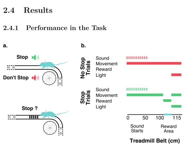
Here we developed a fully automated, head-fixed, delayed response task on a treadmill with a long belt that, given its self-initiation and spatial features, was very engaging and easy to learn for the mice which performed a high number of trials per session with a very good performance. Moreover, our setups allowed us to closely monitor animal behavior, namely licks and movement on the treadmill, giving us access to how the mice were solving the task and guidance in designing the appropriate controls to guarantee that a behavioral strategy was not being used to eliminate the need for keeping a memory through the memory period. These made us confident that, like desired, upon the presentation of an auditory cue, mice kept, throughout the memory period, an active representation that allowed them to perform
2.3. Methods and Materials
2.3
Methods and Materials
2.3.1
Behavioral Apparatus
The results here presented were obtained using three behavioral setups: two of them, used only for behavior, were identical and the third differed only in what was necessary to perform the electrophysiological recordings, namely the possibility of attaching to it a stereotaxic arm, a microscope connected via a flexible moving arm and a light-source - all essential to the insertion of the recording probe. The first two setups were inside the same custom made sound-proof box which was split in two by an internal division, the electrophysiology setup had a dedicated sound-proof box. Each setup was connected to its own dedicated computer with the task being programmed and controlled via Matlab. All setups sensors (rotary encoder, lick detector, belt lap detector) and effectors (speakers, reward valve, intra trial interval (ITI) lights) were controlled via a in-house developed input-output board with a sampling rate of 2082 Hz .
The basic component of each setup was a treadmill formed by two 3D printed wheels and a fabric belt running loosely around both. The back wheel, on top of which the head-fixed mice were placed, had 20 cm of di-ameter and 5 of width; the front wheel was smaller with 10 cm of didi-ameter and the same width. The axes of both wheels were fitted to ball bearings, mounted on laser cut pieces of acrylic, that were fixed, roughly 30 cm apart, on two 50 x 4 x 4 cm parallel aluminum rails. These two rails were fixed centered on the top face of a hollow cube also made with the same 50 x 4 x
4 cm rails. The belt, with 130 x 5 cm, which covered the top of both wheels and hanged towards the ground, had glued on its surface, approximately 100 cm from one of its ends, a set of bands with a rough texture. The textured surface covered a 15 cm stretch and was used to signal the place where the mice could get reward in the task. To make the animals more comfortable while running a half tube, tunnel like, plastic piece was placed around their body during the task. This was fixed to the setup by a set of optical posts and clamps (Thorlabs) that allowed it to be easily put in place and adapted to each animal position.
To monitor the position of the mice on the treadmill an absolute magnetic encoder (US Digital, MAE3) was fitted to one end of the big wheel axis. A pair of infrared (IR) LED emitter and logic detector were used to monitor each complete lap of the belt. For that they were installed, facing each other, on a custom made piece of acrylic, fixed to one of the top aluminum rails just after the front wheel, that extended towards the floor. In its lower end this piece had the shape of a rectangular prism with approximately 5.2 x 2 x 2 cm with the top and bottom sides open to allow the belt to run through the middle. A 0.4 cm hole was punctured in the belt aligned with the IR emitter and detector. The rectangular prism also served the purpose of maintaining the belt aligned with the wheels.
Mice were head-fixed in the setup to a custom made, 0.2 cm thick, alu-minum head-plate. The head-plate was situated on top of the back wheel with each of its lateral edges connected to one of the parallel rails by two optical posts. The posts in each side were connected in a 90◦ angle allowing to change the position of the head-plate both in the dorsal-ventral (DV)
2.3. Methods and Materials
and anterior-posterior (AP) axis relative to the wheel. The head-plate had a hole that, when the mice were head-fixed, gave direct access to the center of the head-implant allowing for the easy insertion of the recording probes. It also allowed the fixation, on its lower face, of speakers in several different positions and on its upper face, facing the animal, of the reward delivery system - lick-port.
To deliver reward to the mice during the task and monitor their licking lick-ports were custom built out of a 3D printed plastic body to which a water delivery spout and a IR LED emitter and logic detector were fitted. For the delivery spout we used an 18 gauge metal needle with the tip cut and polished. The emitter and detector were fitted and glued to specific parts of the plastic body so that the IR beam crossed the front of the delivery spout allowing us to monitor when the animals licked. The lick-port was fixed to the anterior upper face of the head-plate through a set of mini optical posts and clamps (Thorlabs) allowing for a precise positioning by moving it in the AP and DV axis relative to mice’s mouth.
To signal ITI, and illuminate the setups while off-task, two boards of white LED stripes, with adjustable intensity, were attached to the lateral rails of the base cube at the height of the mice when running on the treadmill.
2.3.2
Behavioral Task
2.3.2.1 Experimental Subjects
All mice used in the experiments here presented were male from the C57BL/6 strain. Mice selected to be implanted, and start the training pro-tocol, were typically 3 months old with a weight around 24 gr.