O R I G I N A L A R T I C L E
Use of wound dressings to enhance prevention of pressure
ulcers caused by medical devices
Joyce Black1, Paulo Alves2, Christopher Tod Brindle3, Carol Dealey4, Nick Santamaria5, Evan Call6 & Michael Clark7
1 Nursing, University of Nebraska Medical Center, Omaha, NE, USA 2 Catholic University of Portugal - Institute of Health Sciences, Porto, Portugal 3 Virginia Commonwealth University (VCU) Medical Center, Richmond, VA, USA
4 Tissue Viability, University of Birmingham and University Hospital, Birmingham NHSFT, UK
5 Nursing Research, Translational Research, University of Melbourne & Royal Melbourne Hospital AU, Melbourne, Australia 6 Weber State University, Salt Lake City, UT, USA
7 Tissue Viability, Birmingham City University, Birmingham, UK
Key words
Consensus; Medical device; Pressure ulcers
Correspondence to
Associate Prof. J Black, PhD, RN, CWCN, FAAN
College of Nursing
University of Nebraska Medical Center Omaha
NE 68198-5330 USA
E-mail: jblack@unmc.edu doi: 10.1111/iwj.12111
Black J, Alves P, Brindle CT, Dealey C, Santamaria N, Call E, Clark M. Use of wound dressings to enhance prevention of pressure ulcers caused by medical devices. Int Wound J 2015; 12:322–327
Abstract
Medical device related pressure ulcers (MDR PUs) are defined as pressure injuries associated with the use of devices applied for diagnostic or therapeutic purposes wherein the PU that develops has the same configuration as the device. Many institutions have reduced the incidence of traditional PUs (sacral, buttock and heel) and therefore the significance of MDR PU has become more apparent. The highest risk of MDR PU has been reported to be patients with impaired sensory perception, such as neuropathy, and an impaired ability for the patient to communicate discomfort, for example, oral intubation, language barriers, unconsciousness or non-verbal state. Patients in critical care units typify the high-risk patient and they often require more devices for monitoring and therapeutic purposes. An expert panel met to review the evidence on the prevention of MDR PUs and arrived at these conclusions: (i) consider applying dressings that demonstrate pressure redistribution and absorb moisture from body areas in contact with medical devices, tubing and fixators, (ii) in addition to dressings applied beneath medical devices, continue to lift and/or move the medical device to examine the skin beneath it and reposition for pressure relief and (iii) when simple repositioning does not relieve pressure, it is important not to create more pressure by placing dressings beneath tight devices.
Introduction
Pressure ulcers (PUs) traditionally occur on soft tissue and bony prominences that are exposed to pressure, shear and microclimate changes. However, PUs can also be caused by medical devices attached to or nearby the patient and in uncommon places such as the urethra, ears, upper back, neck and knees (1). Medical device related (MDR) PUs are defined as pressure injuries associated with the use of devices applied for diagnostic or therapeutic purposes wherein the PU that develops has the same configuration as the device. Pressure injuries result from a variety of reasons: the characteristic of the materials used to construct the device, difficulties in adjust-ing or securadjust-ing it to the patient’s body, prolonged pressure in the same place, and pressure forces causing local oedema (2).
While the exact mechanical forces leading to the ulcerations from these devices have not been fully elucidated, it is logical
Key Messages
• MDR ulcers can occur from any medical device • MDR ulcers can become full thickness pressure ulcers • MDR ulcers are reasonably preventable with thin
dress-ings under the device
• skin beneath a medical device must be inspected every shift
• faulty medical devices should be returned to the manu-facturer
to believe that devices secured tightly to the patient’s body can be a source of pressure and if the patient develops oedema in the tissues after the device was initially secured, pressure can be increased. In addition, devices that increase sweating beneath them can be recognised as a source of skin microclimate changes.
Incidence of medical device related PU
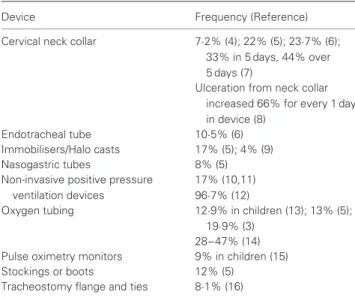
While the development of PUs on the sacrum and heels has been tracked for decades, the presence of PUs beneath medical devices has only recently been identified. The incidence or facility-acquired rates of MDR PUs are not widely reported. However, many institutions have reduced their number of traditional PUs (sacral, buttocks and heel) and therefore the significance of MDR ulcers has become more apparent (Figure 1). The largest study found (N= 104 266), reported that medical devices led to 19·9% of ulcers on the ear, 14·3% of ulcers on the sacrum/coccyx, 10·2% of ulcers on the heel and 8·8% of ulcers on the buttocks (3). The specific devices involved were not included in the report of these findings. Reported incidences or agency-acquired rates of MDR PUs from the literature along with the known device leading to the ulceration are shown in Table 1.
Predictors of risk for MDR PU
The mere presence of a medical device (17,18) or the inser-tion site of the device (19) creates the risk for ulcers. How-ever, there are certain devices and certain patients who are at increased risk. Ackland and colleagues (8) examined the records of 299 subjects in critical care who were wearing Philadelphia collars and were awaiting medical clearance of cervical spine injury. Cervical collar PU development was pre-dicted by ICU admission (P= 0·007), mechanical ventilation (P= 0·005), the necessity for cervical MRI (P < or = 0·001) and time to cervical spine clearance (P < or= 0·001). Time to cervical spine clearance was the major indicator; such that the risk of ulceration increased by 66% for each additional day of wearing the cervical collar. Powers et al. (20) conducted a prospective, descriptive study of 484 patients wearing the Aspen collar. They also concluded that number of days with the cervical collar were found to be a significant predictor of skin breakdown (P < 0·0001). The presence of braided or beaded hair can also contribute to PU development (20).
The ear has been commonly reported as a location for PUs (2,3,5,14,21). Some of the devices that encompass the ear are oxygen tubing and oxygen straps. Pulse oximetry devices are often clipped on to the ear helix or lobe in patients who are vasoconstricted and when finger probes are inaccurate. One study reported the actual pressure exerted on the ear lobe from ear pulse oximetry devices at 20·7 mmHg; the significance of this finding is unknown (22).
Incidence of PUs on the face and nasal bridge of patients using non-invasive positive pressure ventilation (NIPPV) masks has been reported as at least 17% (10,11). NIPPV masks can be difficult to fit to the patient’s face, and without proper fit, the device does not work correctly or not work at all. If the mask causes discomfort, the patient will often opt not to wear it (23). Treatment was curtailed in over 50%
of patients using masks that caused discomfort (11). Oxygen delivery devices are the most common devices leading to pressure ulceration in children (24).
Patients at highest risk of MDR PU have been reported to be those with impaired sensory perception, such as neuropathy, and an impaired ability to communicate discomfort, for example, oral intubation, language barriers, unconsciousness or non-verbal state (5). Patients in critical care units typify the high-risk patient and they often require more devices for monitoring and therapeutic purposes. A secondary analysis of 2079 cases of patients in critical care, step down and surgical units concluded that patients with medical devices were 2·4 times more likely to develop a PU of any kind (2). Inspection of skin beneath medical devices may be cumbersome, because the medical device may be an essential component of the patient’s medical care. Pain from PUs beneath medical devices may also go unreported in unconscious patients. Another risk factor for PUs from medical devices is the tightness of securement method. Devices to support the airway (endotracheal tubes and tracheostomy tubes) (25) tend to be secured tightly. The presence of a medical device is the most frequently cited risk factor for PU development in children, with incidence rates as high as 50% (24). Devices associated with PU development are listed in Table 2.
Skin moisture due to diaphoresis or secretions under the medical device may macerate the skin, making it susceptible to PU formation (26). Moisture is probably a cofactor for their development, making the skin less resilient in the presence of secretions or other humidity. Oedema under the device stretches the skin, making it more fragile and prone to pressure injury. Even if the device initially fits properly, patients may develop oedema after securing of the device, thereby increasing the tissue tension (27). Blood vessels in oedematous deep tissue are compressed from the external pressure of the oedematous fluids, and oxygen transport from capillaries to cells is also impaired in oedematous tissue (2).
Medical devices can be made from a wide variety of mate-rials, such as plastic, rubber, metals, plastics, composites, ceramics or silicone. Manufacturers’ improvements in design are often to improve comfort, but many devices are made of rigid materials or are secured with rigid material. The rigidity and inelasticity in the device or securement device causes friction, rubbing and increased pressure in tissues (10). Cervical collars are often very confining and lead to high occipital pressure (28). PUs may also arise because of the incorrect use, poor positioning or fixation of the equipment (29). Fixation or application of equipment using materials such as adhesive tapes or braces to adjust or secure the device may irritate susceptible skin, especially if oedema develops around the device (2). In addition, bad selection of poor equipment (17), incomplete evaluation of the skin (3) or problems with ill-fitting devices (5) may also be reasons to increase developing of these lesions.
Expert panel conclusions
In response to the increasing concern about MDR ulcers, the expert panel reviewed published data on devices and their prevention. The method used by this panel is described
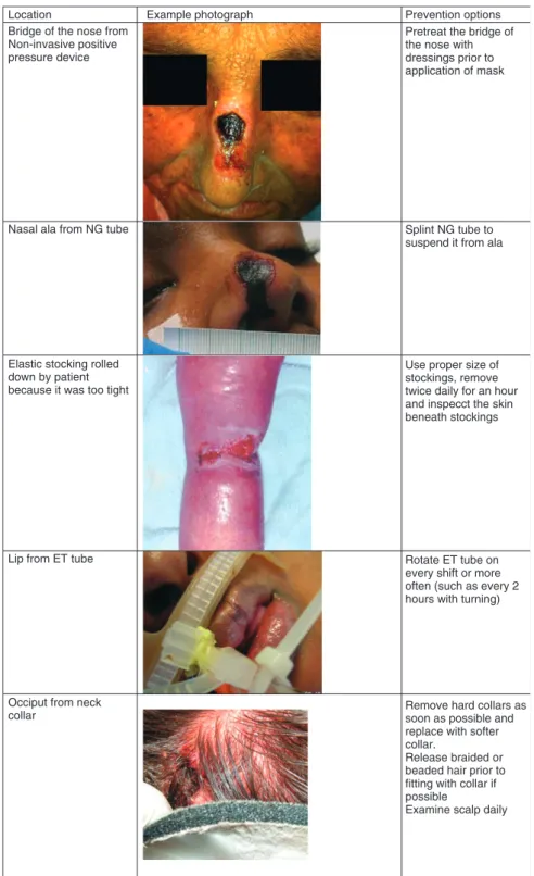
Location
Bridge of the nose from Non-invasive positive pressure device
Nasal ala from NG tube
Elastic stocking rolled down by patient because it was too tight
Example photograph Prevention options
Pretreat the bridge of the nose with dressings prior to application of mask
Splint NG tube to suspend it from ala
Use proper size of stockings, remove twice daily for an hour and inspecct the skin beneath stockings
Lip from ET tube Rotate ET tube on
every shift or more often (such as every 2 hours with turning)
Occiput from neck
collar Remove hard collars assoon as possible and
replace with softer collar.
Release braided or beaded hair prior to fitting with collar if possible
Examine scalp daily
Figure 1 Examples of medical device-related
pressure ulcers.
by Black et al. (30). The panel recognises that we are not an endorsed or elected body such as the National Pressure Ulcer Advisory Panel (NPUAP), the European Pressure Ulcer Advisory Panel (EPUAP) or the Pan Pacific Pressure Injury Alliance. The purpose of this article is to inform practitioners of our findings. Each of the statements that follow are written based on the methodology adopted in the recent PU guidelines (31), in that they have a strength of evidence and a supporting body of literature.
Consider applying dressings that demonstrate pressure redistribution and absorb moisture from body areas in contact with medical devices, tubing and fixators [strength of evidence (of recommendation), (SOE)= B] Three studies were found that reported on the effect of dress-ings placed between the device and the patient’s skin. Weng (12) conducted a quasi-experimental study with three groups of patients using NIPPV masks comparing time-to-nasal ulceration with two dressings and a control group without
Table 1 Type of device and frequency of ulceration
Device Frequency (Reference)
Cervical neck collar 7·2% (4); 22% (5); 23·7% (6); 33% in 5 days, 44% over 5 days (7)
Ulceration from neck collar increased 66% for every 1 day in device (8)
Endotracheal tube 10·5% (6)
Immobilisers/Halo casts 17% (5); 4% (9)
Nasogastric tubes 8% (5)
Non-invasive positive pressure ventilation devices
17% (10,11) 96·7% (12)
Oxygen tubing 12·9% in children (13); 13% (5);
19·9% (3) 28–47% (14) Pulse oximetry monitors 9% in children (15)
Stockings or boots 12% (5)
Tracheostomy flange and ties 8·1% (16)
dressings. One group had a film dressing, another group had a hydrocolloid dressing, and another was a control with no dressing. Ulceration rate was 96·7% in the control group, 53·3% in the group with film dressings and 40% in the group with hydrocolloid dressings. The skin was inspected every 30 minutes and the time-to-ulceration differed between groups with the shortest time in the control group (1111 minutes) and longest time in the hydrocolloid group (3272 minutes). Boesch and colleagues (16) reported a decrease in PU incidence from a baseline of 8·1% to 3·4% after the introduction of a bun-dled change programme including education and Mepilex® Lite™(Mohnlycke Health Care; Gothenburg, Sweden) dress-ing under the tracheostomy tube flanges. Turgania et al. (14) reported a reduction in the incidence of ulcers on the ear due to nasal cannula from 37% to 0% with the use of padding on the ears. A comparison of foam dressings to other types of dressings has not be reported; however, the attributes of foam dressings would appear to have benefit in these patients by reducing pressure and absorbing moisture, where hydro-colloids and films cannot absorb moisture.
Other studies examined products to hold the medical device away from the body orifice. Bite blocks were used to prevent oral PUs (32), splints for nasotracheal tubes (33) and silicone paste moulded to the bridge of the nose (11). The incidence of PUs beneath tracheostomy tubes or ties and/or the connection to the ventilator dropped to zero after the tracheostomy tube length was extended (16).
In addition to applying dressings beneath medical devices, continue to lift and/or move the medical device to examine the skin beneath it and reposition for pressure relief (SOE= C)
This recommendation is from expert opinion. It is appreciated that the use of a dressing on the skin beneath a medical device is not a replacement for other prevention strategies. Skin should be inspected according to agency policy or accepted standards of care. Skin must be inspected for signs of pressure due to the device, which cannot be seen while the device is still
Table 2 Medical devices associated with pressure ulcer formation
Medical device Anatomical site
Endotracheal tubes Lips
Tongue
Nasotracheal tubes Nose
Nares Nasal bridge
Nasal cannulas Ears
Oxygen tubing Nose
Cervical collars Neck
Occiput Clavicle Chin Mandible
Oximetry probes Fingers
Ears Nose
NIPPV/BIPAP Forehead
Nose
Tracheostomy tubes Front of the neck
Tracheostomy braces Around/back of the neck
Stockings/compression devices Behind knees Achilles tendon Lower extremities
Immobilisers Ankles
Wrists
Faecal containment Perianal
Buttocks
Foley catheters Urethra
Upper thighs Buttocks Perianal Splints Heels Braces Arms Wraps Elbow Straps Ankles
Abdominal binders Wrists
Neck Ribs Skin folds
External fixators All locations where the device is
in contact with skin Halo rings Vest brace Catheters Tubing Orthotics Drains Stopcocks G/J tubes JPs T-tubes Ostomy clips Bedpans
BIPAP, bi-level positive airway pressure; NIPPV, non-invasive positive pressure ventilation.
in place. For example, an endotracheal tube must be moved to inspect the lips for pressure injury. In obese patients, the medi-cal device may become hidden it the skin folds, and the nurses need to be diligent to account for all medical devices in these patients. Three-quarters (74%) of device-related PUs were not identified until they were stage III, stage IV or unstageable compared with 54% of non-device-related PUs. Twenty per-cent of non-device-related PUs were first identified when they were stage I, compared with only 5% of device-related ulcers (5). The dressing must be removed to visualise the skin, and then reapplied before replacing the medical device. An impor-tant consideration is the ability of dressing to be removed without injury to the skin during repeated assessments. The bridge of the nose is thinly covered with skin and could be easily injured with highly adhesive dressings. If possible, avoid placing a medical device back onto pressure-injured skin.
More than 50% of the medical devices that contributed to these pressure-related skin injuries were SpO2 probes, arti-ficial airways or bi-level positive airway pressure (BiPAP) masks. The mechanism of device-related pressure injury is similar to that of PUs caused by immobility, but prevention differs. Prevention includes rotating SpO2 probe sites, avoid-ing securavoid-ing the endotracheal tube close to the oral commis-sure, and inserting pressure-relieving material between oxygen masks and the nasal bridge (34).
Agencies may have to determine which member of the team, for example nurses or respiratory therapists (or both) are best suited to move devices and inspect the skin beneath them. Some medical devices are not meant to be moved, such as a cast. In those situations, follow accepted practice for care of patients when skin cannot be seen beneath the device. Agencies will also have to assist the staff to determine the proper size for a device by placing product selection guides in patient-care areas. Support stockings are often applied prior to surgery, and then become too tight when the limb becomes swollen after surgery. Similarly, some medical devices are applied prior to fluid resuscitation (e.g. sepsis) and the securement product, such as tape or straps, becomes tighter as fluid is shifted into interstitial spaces. The early signs of pressure from the medical device and the securement device should be identified and adjustments made in the device or straps to prevent injury.
When simple repositioning does not relieve pressure, it is important not to create more pressure by placing dressings beneath tight devices (SOE= B)
Two studies were found that reported on the effect of thin dressings placed between the device and the patient’s skin. Weng’s (12) quasi experimental study reported that the use of even a thin (film/hydrocolloid) dressing applied to the nose reduced the incidence of nasal bridge PUs by 43·4% and 56·7%, respectively compared to an ulceration rate of 96·7% when no dressing was used. Boesch and colleagues (16) also saw a 4·7% decrease in ulcers under tracheostomy flanges with a bundled change programme including Mepilex®Lite™under the tracheostomy tube flanges. Gunlemez et al. (35) reported
that the use of silicone gel sheeting dressings reduced MDR ulcers due to oxygen delivery devices (P < 0·05)
In these studies, thin dressings appear to reduce the magni-tude of the pressure. Inserting thick dressings for prevention has not been reported, but the authors offer the opinion that more pressure could be created with a thick dressing.
Reporting MDR ulcers
Root cause analyses in these cases indicated that staff were sometimes uncertain about whom to contact about an ill-fitting device, were unsure of what a well-fitted device should look like against the skin, or were not aware of the potential for oedema to affect fit of devices or risk of PU development (2,5). Common characteristics included impaired sensory perception, such as neuropathy, and an impaired ability for the patient to communicate discomfort, for example, oral intubation, language barriers, unconsciousness or non-verbal state. The lack of clarity around best practices for skin inspection with the use of devices may have been a contributing factor in device-related PUs not being identified until they had become more advanced (5,36). Wounds from medical devices are PUs; they should not be classified as wounds from underlying disease states such as arterial insufficiency or be overlooked in institutional reporting. MDR ulcers should be reported separately from other PUs (those on the sacrum, buttocks and heels, for example and not associated with the use of a medical device in the same body area). Further policy work is needed on how to report PUs from medical devices on the mucous membranes that leave no scar and heal quickly. These wounds are PUs but do not appear to cause permanent damage to the patient. A stratification system on severity or likelihood of healing for MDR ulcers would aid in the analysis and reporting issues. Facilities required to do root or common cause analyses on every ulcer can find the process burdensome for some of the quickly healed ulcers on mucous membrane. It is important not to mistake the idea of a lack of scar to equate to an insignificant injury. Any open wound can worsen and methods to reduce the number of these wounds should not be overlooked.
The name of the device that led to the PU should also be recorded in order to keep track of the device causing harm. For example, it may not become apparent that the same medical product is causing harm until the data from the entire hospital system are reviewed. The authors believe that when a pattern of MDR PUs is apparent, the information should be conveyed to the manufacturer and regulatory bodies. If hospital staff continually accepts the development of MDR ulcers as a quality improvement problem, and never report them to the manufacturer, opportunities for improved safety profiles in product design are missed.
Conclusion
PUs from medical devices are increasing in frequency, and it has been said MDR PUs are an epidemic. High-risk patients should be identified during risk assessments, skin inspection beneath devices must occur at every shift and the devices
should be moved as much as possible (for example moving the endotracheal tube and pulse oximetry probe from side to side with each body turn). Applying protective dressings to pad the skin is imperative; most of these ulcers are completely preventable. However, medical professionals can only work with the devices provided. The solution also depends on the development of safer materials that do not increase the risk of developing a PU.
Acknowledgement
The work of this panel of experts was supported by M¨olnlycke Health Care, Gothenburg, Sweden.
References
1. Fletcher J. Device related pressure ulcers made easy. Wounds UK 2012;8:1–4.
2. Black JM, Cuddigan JE, Walko MA, Didier LA, Lander MJ, Kelpe MR. Medical device related pressure ulcers in hospitalized patients.
Int Wound J 2010;7:358–65.
3. Van Gilder C, Lachenbruch C, Harrison P, Davis D. Overall
results from the 2011 international pressure ulcer prevalence survey.
Batesville: Hill-Rom, 2012.
4. Powers J, Daniels D, McGuire C, Hilbish C. The incidence of skin breakdown associated with use of cervical collars. J Trauma Nurs 2006;13:198–200.
5. Apold J, Rudrych D. Preventing device-related pressure ulcers: using data to guide statewide change. J Nurs Care Qual 2012;27:28–34. 6. Watts D, Abrahams E, MacMillan C, Sanat J, Silver R, VanGorder
S, Waller M, York D. Insult after injury: pressure ulcers in trauma patients. Orthop Nurs 1998;17:84–91.
7. Davis JW, Parks SN, Detlefs CL, Williams GG, Williams JL, Smith RW. Clearing the cervical spine in obtunded patients: the use of dynamic fluoroscopy. J Trauma 1995;39:435–8.
8. Ackland HM, Cooper DJ, Malham GM, Kossmann T. Factors predicting cervical collar-related decubitus ulceration in major trauma patients. Spine 2007;32:423–8.
9. Walker J. Pressure ulcers in cervical spine immobilisation: a retro-spective analysis. J Wound Care 2012;21:323–6
10. Jaul E. A prospective pilot study of atypical pressure ulcer presen-tation in a skilled geriatric nursing unit. Ostomy Wound Manage 2011;57:49–54.
11. Jones DJ, Braid GM, Wedzicha JA. Nasal masks for domiciliary positive pressure ventilation: patient usage and complications. Thorax 1994;49:811–2.
12. Weng MH. The effect of protective treatment in reducing pressure ulcers for non-invasive ventilation patients. Intensive Crit Care Nurs 2008;24:295–9.
13. Groeneveld A, Anderson M, Allen S, Bressmer S, Golberg M, Magee B, Young S. The prevalence of pressure ulcers in a tertiary care pediatric and adult hospital. J Wound Ostomy Continence Nurs 2004;31:108–20.
14. Turgania MA, Clark L, Martini C, Miller P, Turner B, Jones S. Incidence, correlates and interventions used for pressure ulcers of the ear. Medsurg Nurs 2011;20:241–7.
15. Noonan C, Quigley S, Curley M. Skin integrity in hospitalized infants and children: a prevalence survey. J Pediatr Nurs 2006;21: 445–53.
16. Boesch RP, Myers C, Garrett T, Nie A, Thomas N, Chima A, McPhail GL, Ednick M, Rutter MJ, Dressman K. Prevention
of tracheostomy-related pressure ulcers in children. Pediatrics 2012;129:e792–7.
17. Ong JC, Chan FC, McCann J. Pressure ulcers of the popliteal fossae caused by thromboembolic deterrent stockings (TEDS). Ir J Med Sci 2011;180:601–2.
18. Hogeling M, Fardin SR, Frieden IJ, Wargon O. Forehead pressure necrosis in neonates following continuous positive airway pressure.
Pediatr Dermatol 2012;29:45–8.
19. Moreiras-Plaza M. Abdominal wall skin pressure ulcer due to a peritoneal catheter. Periton Dialysis Int 2010;30:257–8.
20. Dixon M, Ratliff C. Hair braids as a risk factor for occipital pressure ulcer development: a case study. Ostomy Wound Manage 2011;57:48–53.
21. Schl¨uer AB, Cignacco E, M¨uller M, Halfens RJ. The preva-lence of pressure ulcers in four paediatric institutions. J Clin Nurs 2009;18:3244–52.
22. Goodell T. An in vitro quantification of pressures exerted by earlobe pulse oximeter problems following reports of device-related pressure ulcers in ICU patients. Ostomy Wound Manage 2012;58:30–4. 23. Gregoretti C, Confalonieri M, Navalesi P, Squadrone V, Frigerio
P, Beltrame F, Carbone G, Conti G, Gamna F, Nava S, Calderini E, Skrobik Y, Antonelli M Evaluation of patient skin breakdown and comfort with a new face mask for non-invasive ventilation: a multi-center study. Intens Care Med 2002;28:278–84.
24. Baharestani M. Pressure ulcers in pediatric populations. In: Pieper B, with the National Pressure Ulcer Advisory Panel (NPUAP), editors.
Pressure ulcers: prevalence, incidence and implications for the future.
Washington, DC: NPUAP, 2012:151–71.
25. Baldwin KM. Incidence and prevalence of pressure ulcers in children.
Adv Skin Wound Care 2002;15:121–4.
26. Maklebust J. Pressure ulcers: etiology and prevention. Nurs Clin
North Am 1987;22:359–77.
27. Callaghan S, Trapp M. Evaluating two dressings for the prevention of nasal bridge pressure sores. Prof Nurse 1998;13:361–4. 28. Jacobson T, Tescher A, Miers A, Downer L. Improving
prac-tice efforts to reduce occipital pressure ulcers. J Nurs Care Qual 2008;23:283–8.
29. Mulgrew S, Khoo A, Newton RM, Rajan R, Kumar K. Pressure necrosis secondary to negative pressure dressing. Ann R Coll Surg
Engl 2011;93:e27–8.
30. Black J, Clark M, Dealey C, Brindle CT, Alves P, Santamaria N, Call E. Dressings as an adjunct to pressure ulcer prevention; Consensus panel recommendations. In press, 2013.
31. National Pressure Ulcer Advisory Panel and European Pressure Ulcer Advisory Panel. Prevention and treatment of pressure ulcers:
clinical practice guideline. Washington DC: National Pressure Ulcer
Advisory Panel, 2009.
32. Zaratkiewicz S, Teegardin C, Whitney JD. Retrospective review of the reduction of oral pressure ulcers in mechanically ventilated patients: a change in practice. Crit Care Nurs Q 2012;35:247–54. 33. Huang T, Tseng C, Lee T, Yeh J, Lai Y. Preventing pressure sores
of the nasal ala after nasotracheal intubation: From animal model to clinical application. J Oral Maxillofac Surg 2009;67:543–51. 34. Curley MAQ, Quigley SM, Lin M. Pressure ulcers in pediatric
intensive care: incidence and associated factors. Pediatr Crit Care
Med 2003;4:284–90.
35. Gunlemez A, Isken T, Gokalp AS, Turker D, Arisoy EA. Effect of silicon get sheeting on nasal injury associated with nasal CPAP in preterm infants. Indian Pediatr 2010;47:265–7.
36. Molano Alvarez E, Murillo Perez Mdel A, Salobral Villegas MT, Dom´ınguez Caballero M, Cuenca Solanas M, Garc´ıa Fuentes C. Pres-sure sores secondary to immobilization with cervical collar: a com-plication of acute cervical injury. Enferm Intensiva 2004;15:112–22.