Review Article
MITOSES IN CUTANEOUS MALIGNANT
MELANOMAS:
Relevance in the prognosis
Sara Completo Marques
Orientador: Prof. Dr. Luís Soares de Almeida
Universidade de Lisboa, Faculdade de Medicina de Lisboa
Clínica Universitária de Dermatologia
2 Index Acknowledgments ... 3 Abstract ... 4 Resumo ... 5 Introduction ... 6 Methods ... 6 Results ... 6
Malignant Melanoma - General Considerations ... 6
Diagnosis ... 9
Prognostic factors ... 9
Tumor Mitotic Rate (TMR) as a prognostic factor ... 11
Do mitoses always mean malignancy? ... 14
Sentinel Lymph Node Biopsy ... 16
Immunohistochemistry ... 18
Discussion ... 20
Conclusion ... 23
3 Acknowledgments
First of all, I would like to thank Prof. Dr. Luís Soares de Almeida for accepting to guide me along this path of almost 2 years and for suggesting this interesting and non-unanimous theme, which let me dive in this endless sea of details that can make a difference, gathering two areas of my deepest interest. Prof. Soares de Almeida was the best advisor I could have wished for and a truthfully mastermind in this vast area. My most sincere regards for, during all this time, never letting me out of sight and for always being promptly available for anything I needed, constantly pushing me to a higher level. Only a real Professor can accomplish that!
I also want to thank Prof. Dr. Paulo Filipe for his brilliant advices and suggestions that allowed me to improve my article’s quality.
Moreover, I need to thank Dr Arno Rütten, from Dermatopathologie Friedrichshafen, Germany, for his thoughtful discussion on this review.
Lastly, but not least, I would like to thank my friends and family, specially my parents and Ricardo, for the unconditional love and support, without which I could never survive this long and hard year, even though sometimes I’m not able to show my gratefulness in the clearest way.
4 Abstract
Malignant Melanoma is considered the most dangerous skin neoplasia for its potential to invade distant tissues and its associated mortality. Its incidence is considered epidemic.
Mitotic rate was recently introduced in the AJCC guidelines as the 2nd most important prognostic factor for thin stage I melanomas, after tumoral thickness, as it strongly correlates with a lower survival rate. Together with ulceration, this variable changes the classification of thin tumors (≤1mm) from T1a to T1b, if at least one mitosis/ mm2 is present. This implies an alteration in patients’ management and treatment. It has been a target of discussion for its accuracy among clinicians and interobserver disparities. The cutoff of one mitosis as also been targeted, since mitotic figures can be found in benign lesions, particularly when immunohistochemistry is used.
Mitotic rate has been used to refer patients to sentinel lymph node biopsy, which has also been a disagreement point. Immunohistochemistry in mitoses detection is still under investigation but it is proved to facilitate the observation, although having some objections.
Standardization is essential to a more reliable method and adequate patient’s maneuver.
Key Words: Melanocyte; Melanoma; Mitosis; Mitotic index; Mitotic rate; Prognosis; Sentinel lymph node biopsy; Immunohistochemistry.
5 Resumo
Melanoma Maligno é considerado a neoplasia da pele com maior perigo, pelo seu potencial de invasão de tecidos distantes e mortalidade associada. A sua incidência é considerada epidémica.
O índice mitótico foi recentemente introduzido nas recomendações da AJCC como o 2º factor de prognóstico mais importante para os melanomas finos de grau I, logo após a espessura tumoral, por se correlacionar com uma diminuição da taxa de sobrevida. Juntamente com a ulceração, esta variável altera a classificação dos tumores finos (≤1mm) de T1a para T1b, se pelo menos uma mitose/mm2 estiver presente. Isto implica uma alteração na gestão e tratamento dos doentes. Tem sido um alvo de discussão devido à fiabilidade entre clínicos e disparidades entre observadores. O “cutoff” de uma mitose tem também sido visado, já que as imagens de mitoses podem ser encontradas em lesões benignas, particularmente se forem usadas técnicas de imuno-histoquímica.
O índice mitótico tem sido utilizado para referenciar doentes para biópsia de gânglio sentinela, o que também tem sido um ponto de discórdia. A utilização de imuno-histoquímica na deteção de mitoses continua sob investigação, no entanto está provado que facilita a observação, embora haja algumas objeções a ter em conta.
Uniformizar a técnica é essencial para um método mais fiável e uma abordagem adequada dos doentes.
Palavras-chave: Melanócito; Melanoma; Mitose (s); Índice Mitótico; Prognóstico; Biópsia de Gânglio Sentinela; Imuno-histoquímica.
6 Introduction
A review of the bibliography was carried out about a mediatic theme that is Malignant Melanoma. Mitotic rate was chosen for its integration as a prognostic marker in the last AJCC guidelines published in 2009. It is a thematic that involves discussion and disagreement among clinicians and, particularly, pathologists.
Therefore, we agglomerated and discussed the theme exposed in recent bibliography, conjugating it with previous knowledge and supposedly acquired facts, in order to achieve a deeper acquaintance in how to deal with mitoses in malignant melanoma’s prognosis, the process of their acquirement and the consequences they can lead to. We end by suggesting that new studies are accomplished to clarify the methods for mitotic rate applications in this skin neoplasia.
Methods
The review of the literature was carried out using “Pubmed” and “repositorio.ul” for articles between 2000-2016 and afterwards between 1945-2016. The terms used for the search included: melanoma, histopathology, mitosis (or mitoses), mitotic index (or mitotic rate), benign nevus (or nevi), prognosis, sentinel lymph node biopsy and immunohistochemistry.
The most relevant abstracts were assessed for information integrating mitotic rate and malignant melanoma, the process for their acquisition and their possible consequences.
Additionally, related articles and the reference lists of the selected ones were also used as sources for further potential information.
Results
Malignant Melanoma - General Considerations
Melanocytes, cells from which melanocytic lesions develop, originate from ectoderm in neural crest, sharing some characteristics with neuronal cells. Melanocytes in humans exist in multiple regions of the human body besides the skin and hairs, namely iris, inner ear, adrenal gland, gastrointestinal, genitourinary and even central nervous system. For this reason, malignant melanoma (MM) can occur in any of these
7
places. Some studies discovered distinct properties and DNA modifications depending on the anatomical location of the lesion, no matter what its histologic group was 1,2.
MM is a malignant neoplasia derived from melanocytes, usually beginning in the epidermis. While confined to epidermis, MM is considered in situ and it lacks potential for metastasis; this phase is called radial growth. Afterwards it might penetrate dermis, initiating the vertical growth and acquiring the capacity to metastasize into distant organs, since in opposition to epithelium, these tissues are highly vascularized, with blood and lymph vessels 1,2.
It is considered the most malignant skin neoplasia for its potential to invade, not only locally but mainly distant tissues, by vascular or lymphatic dissemination 2,3.
MM originate more commonly de novo, directly from a normal melanocyte or, on the contrary, 20-25% may transit from a previous benign lesion, including congenital nevi, or even from an area of an old sunburn or previous radiotherapy 2,4. They can also appear in association to HIV infection, lymphoma or after transplant 2.
Melanoma is considered the 7th more common cancer in the USA 5, accounting for approximately 5% of all new cancers in 2014 6. Regarding skin cancers, it corresponds to less than 7% of all but accounts for about 75% of deaths from these malignancies 1.
According to recent studies, its incidence has been increasing in a large scale for the last few decades, being even considered by some an epidemic cancer, nowadays reaching a rate of almost 40 per 100.000 in Australia2 and 26,3 per 100.000 in the USA7. It was estimated that almost 74.000 new cases of MM appeared in 2015 and there were around 10.000 new deaths caused by this cancer 8. In Europe, MM’s incidence was predicted to be around 12-16:100.000 inhabitants, in women and men respectively. However, it depends if it is in the north or south, being higher in Central and Eastern European countries 9. In Mediterranean countries incidence varies from 3 to 5:100.000 per year and in the Nordic ones from 12 to 25:100.000 (and rising) 10. There is no accurate estimation for Portugal individually but we integrate the Mediterranean countries and correlate with the other European southern countries such as Spain and Italy. Melanoma is 20 times more common in Caucasians, mostly in sun exposed skin and, particularly in areas of intermittent sun exposure, namely trunk in men and legs in women, and after sunburns in childhood 2,11.
The epidemic concept introduced in the previous paragraph is a discussion target between clinicians. Some argue it is in part due to overdiagnosis by more free
8
screenings, in context of increased alert among the population, aided by media cover. Also, it may be a result of a more concerned reporting after detecting and excising a lesion or, on the other hand, on a propensity to diagnose as malignant doubtful lesions, mislead by fear of missing some dangerous lesion. Still, some admit there might have been a truly increment in MM incidence considering two facts: the preferential incidence in the elderly and the higher average life expectancy of the population. In the end, the main reason pointed is the hard distinction from other benign pigmented lesions
12
.
Recent studies prove that more than one half of MM are considered thin, with Breslow depth <1mm and a very good prognosis: 10-year survival of 83,1-96,5% 7,13.
Incidence varies according to risk factors, which can be divided in personal and environmental, the latter especially during childhood (Tables 1 and 2) 2.
Every type of melanoma can develop metastases, even the thin ones that supposedly had a great outcome. That is why scientists and clinicians have been trying to understand which characteristics, namely histologic ones, can be associated with worse prognosis 2.
Table 1: Personal Risk Factors Age
Phototype/ Sun susceptibility Personal/ Family history of MM
UV-rays exposition (chronical: lentiginous MM; intermittent: superficial spreading)
Precursor lesions
(number of dysplastic, congenital and common nevi)
Trauma
Table 2: Environmental Risk Factors
Lower latitude Higher altitude
9
Diagnosis
Still today, MM diagnosis relies in the correlation among clinic and microscopic Hematoxylin-Eosin (H&E) stained observations, representative of the whole tumor 2.
Initially the lesion has homogeneous color, regular borders and smooth superficies. It is indispensable to promptly set differential diagnosis between other dark skin tumors, even pigmented basal cell carcinomas 1,2. Later on, if the lesion is a MM, it will probably change and gain various tones, irregular borders and rough superficies, presenting atrophic, ulcerated or even warty zones. Usually ABCDE rule is used to help the interpretation of these malignant lesions. The letters of the acronym correspond to “Asymmetry” of the lesion, “irregular Borders”, “different Colors”, “Diameter larger than 6 mm” and “Evolving and rapid growth” 14,15. However, some critics have been made to diameter, as it looks arbitrary; smaller lesions can also be diagnosed as MM. It has been considered changing to “Dark” instead 14.
Besides, other minor signs are used, such as pruritus, hypopigmentation, inflammation, hemorrhage, ulceration, or just the fact that is a lesion distinct from all other nevi in that person – “ugly duckling signal” 2.
Usually MM arises in people who are around 40 years old or more, with lower phototypes. However, the greatest impact in mortality is seen at the average age of 20 to 40 years old 8. Exceptionally, they can appear during childhood or puberty 2. They tend to grow and invade the dermal-epidermal junction, gaining power to spread and metastasize through blood or lymphatic system 16.
Prognostic factors
Usually, malignant tumors are staged evoking TNM classification: primary Tumor (T), lymph Node metastasis presence and number (N) and presence of local or distant Metastasis (M), separating it in I-II when exclusively cutaneous, III with loco-regional metastases and IV when distant metastases exist. This is a classification useful to make clinical decisions, having treatment and prognosis in mind. It is of high importance to have a standardized staging for tumors, allowing to offer the best diagnosis and therefore treatments to cancer patients 17.
Although clinical presentation is useful for MM prognosis, histological study is indispensable 18.
10
Since at least 4 decades, classification of primary cutaneous melanomas was made considering invasion depth into the dermis/ subcutis or Clark level, stratifying tumors between T1 and T4 levels. Clark level system divides in 5, from level I when the lesion is confined to the epidermis and its adnexal structures like follicles and glands (in situ MM) to level V with adipose tissue invasion (Figure 1) 19,20.
Other important variables in the prognosis include 2,21:
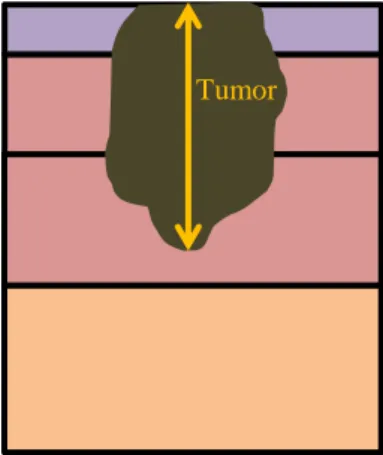
Breslow thickness: MM depth in mm, vertical measure between the high granular zone of epidermis above the tumor (or base of ulceration), until the deepest tumor cell below the epidermis (Figure 2).
Ulceration: complete non-traumatic loss of the epidermis created by the tumor located underneath; undoubtedly worsens the prognosis.
Vertical growth phase (presence or absence).
Epidermis Papillary dermis Reticular dermis Subcutaneous tissue I II III IV V
Figure 1: Clark Level I-V
Figure 2: Breslow thickness in mm
11
Regression: usually the area is clinically characterized by depigmentation. It occurs after some areas of the lesion are destroyed by memory lymphocytes, shifting melanocytes for scar tissue, telangiectasias and/ or melanophages. This could mean the tumor was previously thicker, which would supposedly worsen the prognosis. However, in a recent study, regression was associated with tumor-directed T-cell response, which improved prognosis 22.
Tumor-infiltrating lymphocytes (TIL): presence of lymphocytes and/ or plasma cells around or inside the tumor. Dense TIL infiltrate has been associated with an excellent prognosis, independently of other adverse variables 23.
Vascular or lymphatic invasion.
Tumor Mitotic Rate (TMR) as a prognostic factor
In 2009 the melanoma staging system was substantially revised and some changes with consequences related to the approach to patients were published by the American Joint Commission on Cancer Staging Committee (AJCC). Mitotic rate (MR) was added as a new element, substituting Clark level, after demonstrated to be an important and independent prognostic factor. Mitotic index is, therefore, a quantitative measure of cellular proliferation in primary melanoma. Histologically, it is defined as the number of melanocytic dermal mitoses per mm215,19,21,24–26.
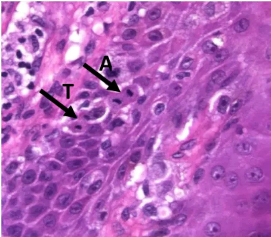
Mitoses can be defined by an absence of the nuclear membrane in the end of prophase and by the presence of condensed chromosomes (chromatin’s extensions), either clotted in the beginning of metaphase, arranged in a plane in metaphase or anaphase, or even in separate clots in telophase 24,27. There is also an absence of a central clear zone in the dense nucleus and a clear basophilic cytoplasm (Figure 3) 28.
Figure 3: Mitoses in Anaphase (A) & Telophase (T)
A
T
12
It is important to differentiate dermal from epidermal component in this analysis, since mitoses in epidermis only have diagnostic value, being irrelevant to prognosis 29.
However, for at least 30 years, it has been known that a high proliferative rate corresponds to an adverse prognosis 20. Recently, it was demonstrated that an increased mitotic rate correlated strongly with lower survival rates, independently of the tumor thickness 21,26,30,31. However, it also correlated with increasing tumor thickness and ulceration, meaning thicker MMs have higher MRs 26,28,32. Some tried to show a connection with sentinel lymph node positivity 30.
MR was then considered the second most powerful predictor of survival, after Breslow tumor thickness, particularly among patients with thin melanomas, even exceeding ulceration 3,17,19,21,26 . It means the tumor is growing and can metastasize faster, behaving more aggressively 3,26. In fact, the AJCC classification referred mitotic rate was only relevant for staging thin melanomas 19. Nonetheless there are studies showing it is also appropriate for treatment decisions in thick tumors 17.
The most significant correlation with survival was identified at a threshold of at least one mitotic figure per square millimeter, especially for thin melanomas. When mitoses are not found, it is recommended to report as 0 mitoses/mm2, instead of <1mitosis/mm2 26. It was also proved that mitotic index is highly predictive when it comes to lymph node macrometastasis and sentinel node micrometastasis, as well as distant metastases 3,17.
For patients with T1 melanoma the three most powerful predictors of survival are now considered tumor thickness of Breslow, mitotic rate/ index and ulceration. These three characteristics should be present in the pathology report after lesion biopsy 21. Depth of invasion (level of Clark) was proved not to be statistically relevant when the last two were included in the analysis. Accordingly, it was defined that mitotic rate would replace Clark level of invasion as a primary criterion for defining T1b melanoma, due to ineffectiveness of Clark level in a lesion without mitogenicity 16,21,33.
In this context, T1a melanomas are restricted to those ≤1.0mm of thickness, without ulceration or a mitotic rate of 0/mm2. On the other hand, T1b is defined as those ≤1.0 mm of thickness and, at least, one mitotic figure per mm2
or tumor ulceration17,19,26,34. Clark level is only used for staging when mitotic rate cannot be accurately measured, regarding thin melanomas, and, optionally, in thicker melanomas19–21.
13
Therefore, MR, together with ulceration, currently distinguish between T1a and T1b stage, and thus between IA and IB in TNM scale 21. According to this classification implemented in 2009, in a non-ulcerated lesion, a single dermal mitotic figure is sufficient to change the tumor prognosis and patient management 13,17,19,33,35. The most immediate impact in 10-year survival prognosis for patients with thin melanomas is the decrease from 95% to 88% in T1a and T1b, respectively 19,35,36.
In regard of T2 melanomas, there is still a lot to prove. There are some recent studies trying to understand if elevated mitotic index is related to sentinel lymph node positivity, recurrence of the disease or even its independence from other factors. Baker et al in 2015 showed MR ≥1/mm2 remains an important risk factor in this group of relatively thicker melanomas. In his study, MR was related to disease recurrence in some specific conditions, such as no ulceration. Moreover, it was associated with lower time until recurrence in patients with negative sentinel lymph node analysis. It appeared MR lost significance in predicting sentinel lymph node positivity as the thickness increased, hence having a higher impact for T1 and some T2 melanomas. However, undoubtedly, MR was strongly associated with death risk, even in thicker tumors. Baker et al showed higher MR associates with worse survival (proved for index higher than 20 mitoses/ mm2, although only a small amount of patients have an index this high). The influence of higher MR in recurrence and overall survival is independent from age 6.
In this context, it is important to understand how mitoses are counted. It is recommended by the AJCC that this rate is determined by the “hot spot approach”. Frishberg, D. (2001) and Weyers, W. (2009) described the so-called “hot spot” technique as the area in the vertical growth phase containing most mitotic figures. After counting the mitoses in the hot spot, it is extended to adjacent fields until an area corresponding to 1 mm2 is achieved. If no “hot spot” is found, and mitoses are randomly scattered throughout the lesion, several randomly chosen areas should be considered, summed, and the mitotic rate listed as the average. In tumors with invasive component of ≤1 mm, a rate per mm2
may be extrapolated 20.
In practice, usually 3-5 high power fields are evaluated, in a 40x magnification, depending on the size of microscopy ocular field, and then mitotic rate is presented in absolute numbers. Should integrate the accounting only dermal mitoses and unequivocally from melanocytes, without exhaustive section of the melanoma 16,33. This method is said to be time consuming and prone to variability between observer 17,30,31.
14
Difficulties identifying melanocytic mitotic figures include the following: discrimination of melanocytes from keratinocytes can be difficult in the dermo-epidermal junction; in heavily inflamed lesions, lymphocytic infiltrate intermixes with melanocytes and they can hardly be distinguished from each other; in heavily pigmented lesions mitotic figures can easily be overlooked in H&E staining; in thin melanomas ≤1mm2
the space is limited and it becomes complicated to discern which cell is undergoing mitosis 17,30.
It is also important to be aware of how extensively one should examine the tumor section, particularly in thin melanomas, group in which the great majority report only 1 or 2 mitoses per square millimeter. Searching few sections may lead to subclassification of a pT1b tumor into the pT1a category. On the other hand, when a thorough examination of the whole tumor is performed, there is no certainty about the relevance of one mitotic figure found in those conditions 33,35.
AJCC guidelines do not recommend an exhaustive search for mitoses 26,34, supposedly limiting the sections in two 13. It is also discouraged to preserve tissue for further testing, namely molecular 32. There is a fluctuation in the number of mitotic figures across various sequential sections, enhancing the importance of evaluating more than just one section per lesion. In 2014 a study suggested 3 to 5 sections improved the accuracy of mitotic index detection 32. In 2015, Shin & Tallon ought to prove which range of tumor tissue samples had more significance for the staging criteria, reaching the number of 3. They also came across data that showed wide differences in number of tissue samplings cut for various pathologists, between or even within centers, concluding there is no standardization for this assessment, which can, again, lead to completely different prognosis and clinical patient treatment 35.
Do mitoses always mean malignancy?
Various studies have revealed there is discordance among different pathologists when it comes to distinguish between thin melanoma, dysplastic and atypical nevi and even ordinary benign lesions. Often this distinction is not so easy to make between MM and other pigmented lesions, which can present themselves with characteristics typically seen in malignant ones 12. This is problematic regarding the consequences to patients. Those who erroneously got the melanoma diagnosis have follow-up programs for years, pulling-out life quality. On the other hand, those who are not diagnosed, may not get
15
access to the optimal treatment for their cancer, namely sentinel lymph node biopsy (SLNB), wide excision and/or adjuvant therapy, increasing the probability for a catastrophic outcome 5.
Back in 1949, Lund and Stobbe studied 200 nevi. They found mitotic figures in 6,5% of the nevi and almost 80% (10 in 13 nevi) appeared in the first two decades of life 37. Usually, there is a higher dermal melanocytic mitotic rate in younger people 7,38.
In 2009, Jensen et al. also agreed that, as nevi can grow, mitoses are expected to happen. They studied 157 randomly selected conventional nevi and 4% of them exhibited mitotic figures 39.
In a study about spitzoid lesions the authors concluded mitoses were rare in spitzoid nevi, although being present and even atypical in some cases. Moreover, deep-dermal mitoses were found, not only in spitzoid melanoma (malignant Spitz tumor) but in atypical spitzoid tumor as well, being the latter a transitional lesion 40. Some characteristics clearly associated with worse prognostic for MM, such as positive sentinel lymph node, are related to an indolent course in spitzoid lesions, including atypical Spitz tumor. Moreover, factors often used to predict the tumor behavior, are not useful in these lesions 41.
Another study performed in 2014 came across the idea that in deep penetrating nevi, even though mitoses are rare, some sparse figures (0 – 1.2/mm2) can appear. This lesion doesn’t have a regular and uniform morphology, frequently showing histological characteristics of MM, namely cytologic atypia and random nuclear pleomorphism, especially in the deepest regions 42.
Some other studies showed higher MR in benign lesions than in thin melanomas, as well as an increase to around 43% of benign nevi with mitoses when using immunohistochemistry techniques 13.
Culpepper, Granter & McKee (2004) wrote nevi apparently benign are commonly mitotically inactive. Nonetheless they may have a sporadic mitotic figure and it should not worry the observant, considering mitosis was incidental and occasional, without clinical impact, since all the other characteristics of the lesion pointed to innocence (symmetry, maturation, no nuclear transformation). A mitotic figure can be present in case the nevus is growing, which happens in benign lesions, although slower and in a lower rate, when comparing to malignant tumors. However it can also happen in a developing melanoma, associated with a previous existent common nevus. In this case
16
the question arises: should one consider this lesion a malignant melanoma and categorize it as T1b due to that one mitosis, without taking into account all the other features which might point to benignity? 4 We would promptly answer: “No”.
Usually melanoma has more mitoses and higher cellular and nuclear atypia, contrasting with benign lesion, where mitotic figures are rare and atypia is absent. That is why borderline melanocytic lesions are the most difficult to catalog, even for experienced pathologists, as they tend to mix up all these aspects used to differentiate benign from malignant tumors 42. For instance, dysplastic nevi sometimes considered “severely dysplastic” are usually what should be called MM or MM in situ, arising from a benign lesion 43.
Regarding mitoses, it is now obvious they are not capable of distinguish certainly between benign and malignant lesions. Even hyperplastic skin may show mitoses, feature usually associated with malignant tumors 40. If lesions are exhaustively observed, more mitotic figures will be detected, even in conventional nevi and thin melanomas 7,20.
Sentinel Lymph Node Biopsy
Sentinel Lymph Node Biopsy (SLNB) has been used to determine prognosis for doubtful lesions, which have been linked to high probability of metastases. Some even argue lymph node positivity “is the most important independent prognostic factor for survival in primary melanoma ≥1mm in thickness” 21,22,25,44. The fact is, although it has not been proved that early surgical removal of invaded lymph nodes increases long term survival, it contributes to a precise staging of the lesion, clarifying prognosis and adapted treatment 44.
Being important for prognosis, mitotic rate may as well be useful to determine which MM should undergo SLNB, being the lesions as better stratified as possible 25.
SLNB is a unique technique to detect lymph node micrometastases and it has a much lower morbidity and adverse effects, when compared to elective lymphadenectomy 44. It may additionally be helpful for future research regarding new therapeutic techniques 13.
SLNB has been preferentially performed in “intermediate to thick” tumors (≥1mm2
17
effective in thin melanomas, when additional risk factors are present. These factors include ulceration and mitotic rate, which change the classification to T1b if present 7,18. Thin melanomas mitotically inactive (0 mitoses per mm2), which corresponds to the majority, have low probability of sentinel lymph node positivity. This percentage tends to rise with increasingly MR 13.
In melanomas with depth ≤0,75mm the incidence of positive SLNB is less than 5%. When depth is between 0.75–1.0 mm, the rate of positive sentinel nodes biopsy is around 5% and the prognosis for these 5% of the patients is much poorer than in patients with a negative sentinel node biopsy. In these tumors cluster, when mitotic rate of 1/mm2 or higher is present, SLNB to search for lymph node micrometastasis may be proposed to the patient, especially if other risk factors are present 7,21,35. These risk factor for biopsy positivity comprehend: vascular and lymphatic invasion, positive margins and youngsters 21.
MR has, therefore, been used as indicative to proceed with SLNB, even before it was added to the AJCC staging system. It is used due to a proved relation of positivity amidst the two factors in previous studies. Contradictorily, recent studies have increasingly shown the data is currently insufficient to determine the risk of clinically occult lymph node metastasis, solely by using MR 7,13 and some determine it should not therefore be indicative to proceed 46. There are various reasons for these two visions. One example is: SLNB may be positive in cases where the tumors had higher Breslow thickness, which may overshadow the MR prognostic value. It was discovered MR is highly predictive of SLNB positivity in intermediate thickness MM (1-4 mm). The need for more studies proving this relation is unanimous 44,46.
Previous studies have shown no survival improvements when SLNB is performed, namely in thin melanomas 7,33,44. Currently it is not advisable to perform this procedure in melanomas ≤0,75mm deep, staged as IA or IB 13
, given the low yield and cost effectiveness 7. In the 4th Interdisciplinary Melanoma/ Skin Cancer Centers Meeting it was suggested by Daniel Coit that SLNB costs around 10,000 dollars per patient to identify one unpreventable death in consequence of metastatic melanoma 7.
Moreover it is important to remember that, besides the monetary implications, this procedure is not free of complications, namely because nearly 80% of the patients who undergo this procedure have negative biopsies. Common complications include:
18
infection, hematoma formation, sensory abnormalities, lymphedema, thrombophlebitis
7,44
.
Currently SLNB is performed in non-thin MM (Breslow thickness >1mm), as it was proved to relate with a risk of 10% of nodal invasion 25.This risk is around 5% in thinner MM 35, so this procedure is only considered when more risk factors are present, as pointed before. For instance, when ulceration is present, it is recommended by the AJCC that even tumors thinner than 0,75 mm should be referred for SLNB, as this complicates the correct measurement of Breslow thickness 44.
Although some patients with T1b MM are suggested to undergo SLNB, it should be made considering all the adverse variables present. It is not mandatory, not even recommended, that all T1b MM patients are subjected to such technique 25,26.
Concluding, as recommended by the AJCC, mitotic rate by itself should not be indicative for the selection of patients to undergo SLNB 44.
Immunohistochemistry
According to AJCC, the determination of mitotic figures for TNM is only supposed to use H&E stain, whereas it was proved to have large interindividual reproducibility 17.
Nonetheless, there are some mitoses’ (and consequently proliferation’s) specific markers, namely antigen Ki-67 and M-phase antibodies (phosphorylated histone H3 - anti-pHH3; mitotic protein monoclonal 2 - MPM-2), which might show higher counts of mitotic figures than H&E 2,17,47.
Antigen Ki-67 is a sensitive marker of proliferation and cell growth that stains positive in 13-30% of the cells in MM but in less than 5% in benign melanocytic lesions. It is even considered a statistically significant and independent prognostic factor. However, it is not specific for mitoses in melanocytic cells and, therefore, it might overestimate mitotic index when other proliferative cells are present, as can happen with a vast inflammatory infiltrate around the tumor. In these cases, a double staining with Ki-67 and, for instance, MART-1 (melanoma antigen recognized by T cells) might be used to specifically stain mitoses in melanocytic lineage 2,47,48.
Monoclonal antibody for pHH3 identifies mitoses in all stages, including prophase, and has been applied in other tumors, like meningiomas or astrocytomas, for
19
the same purpose. Mitotic figures were easily observed at 10x or 20x magnification, instead of 40x in H&E-stained sections (Figure 4 (a), (b)) 30.
Casper et al 30 showed, comparing to H&E-stained sections, a mean increase of 243% mitotic figures in tissue sections stained using anti-pHH3, in which 100% MM had mitoses present 13,30. These results present an intensification of sensitivity and sensibility for mitoses, with reduced observation time, when compared with H&E, where some would go unidentified. Therefore, in studies using anti-pHH3 stain, a MR ≥2 mitoses/mm2
corresponded to ≥1/mm2 in H&E, therefore defining T1b stage 13,35. Probably, with increased use of immunohistochemical markers, T1b stage as we know it would lose its significance 35.
Advantages of using immunohistochemistry directed to mitoses, namely anti-pHH3, are listed in Table 3 38,48,49.
Table 3: Advantages of
immunohistochemistry (Anti-pHH3) Easier distinction of mitotic figures from
apoptosis
Pattern of distribution uncovering mitotic hot-spots
Reduced average time needed to establish the mitotic count
Figure 4 (a), (b): Mitoses in various stages (Anti-pHH3) stain
20
One of the objections addressing anti-pHH3 was the identification of mitoses that did not belong to melanocytes or confusion between epidermis-dermis junctional mitoses from superficial dermal ones. These mistakes will increase in a large scale the number of mitoses caught, turning uncertain the prognostic impact 17,18,30.
These difficulties can be bypassed with a dual staining set, using for instance melanoma-associated antigen recognized by T-cells (Melan-A), marking melanocytes30. Recent reports used this dual staining and a correlation with prognosis was positively suggested 18,36. In a small study, Ottmann et al 18 showed that the increase in mitoses detection using anti-pHH3 disappeared when Melan-A immunostaining was added. This means the large increase in mitoses counting was, in this case, due to mitoses in other cells than melanocytes. They could not prove mitoses found by anti-HH3 influenced staging or prognosis for the lesion 18.
Therefore, the conclusion is the routinely investigation for mitotic rate, particularly in thin melanomas, should be performed using H&E, taking into account only the dermal figures, as suggested by the AJCC. Anti-pHH3, always used as a double staining with a melanocytic marker such as Melan-A, should be restricted to staging difficult cases, particularly when H&E stains are equivocal 18,47.
Discussion
It has been repeatedly shown that after MR introduction on the staging system in 2009, a considerable percentage of patients classified with T1a MM shifted to T1b. Consequently, prognosis is considered worse and, for that reason, increasingly more patients are subjected to invasive techniques, namely SLNB, changing the previous conduct towards patients 25.
It is still of vast importance to distinguish some details such as the significance of epidermal vs. dermal mitosis and their depth in the layers, as well as their morphology. Additionally it comes to our attention that not every melanoma included in T1a behaves similarly, existing characteristics that differentiate them from one another in a high degree 7.
One of the greatest defaults was that there are not enough prospective studies to dictate the reliability of the hot spot approach to mitotic rate in MM. Therefore, recent studies have invested in this matter and showed high reproducibility among pathologists, when the hot spot method is used 13,25,26. However, it is still doubtful how
21
thorough the section should observed and even how much sections should be made. It is recommended to search 5 contiguous fields but this pops up the question: “is this truly representative of the MR within the entire lesion?” Regarding one single melanoma, MR can be highly altered, just out of chance7.
Actually, in a high percentage of cases, mitotic rate could not be accurately measured and got different appreciations by different observers 17,20,33. This can depend for example on how much time one invests in that section’s microscope observation, leaving the diagnosis to be a matter of chance instead of standardized scales 17,20. Normally mitoses can be observed in 30 to 120 minutes, being metaphase even shorter; in MM with atypical highly-ploidy cells, metaphase might be longer, which can increase MR50. There are, in fact, some challenges interpreting mitotic figures and that can be due to a various number of factors, namely, difficulty determining junctional or dermal mitosis, small and easily missed mitotic figures, confusion with another cell mitosis such as endothelial or keratinocytes (which are the most common cells in epidermis), with pyknotic nuclei or even with the population of infiltrative lymphocytes (inflammation) or numerous melanophages 17,20,32,50. Additionally, staging can be affected by how tissue sampling is collected and fixated, the system used to score mitoses, the number of fields covered and even the quality of the material for the analysis 13,24,25,50.
When we are referring to a highly proliferative melanoma, even though some mitotic figures might not be actually from melanocytes, it does not make a huge difference. On the other hand, in a thin melanoma, mitoses are usually present in just one figure, demanding a careful and expandable search. In the latter it is much more difficult to discriminate whether that one mitotic figure found in so many fields is sufficient for changing classification for T1b 17,19,50. It has been reported to be an overestimating risk factor 6, because if the only mitotic figure present is misinterpreted (which can happen), it is changing, erroneously, the tumor from T1a stage to T1b and, consequently, changing patient’s management 7,13,33
.
In the case of Breslow depth, a micrometer is used to reduce interobserver variability. However, in determination of mitotic rate there is still not a tool to help reduce interobserver variability or precisely identify mitotic figures 32. It has to be studied the way to make hot spot technique and mitotic detection less abstract.
22
Additionally it has to be reviewed if one mitotic figure should change MM classification, especially since we can also find mitoses in hyperplasia processes or in common benign melanocytic nevi, in almost 50% if a specific immunohistochemical marker is used 7,13,33,50. Every process that grows is expected to show mitoses if one search for long enough. The mitotic figure itself is not a synonym of malignancy and, most often, it does not mean a poorer prognosis 7.
Thus, it is clear there is not one characteristic in itself capable of differentiating among pigmented benign and malignant lesions 5.
Besides, melanomas are malignant tumors supposedly growing every day. With immunohistochemical markers, nearly 100% of them have mitoses present. With this documented, it turns hard to understand why one mitotic figure, “possibly the only one in hundreds of sections” 50
can determine the change of classification to a poorer prognostic level and promote invasive and expensive procedures, for instance SLNB30,50. In fact, detecting a single mitotic figure in such a big area has been considered arbitrary 20.
As we could analyze, T1b stage, when together with other risk factors, is indicative for undergoing SLNB 25,33. Melanomas with Breslow thickness 0,5mm or lower are unlikely to show positivity for malignant cells in lymph node analysis and, even for those between 0,76-1,00mm, if only one mitosis is present, they are considered of low risk for nodal metastases. Therefore, not all patients with T1b MM should be submitted to this invasive technique 7,13.
Doctors and pathologists need to choose which patient from IB stage should undergo SLNB 13, taking into account the low cost effectiveness this procedure has shown 7.
Therefore, the extreme importance of correlating histological results with morphological and clinical context of the patient (including previous trauma, sun exposure, personal and family history, age or site of the lesion) before any practical decisions must be highlighted. Also it is extremely important we always recall the exceptions, for instance, neonatal and children’s melanoma, which is improbable but not impossible to appear 4.
There are several methods to help clinicians diagnose melanoma, including cytological features, in which mitoses can be integrated. However, none of these methods are exclusive to melanoma and many can also be found in melanocytic benign
23
lesions, as conventional nevi. However, usually, they are less pronounced than generally seen in malignant lesions. For instance, dermal mitoses have been found to be histological characteristics of some melanomas similar to benign nevi, such as naevoid melanomas 51.
Also, prognosis is dependent on other characteristics besides the primary tumor itself (tumor thickness, ulceration, and mitosis), such as the hosts’ immune response22,52; for example, the presence of tumor-directed T-cell responses are associated with a favorable outcome in metastatic melanoma 22. Moreover, we should take into consideration that, like in any other condition, there are factors other than those referred in AJCC influencing patient prognosis, namely general state of health, psychological stability or even social network 20.
This is why diagnosis can not be based just on checklists but, instead, it should be a rigorous and critical analysis of every feature of a lesion and the whole patient 51.
Conclusion
Clinical and histological diversity in primary cutaneous MM seems to be associated with a heterogeneity not only phenotypical but genotypical as well. Investigation to separate patients into different groups may be interesting, in order to individualize both prognosis and treatment 52.
A new prognostic variable is of practical use when it is significant statistically, easily assessed, reliably detected and with a great impact in the prognosis 20.
MR is considered objective in characterizing MM tumors for their own biology. Therefore, an urgent uniformity for the method of measurement and effort required to find mitosis is needed, to render the staging more accurate and reliable among diverse technicians 26,35, considering one mitotic figure can have the absolute power of shifting patient’s management, surgically and medically 30
.
For the future, some more studies need to be accomplished to specifically determine mitotic rate power and standardized use of immunohistochemical stains, defining the consequences in the attitude towards patients. An automatized recognition of mitotic figures with image analysis systems might be a way to go, even though some of the errors currently made could happen and should be overcome before implementing this new system.
24 References
1. Carlson J, Ross J, Slominski A. New technologies in dermatopathology that help to diagnose and prognosticate melanoma. Clin Dermatology, Elsevier. 2009;27:75-102.
2. Soares Almeida L. Melanoma maligno – variedade desmoplásica. Características histológicas e imuno-histoquímicas com valor diagnóstico e prognóstico. Tese
Doutor em Med Lisboa Fac Med Univ Lisboa. 2009.
3. Donizy P, Kaczorowski M, Leskiewicz M, et al. Mitotic rate is a more reliable unfavorable prognosticator than ulceration for early cutaneous melanoma: a 5-year survival analysis. Oncol Rep. 2014;32(6):2735-2743. doi:10.3892/or.2014.3531.
4. Culpepper K, Granter S, McKee P. My approach to atypical melanocytic lesions.
J Clin Pathol. 2004;57(11):1121-1131. doi:10.1136/jcp.2003.008516.
5. Shoo A, Sagebiel RW, Kashani-Sabet M. Discordance in the histopathologic diagnosis of melanoma at a melanoma referral center. Am Acad Dermatology. 2010;62(5):751-756. doi:10.1016/j.jaad.2009.09.043.
6. Baker JJ, Meyers MO, Deal AM, Frank JF, Stitzenberg KB, Ollila DW. Prognostic significance of tumor mitotic rate in T2 melanoma staged with sentinel lymphadenectomy. J Surg Oncol. 2015;111(6):711-715. doi:10.1002/jso.23880.
7. Litzner BR, Etufugh CN, Stepenaskie S, Hynan LS, Cockerell CJ. Mitotic Rate in Cutaneous Melanomas ≤1 mm in Thickness. Am J Dermatopathol. 2012;34(8):1. doi:10.1097/DAD.0b013e3182604d96.
8. Siegel R, Miller K, Jemal A. Cancer statistics , 2015 . A Cancer J Clin. 2015;65(1):5-29. doi:10.3322/caac.21254.
9. Forsea A, del Marmol V, Stratigos A, Geller A. Melanoma Prognosis in Europe: Far from Equal. Br J Dermatol. 2014;171(1):179-182. doi:doi: 10.1111/bjd.12923.
10. Dummer R, Hauschild A, Lindenblatt N, Pentheroudakis G, Keilholz U. Cutaneous melanoma : ESMO Clinical Practice Guidelines for diagnosis , treatment and follow-up. Ann Oncol. 2015;26(5):126-132. doi:10.1093/annonc/mdv297.
25
Clinicopathological analysis of 1571 cutaneous malignant melanomas in Valencia, Spain: factors related to tumour thickness. Acta Derm Venereol. 2006;86(1):50-56. doi:10.2340/00015555-0004.
12. Weyers W. The “epidemic” of melanoma between under- and overdiagnosis. J
Cutan Pathol. 2012;39(1):9-16. doi:10.1111/j.1600-0560.2011.01831.x.
13. Kirkland EB, Zitelli J a. Mitotic Rate for Thin Melanomas. Dermatologic Surg. 2014;40(9):937-945. doi:10.1097/01.DSS.0000452619.94264.ff.
14. Goldsmith S, Solomon A. A series of melanoma smaller than 4mm and implications for the ABCDE rule. J Eur Acad Dermatology Venerol. 2007;21:929-934.
15. Shen S, Wolfe R, McLean CA, Haskett M, Kelly JW. Characteristics and associations of high-mitotic-rate melanoma. JAMA dermatology.
2014;150(10):1048-1055. doi:10.1001/jamadermatol.2014.635.
16. Elder D. Prognostic models for melanoma. J Cutan Pathol. 2010;37(1):68-75. doi:10.1111/j.1600-0560.2010.01508.x.
17. Garbe C, Eigentler TK, Bauer J, et al. Histopathological diagnostics of malignant melanoma in accordance with the recent AJCC classification 2009: Review of the literature and recommendations for general practice. J Ger Soc Dermatology. 2011;9(9):690-699. doi:10.1111/j.1610-0387.2011.07714.x.
18. Ottmann K, Tronnier M, Mitteldorf C. Detection of mitotic figures in thin melanomas—Immunohistochemistry does not replace the careful search for mitotic figures in hematoxylin-eosin stain. J Am Acad Dermatol. 2015;73(4):637-644. doi:10.1016/j.jaad.2015.07.007.
19. Balch CM, Gershenwald JE, Soong S -j., et al. Final Version of 2009 AJCC Melanoma Staging and Classification. J Clin Oncol. 2009;27(36):6199-6206. doi:10.1200/JCO.2009.23.4799.
20. Weyers W. The magic of numbers: malignant melanoma between science and pseudoscience. Am J Dermatopathol. 2011;33(4):371-378. doi:10.1097/DAD.0b013e3181faab45.
21. Bichakjian CK, Halpern AC, Johnson TM, et al. Guidelines of care for the management of primary cutaneous melanoma. J Am Acad Dermatol. 2011;65(5):1032-1047. doi:10.1016/j.jaad.2011.04.031.
26
Protective Factor for Lymph Node Micrometastasis. JAMA Dermatology. 2015;151(12):1291. doi:10.1001/jamadermatol.2015.2236.
23. Azimi F, Scolyer RA, Rumcheva P, et al. Tumor-infiltrating lymphocyte grade is an independent predictor of sentinel lymph node status and survival in patients with cutaneous melanoma. J Clin Oncol. 2012;30(21):2678-2683. doi:10.1200/JCO.2011.37.8539.
24. Azzola MF, Shaw HM, Thompson JF, et al. Tumor mitotic rate is a more powerful prognostic indicator than ulceration in patients with primary cutaneous melanoma: an analysis of 3661 patients from a single center. Cancer. 2003;97(6):1488-1498. doi:10.1002/cncr.11196.
25. Caldarella A, Fancelli L, Manneschi G, Chiarugi A, Nardini P, Crocetti E. How staging of thin melanoma is changed after the introduction of TNM 7th edition: a population-based analysis. J Cancer Res Clin Oncol. 2015:1-4. doi:10.1007/s00432-015-2007-1.
26. Thompson JF, Soong S-J, Balch CM, et al. Prognostic significance of mitotic rate in localized primary cutaneous melanoma: an analysis of patients in the multi-institutional American Joint Committee on Cancer melanoma staging database. J
Clin Oncol. 2011;29(16):2199-2205. doi:10.1200/JCO.2010.31.5812.
27. Van Diest P, Baak J, Matze-Cok P, et al. Reproducibility of mitosis counting in 2,469 breast cancer specimens: results from the Multicenter Morphometric Mammary Carcinoma Project. Hum Pathol. 1992;23:603-607.
28. Vollmer RT. A Probabilistic Analysis of Mitotic Counts in Melanoma. Am J Clin
Pathol . 2014;141 (2 ):213-218. doi:10.1309/AJCPFOV67CLZVPZZ.
29. Elder D. Pathological Staging of Melanoma. In: Thurni M, Marincola F, eds.
Molecular Diagnostics for Melanoma: Methods and Protocols, Methods in Molecular Biology. Vol 17. 14th ed. New York; 2014:325-351.
doi:10.1007/978-1-62703-727-3.
30. Casper DJ, Ross KI, Messina JL, et al. Use of anti-phosphohistone H3 immunohistochemistry to determine mitotic rate in thin melanoma. Am J
Dermatopathol. 2010;32(7):650-654. doi:10.1097/DAD.0b013e3181cf7cc1.
31. Attis MG, Vollmer RT. Mitotic rate in melanoma: a reexamination. Am J Clin
Pathol. 2007;127(3):380-384. doi:10.1309/LB7RTC61B7LC6HJ6.
27
serial sections of thin melanomas. J Am Acad Dermatol. 2014;71(6):1204-1211. doi:10.1016/j.jaad.2014.07.056.
33. Larson AR, Rothschild B, Walls AC, et al. Impact of the 2009 AJCC staging guidelines for melanoma on the number of mitotic figures reported by dermatopathologists at one institution. J Cutan Pathol. 2015;42:536-541. doi:10.1111/cup.12517.
34. Piris A, Mihm MC, Duncan LM. AJCC melanoma staging update: impact on dermatopathology practice and patient management. J Cutan Pathol. 2011;38(5):394-400. doi:10.1111/j.1600-0560.2011.01699.x.
35. Shin C, Tallon B. Assessment of tumor mitotic rate in primary cutaneous malignant melanomas 1 mm or less in thickness. J Am Acad Dermatol. 2015;72(3):405-409. doi:10.1016/j.jaad.2014.09.057.
36. McNiff JM. Commentary: Mitotic counts in thin melanomas. J Am Acad
Dermatol. 2015;72(3):410-411. doi:10.1016/j.jaad.2014.12.005.
37. Lund H, Stobbe G. The natural history of the pigmented nevus; factors of age and anatomic location. Am J Pathol. 1949;25(6):1117-1155.
38. Glatz K, Hartmann C, Antic M, Kutzner H. Frequent mitotic activity in banal melanocytic nevi uncovered by immunohistochemical analysis. Am J
Dermatopathol. 2010;32(7):643-649. doi:doi: 10.1097/DAD.0b013e3181d7ce6f.
39. Jensen S, Radfar A, Bhawan J. Mitoses in conventional melanocytic nevi. J
Cutan Pathol Pathol. 2007;34(9):713-715.
40. Diaconeasa A, Boda D, Solovan C, Enescu DM. Histopathologic features of Spitzoid lesions in different age groups. Rom J Morphol Embriol. 2013;54(1):51-62.
41. Gerami P, Busam K, Cochran A, et al. Histomorphologic Assessment and Interobserver Diagnostic Reproducibility of Atypical Spitzoid Melanocytic Neoplasms With Long-term Follow-up. Am J Surg Pathol. 2014;00(00):1-7. doi:10.1097/PAS.0000000000000198.
42. Strazzula L, Senna MM, Yasuda M, Belazarian L. The deep penetrating nevus. J
Am Acad Dermatol. 2014;71(6):1234-1240. doi:10.1016/j.jaad.2014.07.026.
43. Cockerell CJ. Counterpoint: The “dysplastic” nevus. J Am Acad Dermatol. 2015;73(3):515-517. doi:10.1016/j.jaad.2015.05.037.
28
Status in Melanoma : Prognostic Value in a Tertiary Hospital and Correlation with Mitotic Activity ଝ. Actas Demo-Sifilográficas. 2014;105(1):60-68.
45. Coit D. Sentinel Lymph Node Biopsy for Melanoma: A Plea to Let the Data Speak. Ann Surg Oncol. 2014:3359-3361. doi:10.1245/s10434-014-3947-4. 46. Wat H, Senthilselvan A, Salopek TG. A retrospective, multicenter analysis of the
predictive value of mitotic rate for sentinel lymph node (SLN) positivity in thin melanomas. J Am Acad Dermatol. 2015;74(1):94-101. doi:10.1016/j.jaad.2015.09.014.
47. Compton LA, Murphy GF, Lian CG. Diagnostic Immunohistochemistry in Cutaneous Neoplasia: An Update. Dermatopathology. 2015;2(1):15-42. doi:10.1159/000377698.
48. Nasr M, El-Zammar O. Comparison of pHH3, Ki-67, and survivin immunoreactivity in benign and malignant melanocytic lesions. Am J
Dermatopathol. 2008;30(2):117-122. doi:10.1097/DAD.0b013e3181624054.
49. Tapia C, Kutzner H, Mentzel T, Savic S, Baumhoer D, Glatz K. Two mitosis-specific antibodies, MPM-2 and phospho-histone H3 (Ser28), allow rapid and precise determination of mitotic activity. Am J Surg Pathol. 2006;30(1):83-89. 50. Weyers W. “Mitogenicity”-the latest and mosthilarious episode in the slapstick
comedy of melanoma management. Dermatol Pract Concept. 2012;2(3):57-64. doi:10.5826/dpc.0203a11.
51. Urso C, Rongioletti F, Innocenzi D, et al. Histological features used in the diagnosis of melanoma are frequently found in benign melanocytic naevi. J Clin
Pathol. 2005;58(4):409-412. doi:10.1136/jcp.2004.020933.
52. Weiss SA, Hanniford D, Hernando E, Osman I. Revisiting determinants of prognosis in cutaneous melanoma. Cancer. 2015;121:4108-4123. doi:10.1002/cncr.29634.