New additions to the Azorean algal flora, with ecological
observations on rhodolith formations
Edgar F. ROSAS-ALQUICIRA1,2,3, Rafael RIOSMENA-RODRÍGUEZ3, Ruben P. COUTO2,4and Ana I. NETO1,2 (1)CIIMAR (Centro Interdisciplinar de Investigação Marinha e Ambiental), Universidade do Porto,
Rua dos Bragas, 289 - 4050-123 Porto, Portugal
(2)Secção de Biología Marinha, Departamento de Biologia, Universidade dos Azores, Rua Mãe de Deus 58, Apart. 1422, P- 9502, Ponta Delgada, São Miguel, Azores, Portugal
(3)Programa de Investigación en Botánica Marina, Departamento de Biología Marina, Universidad Autónoma de Baja California Sur, Apartado postal 19-B, La Paz B. C. S. 23080 México
(4)CIRN (Centro de Investigação de Recursos Naturais), Universidade dos Açores, Rua Mãe de Deus, Apart. 1422, 9501-801 Ponta Delgada, São Miguel, Azores, Portugal
Corresponding author: E. F. Rosas-Alquicira. Fax: +351296 650 100. E-mail: [email protected]
Abstract: Non-geniculate coralline algae are abundant and ecologically important in the Azorean littoral, although they have been investigated only sporadically. This paper is the first report on the occurrence of Neogoniolithon brassica-florida, Lithophyllum crouanii and Phymatolithon calcareum as rhodoliths in the Azores (the first two in Ilhéu de Vila Franca, São Miguel Island, and the third at Lajes do Pico, Pico Island). At each sampling site area and percent cover of rhodoliths were estimated along their depth distribution range, and for each species rhodolith density, branch density, and sphericity were deter-mined. Rhodoliths were distributed between 2 and 4 m depth, and differences were found in the percent cover between sites, and in rhodolith density, maximum length, branch density and sphericity between species, with higher values for Neogoniolithon brassica-florida. Sphericity differences may result from differences in growth characteristics between species. Résumé : Additions aux algues des Açores et observations écologiques des formations de rhodolithes. Les algues corallines non-géniculées sont abondantes et écologiquement importantes sur le littoral des Açores mais seul un petit nombre d’études a été dédié à ces espèces. Ce papier est le premier signalement de Neogoniolithon brassica-florida, Lithophyllum crouanii et Phymatolithon calcareum aux Açores. Les deux premières espèces ont été trouvées à Ilhéu de Vila Franca (île de São Miguel), et la troisième à Lajes do Pico (île du Pico), et toutes sont des rhodolithes. Les respectives aires et pourcentages de couverture ont été estimés en fonction de la profondeur à chaque site. La densité des rhodolithes, la densité des branches, et la sphéricité ont été déterminées. Les individus étaient répartis entre 2 et 4 m de profondeur, couvrant une aire relative-ment petite à chaque site. Il y avait des différences en pourcentage de couverture entre les sites, et parmi les espèces dans la densité en rhodolithes, la longueur maximale, la densité des branches, et la sphéricité, Neogoniolithon brassica-florida ayant les valeurs plus élevées. La différence de sphéricité est probablement le résultat de caractéristiques de croissance différentes selon les espèces.
Keywords: Neogoniolithon brassica-floridal Lithophyllum crouaniilPhymatolithon calcareuml RhodolithslEcology l Azores
Reçu le 3 novembre 2008 ; accepté après révision le 7 mai 2009. Received 3 November 2008; accepted in revised form 7 May 2009.
Introduction
Taxonomy of non-geniculate coralline algae from the NE Atlantic coast has been well developed (see Adey & McKibbin, 1970; Irvine & Chamberlain, 1994), and rhodolith forming species have received considerable attention as these form extensive communities over a wide depth range. however, it remains an information gap regarding species distribution, ecology, and habitat functionality (Konar et al., 2006).
Non-geniculate coralline algae constitute an important substrate for epiphyte adhesion at the low intertidal and subtidal levels in the Azorean rocky littoral. Although there are only a few papers that report the presence of corallines in the area (see Neto, 1994), a total of 25 non-geniculate species have been previously listed. however, taxonomic problems still exist with unresolved taxa and unconfirmed records.
As far as it is known, rhodolith concentrations in the Azores occur only in two protected bays, Ilhéu de Vila Franca on São Miguel Island and Lajes do Pico on Pico Island. however their species composition is unknown and descriptive morphological and ecological information remains incomplete.
This paper therefore aims to: i) evaluate the distribution and percent cover of rhodoliths in those bays; ii) describe the species composition and iii) determine the density, growth form, branch density and sphericity of each species.
Materials and methods
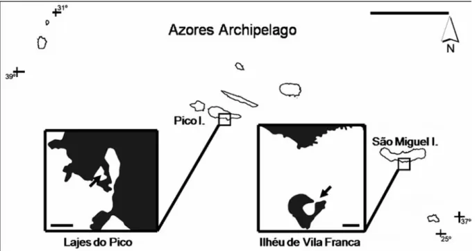
Sampling and field workThe bays of Ilhéu de Vila Franca (São Miguel Island, 37º42’30’’N, 25º26’52’’W, 8000 m2) and Lajes do Pico (Pico Island, 38º23’26’’N, 28º15’06’’W, 5000 m2) are both shallow (2 to 4 m depth) and sheltered (Fig. 1). Their seabed is of sand with sparse rocks.
Sampling was designed to evaluate rhodolith distribu-tion, percent cover, composition and morphology. The total area of rhodolith concentration was measured (length x width) and the maximum and minimum depths within this area were recorded. Sampling took place along four 20 m long parallel transects separated by 10 m. On each transect four points were selected randomly to evaluate rhodolith percent cover using 25 x 25 cm quadrats, following the point intercept method of hawkins & Jones (1992). All rhodoliths inside each quadrat were collected for species identification and morphological characterization.
Laboratory procedures
The collected material was air dried and/or fixed in 4% N-formalin in seawater and incorporated into Phycological herbarium of Departamento de Biologia, Universidade dos Açores, Portugal (AZB).
From each rhodolith collected, permanent slides were made for optical microscopy following the method of
Figure 1. The Azores archipelago (scale bar: 100 km) and location of the study sites (scale bar: 150 m). Figure 1. L’archipel des Açores (Echelle : 100 km), localisation des sites d’étude (Echelle : 150 m).
Riosmena-Rodríguez et al. (1999); fractured fragments were prepared for scanning electron microscopy (SEM) following Woelkerling (1988). Morphological and anatomical observations also followed Woelkerling (1988). For each species, diameter and length measurements were taken from 30 randomly selected epithallial, subepithallial and vegetative cells. Diameter and height or length measurements were taken from 10 randomly selected sporangial chambers and tetrasporangia for each species. All measurements were taken with the software AxionVision LE 4.2 from digital photographs.
The longest (L), shortest (S), and intermediate (I) dimensions of all specimens collected were measured with a Vernier caliper (+/- 0.01 cm) to calculate rhodolith sphericity according to the method of Sneed & Folk (1958). Growth form was recorded and branch density estimated through the number of tips larger than 1 or 2 mm in diameter in five 1 cm2 squares randomly located on the rhodolith surface (Steller & Foster, 1995).
Results
Species account and spatial distributionRhodoliths covered a small area of bays’ seabed, 6.56% (525 m2) at Ilhéu de Vila Franca and 6.12% (306 m2) in Lajes do Pico. Three species were identified: Neogoniolithon brassica-florida (harvey) Setchell et Mason and Lithophyllum crouanii Foslie in Ilhéu de Vila Franca; Phymatolithon calcareum (Pallas) Adey and D.L. McKibbin in Lajes do Pico. Specimens were larger, more numerous and more densely distributed at Ilhéu de Vila Franca than at Lajes do Pico (Table 1). Neogoniolithon brassica-florida (n = 22) was the dominant species at Ilhéu de Vila Franca and Phymatolithon calcareum (n = 7) was the only species present at Lajes do Pico. A single specimen
of Lithophyllum crouanii was seen and collected at Ilhéu de Vila Franca.
All rhodoliths were monospecific and nucleated, with nucleus smaller than 50% of the whole specimen’s volume. Brief species description
Neogoniolithon brassica-florida (harvey) Setchell &
Mason (1943) sensu Penrose (1996). (Table 2, Figs 2 & 3)
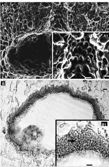
Lumpy growth form (Fig. 2A), ranging from 3.18 to 8.14 cm; a single layer of epithallial cells, 9.04-9.26 (mean = 9.15) μm in diameter and 4.68-4.89 (mean = 4.78) μm in length, with rounded distal walls (Fig. 2B); sub-epithallial cells 8.51-9.15 (mean = 8.83) μm in diameter and 4.36-4.57 (mean = 4.46) μm in length; cell fusions and intercalary trichocytes present (Fig. 2C); internal construction monomerous (Fig. 2D) consisting of a single system of branched, laterally cohering, filaments that collectively contribute to a ventral or central situated core and a peripheral region where portions of core filaments or their derivatives curve outwards towards the thallus surface, with each filament composed of cells 1.26 – 7.54 (mean = 2.74) μm in diameter and 2.39-8.82 (mean = 5.39) μm in length; uniporate conceptacles extremely large and usually visible to the naked eye; tetrasporangial chambers 180-237 μm in diameter and 100-112 μm in height; pore canal lined by several cells oriented more or less parallel to the thallus surface and protruding laterally into the pore canal (Figs 3A, A1); conceptacle roof 8 cells thick; mature tetra -sporangia 28-40 µm in diameter and 40 - 68 µm in length; male chamber 297 μm in diameter and 198 μm in height, spermatangial filaments unbranched, formed on the chamber floor, walls and roof (Figs 3B, B1).
Lithophyllum crouanii Foslie (1898) sensu Chamberlain
et al. (1988) (Table 2 & Figs 4)
Encrusting growth form (Fig. 4A), unbranched, 2.8 cm in size; epithallial cells flattened (Fig. 4B) with 9.51-13.7 (mean = 10.7) μm in diameter and 4.45-6.92 (mean = 5.95) μm in length; subepithallial cells 7.79-11.61 (mean = 9.46) μm in diameter and 6.31-10.5 (mean = 8.83) μm in length; secondary pit connections present (Fig. 4B); internal construction dimerous with postigenous filament cells horizontally aligned (Fig. 4B) measuring 5-8.54 (mean = 6.57) μm in diameter and 5.42-8.97 (mean = 6.94) μm in length; primigenous filaments composed of non-palisade cells (Fig. 4C) 5.83-7.92 (mean = 7.29) μm in diameter and 5.42-12.31 (mean = 8.71) μm in height; tetrasporangial conceptacles uniporate; tetrasporangial chambers globose 80130 μm in diameter and 80100 μm in height; tetra -Table 1 Rhodolith abundance and morphology (mean ±
standard deviation).
Tableau 1. Abondance et morphologie des rhodolithes (moyenne ± écart type).
N. brassica-florida P. calcareum
(Ilhéu de Vila Franca) (Lajes do Pico)
n = 22 n = 7 Rhodolith cover (%) 7.25 ± 1.79 1.50 ± 0.95 Rhodolith density 24.00 ± 8.16 6.00 ± 3.82 (rhodoliths.m-2) Maximum length (mm) 5.10 ± 0.21 1.26 ± 0.73 Branch density (branches.cm-2) 24.00 ± 1.11 1.07 ± 0.74
Species Site Chamber diameter Chamber height Resource information
Neogoniolithon Azores 180 - 237 100 - 112 The present study
brassica-florida Indonesia 650 - 800 175 - 225 Verheij, 1994
Australia 670 - 1050 nd Ringeltaube & harvey, 2000
370 - 600 200 - 360 harvey et al., 2006
590 - 820 370 - 820 Penrose, 1996
Mexican Caribbean 550 - 900 200 - 340 Mateo-Cid & Pedroche, 2004
Lithophyllum Azores 80 - 130 80 - 100 The present study
crouanii Denmark 92 - 116 nd
British Isles
(type specimen) 167 150
British Isles 85 (135) - 180 55 (90) - 120 Chamberlain et al., 1988 120 (140.5) - 161 81 (117.5) - 130
Gulf of Maine 130 (145) - 160 70 (90) - 97
Phymatolithon Azores 274 - 364 170 - 204 The present study
calcareum British Isles 350 nd Woelkerling & Irvine, 1986 230 - 250 117 - 130 Irvine & Chamberlain, 1994 Atlantic coast of France 120 - 250 50 - 60 Irvine & Chamberlain, 1994;
Mendoza & Cabioch, 1998 Atlantic coast of Spain 126 - 190 58 - 89 Adey & McKibbin, 1970
Mediterranean Sea 180 - 245 85 - 140 Basso, 1994
Alaska 80 - 200 80 - 170 Konar et al., 2006
Table 2 Comparative summary of information on tetrasporangial conceptacles for Neogoniolithon brassica-florida, Lithophyllum
crouanii and Phymatolithon calcareum from different geographical areas. Values are given in µm.
Tableau 2. Résumé comparatif d’information sur les conceptacles tétrasporocytes de Neogoniolithon brassica-florida, Lithophyllum
crouanii et Phymatolithon calcareum provenant d’aires géographiques différentes. Les valeurs sont données en µm.
Figure 2. Neogoniolithon brassica-florida, vegeta-tive morphology and anatomy (longitudinal sections: B, C & D). A. Lumpy growth form. B. Rounded distal wall epithallial cells (arrow). C. Intercalar trichocytes (arrow) and cell fusion (double arrow). D. Monomerous growth with a central core (double arrow) and peripheral filaments toward thallus surface (arrow). Scale bar: 1 cm (A) and 10 mm (B-D).
Figure 2. Neogoniolithon brassica-florida,
morphologie et anatomie végétatives (sections longitudinales: B, C & D). A. Forme de croissance grumeleuse. B. Cellules épithéliales de la paroi distales arrondies (flèche). C. Trichocytes intercalaires (flèche) et fusion cellulaire (double flèche). D. Croissance monomérique avec le noyau central (double flèche) et les filaments périphériques vers la surface du thalle (flèche). Echelle : 1 cm (A) et 10 µm (B-D).
sporangial conceptacles roof not protruding above thallus surface and 2 to 4 cells thick; floor of mature chambers usually 6 or more cells below thallus surface; tetra -sporangial conceptacle pore canal lined by cells that project somewhat into the pore canal but do not completely occluded the pore (Fig. 4D); mature tetrasporangia 20-30 µm in diameter and 35-45 µm in length.
Phymatolithon calcareum (Pallas) Adey & D.L.
McKibbin, 1970 (Table 2, Figs 5 & 6)
Lumpy growth form (Fig. 5A), ranging from 1.92 to 3.02 cm; epithallial cells with outermost rounded walls 6.53-9.18 (mean = 7.25) μm in diameter and 5.1-7.21 (mean = 6.17) μm in length; subepithallial cells (Fig. 5B) shorter Figure 3. Neogoniolithon brassica-florida, reproductive
anatomy (longitudinal sections). A. Tetrasporangial conceptacle. A1. Pore canal lined by cells oriented more or less parallel to thallus surface (arrow). B. Male conceptacle. B1. Detail of spermatangial filaments on the chamber roof conceptacle. Scale bar: 10 mm.
Figure 3. Neogoniolithon brassica-florida, anatomie repro-ductrice (sections longitudinales). A. Conceptacle tetrasporangial. A1. Canal de pore entouré de cellules orientées plus ou moins parallèlement à la surface du thalle (flèche). B. Conceptacle mâle. B1. Détail de filaments spermatocystes vers le plafond de conceptacle. Echelle : 10 µm.
Figure 4. Lithophyllum crouanii, vegetative morphology and vegetative/reproductive anatomy (longitudinal sections: B, C & D). A. Encrusting growth form. B. Postigenous filaments cells horizontally aligned, epithallial cells flattened (double arrow) and secondary pit connection (arrow). C. Dimerous construction, with primigenous filaments composed of non-palisade cells (double arrow) and postigenous filaments (arrow). D. Tetrasporangial chamber globose, with floor located more than four cells below thalli surface (arrow) and a pore canal lined by cells oriented more or less parallel to thallus surface (double arrow). Scale bar : 1 cm (A) and 10 µm (B-D).
Figure 4. Lithophyllum crouanii, morphologie végétative, anatomie végétative/reproductrice (sections longitudinales: B, C & D). A. Forme de croissance encroûtante. B. Cellules des filaments postigènes alignées horizontalement, cellules épithéliales aplaties (double flèche) et connection secondaire (flèche). C. Croissance dimérique, avec filaments primigènes pas composées de cellules en palissade (double flèche) et postigènes filaments (flèche). D. Conceptacle avec base située plus de quatre cellules en dessous de la surface des thalles (flèche) et canal de pore entouré de cellules orientées plus ou moins parallèlement à la surface du thalle (double flèche). Echelle : 1 cm (A) et 10 µm (B-D).
than those immediate inward from which they were derived, 4.58-5.32 (mean = 5.32) μm in diameter and 4.62-7.3 (mean = 6.34) μm in length; cells of adjacent filaments linked laterally by cell fusions (Fig. 5C); construction monomerous (Fig. 5D) consisting of a single system of branched, laterally cohering, filaments that collectively contribute to a ventral or central situated core and a peripheral region where portions of core filaments or their derivatives curve outwards towards the thallus surface, with each filament composed of cells 2.5-8.14 (mean = 5.3) μm in diameter and 6-16.92 (mean = 11.21) μm in length; carpogonial conceptacle with mature carpogonial branches (Figs 6A, A1); tetrasporangial conceptacles multiporate (Fig. 6B); tetrasporangial chambers 274-364 µm in diameter and 170-204 µm in height; mature tetrasporangia up to 92.13-96 µm in diameter and 68-72 µm in length.
Figure 5. Phymatolithon calcareum, vegetative morphology and anatomy (longitudinal sections: B, C & D). A. Lumpy growth form. B. Size gradient of the subepithallial cells (epithallial cell -arrow; subepithallial cell - double arrow). C. Cell fusion. D. Monomerous growth with a central core (double arrow) and peripheral filaments toward thallus surface (arrow). Scale bar: 1 cm (A), 10 mm (B, D) and 2.5 mm (C).
Figure 5. Phymatolithon calcareum, morphologie végétative et anatomie (sections longitudinales: B, C & D). A. Forme de croissance grumeleuse. B. Gradient de taille des cellules sub-épithéliales (cellules sub-épithéliales - flèche; cellules subsub-épithéliales- subépithéliales-double flèche). C. Fusion cellulaire. D. Croissance monomérique avec un noyau central (double flèche) et filaments périphériques vers la surface du thalle (flèche). Echelle : 1 cm (A), 10 µm (B, D) et 2.5 µm (C).
Figure 6. Phymatolithon calcareum, reproductive anatomy (longitudinal sections). A. Carpogonial conceptacle. A1. Detail of mature carpogonial branches (trichogyne-arrow). B. Tetrasporangial multiporate conceptacle containing mature tetrasporangia (arrow) with apical plug (double arrow). Scale bar: 10 mm.
Figure 6. Phymatolithon calcareum, anatomie reproductive (sections longitudinales). A. Conceptacle femelle. A1. Détails des rameaux carpogoniaux matures (tricogyne-flèche). B. Conceptacle tétrasporocyte multiporé contenant des tétraspores matures (flèche) avec bouchon apicale (double flèche). Echelle : 10 µm.
Morphology
Rhodoliths of Neogoniolithon brassica-florida and Lithophyllum crouanii were more spherical than those of Phymatolithon calcareum (Fig. 7) and also larger and more densely branched (Table 1).
Discussion
The non-geniculate coralline algae Neogoniolithon brassi-ca-florida, Lithophyllum crouanii and Phymatolithon calcareum are reported for the first time for the Azores archipelago, all growing as rhodoliths.
Despite some differences in cell size, plants collected in the Azores generally correspond to descriptions and
drawings by Adey & McKibbin (1970), Woelkerling & Irvine (1986), Irvine & Chamberlain (1994), Penrose (1996) and Mendoza & Cabioch (1998) for material collected at other locations.
Anatomically Neogoniolithon is much closed to Spongites, the distinction being made only by reproductive features, namely spermatangia position and origin of goni-moblast filaments (Penrose, 1996). The presence in the Azorean specimens of male material allowed a positive determination of Neogoniolithon. Azorean plants posses large conceptacles which protrude conspicuously above the surrounding thallus surface, a feature that according to Penrose (1996) corresponds to N. brassica-florida.
Anatomically Lithophyllum is closed to Titanoderma. Azorean material was identified as Lithophyllum by having primigenous filaments composed of non-palisade cells, following the concept of Lithophyllum sensu stricto (Irvine & Chamberlain, 1994). Distinguishing features of Lithophyllum crouanii from other species of the same genus present in the Atlantic are: i) dimerous internal construction with horizontally aligned postigenous filament cells; ii) globose tetrasporangial chambers; iii) tetra -sporangial conceptacles roof not protruding above thallus surface; and iv) the size of tetrasporangial conceptacles. Lithophyllum corallinae (Crouan & Crouan) heydrich 1897b is the closest species, but has larger tetrasporangial conceptacles and a thallus surface not obviously and more or less completely terraced as a consequence of applanate branch development (see Woelkerling & Campbell, 1992).
Phymatolithon calcareum distinguishes from other species of the genus by having a branched thallus (Irvine & Chamberlain, 1994).
Neogoniolithon brassica-florida is commonly known from the Mediterranean (Mannino et al., 2002), Indian Ocean (Verheij, 1994), Pacific Ocean, Red Sea and S Australia (Penrose, 1996; Ringeltaube & harvey, 2000; harvey et al., 2006). It was recently recorded from the tropical W Atlantic, as Neogoniolithon foslieie (heydrich) Setchell et Mason (Mateo-Cid & Pedroche, 2004). Its occurrence in the Azores represents the first record for this species in this archipelago, the Macaronesian region, and the NE Atlantic Ocean. It may be more widely distributed in the area.
Lithophyllum crouanii is widely distributed throughout the N Atlantic (Irvine & Chamberlain, 1994) and for Canary Islands (Afonso-Carrillo & Sansón, 1999). Its occurrence in the Azores is within the overall distributional range of the species. however, this species is here reported for the first time as a rhodolith forming species.
Phymatolithon calcareum is widely distributed through-out the NE Atlantic: the British Isles (Woelkerling & Irvine, 1986), Ireland (De Grave et al., 2000), France (Mendoza & Cabioch, 1998) and Spain (Adey & McKibbin, 1970; Peña Figure 7. Rhodolith sphericity, Neogoniolithon
brassica-florida (l), Lithophyllum crouanii (+) and Phymatolithon calcareum (*). Each point is the sphericity of one rhodolith (some
times more than one rhodolith, when the index values were the same) (N. brassica-florida, n = 22; L. crouanii, n = 1; P.
calcareum, n = 7). S, I and L correspond to shortest, intermediate,
and longest dimensions (cm) of each individual. Equations shown were used to determine placement along each respective axis. Plants nearest the apex are most spheroidal.
Figure 7. Sphéricité des rhodolithes, Neogoniolithon
brassica-florida (l), Lithophyllum crouanii (+) et Phymatolithon calcareum (*). Chaque point est la sphéricité d’un rhodolithe
(parfois plus d’un rhodolithe, quand les valeurs de l’indice sont les mêmes) (N. brassica-florida, n = 22; L. crouanii n = 1; P.
calcareum n = 7). S, I et L correspondent aux dimensions les plus
courtes, intermédiaires et les plus longues (en cm) de chaque individu. Les équations montrées ont été utilisées pour déterminer les positions le long de chaque axe respectif. Les algues les plus proches de l’apex sont les plus sphériques.
& Bárbara, 2008), in the Mediterranean, e.g. Italy (Basso, 1998) and in the NW Pacific (Konar et al., 2006), both intertidal and subtidally. It has been reported as a rhodolith-forming species for the Canary and Madeira Islands (Afonso-Carrillo et al., 1985). Its occurrence in the Azores is within the overall distributional range of the species.
Interestingly, Lithothamnion corallioides (P. and h. Crouan) P. and h. Crouan 1867, reported for Cape Verde, Canary and Madeira Islands, and recorded as rhodolith-forming species in Madeira (Cabioch, 1974) and in the Canary Islands (Afonso-Carrillo & Sansón, 1999) was not found in the Azores.
The number of rhodolith-forming species in the Atlantic and Mediterranean is variable, ranging from twelve in the NE Atlantic (Adey & McKibbin, 1970; Irvine & Chamberlain, 1994; Adey et al., 2005), six species in the Mediterranean (Basso et al., 1996; Basso, 1998), a single species in the NW Atlantic (Bird & Mc Lachlan, 1992) and three species in the tropical NW Atlantic (Littler & Littler, 2000). In the Azores, the three species reported were not abundant and had a sparse distribution (below 7%) when compared maërl facies or rhodolith beds reported on other locations, e.g. Spain (100%, Peña & Bárbara, 2008), Ireland (5- 50%, De Grave et al., 2000). According to the nomenclature of Steller et al. (2003), rhodolith habitats in the Azores should thus be designated as sand with rhodolith aggregations. The studied sites are very sheltered, which might explain the low abundance of rhodoliths in agreement with Marrack (1999) that suggests water motion as a crucial factor for maintenance of rhodoliths.
Azorean rhodoliths are predominantly lumpy, although differences were observed on rhodolith size and shape. Neogoniolithon brassica-florida rhodoliths were predominantly spheroid, larger and more densely branched than those of Phymatolithon calcareum, which were predominantly ellipsoid. Variations in sphericity are associated with water motion (Bosence, 1991), sediment grain size (Bosence, 1983), depth (Steller & Foster, 1995) and species internal structure and growth patterns (Sneed & Folk, 1958; Basso et al., 2009). Since both studied rhodolith habitats in the Azores are similar in terms of hydrodynamics and depth, it is likely that the differences in rhodolith shape are more related to internal factors such as growth patterns, physiology and type of nucleus.
Phymatolithon calcareum is listed in Annex V of the EU habitats Directive (Council Directive 92/43/EEC) which states that exploitation should be compatible with the maintenance of a favourable conservation status (De Grave et al., 2000). Although exploitation of this species in the Azores is unlikely, conservation measures should apply since its distribution is known only from a single bay at Lajes do Pico.
Acknowledgements
The authors wish to thank Drs. Julio Afonso-Carrillo, José M. N. Azevedo, Francisco Wallenstein, Gustavo Martins and Nuno Álvaro for general discussions and help with data treatment; Dr. Jorge Medeiros for help with the electronic microscopic observations; Camille Fontaine for helping with the French version of the abstract and Figure captions; the anonymous reviewers and Dr. Daniela Basso (Univ. Milano-Bicocca); Dr. Ian Tittley and Francisco Wallenstein for the English revision of the manuscript. This work was partly supported by CIRN (Centro de Investigação de Recursos Naturais; University of the Azores). Edgar Rosas-Alquicira was supported by the Programme AlBan, the European Union Programme of high Level Scholarships for Latin America (through scholarship E05D060221MX) and CONACYT doctoral scholarship 176162. Ruben Couto was supported by a PhD Grant (M3.1.1/I/014A/2005) from DRCT (Direcção Regional da Ciência e Tecnologia). Rafael Riosmena-Rodríguez acknowledges the support of SEMARNAT CONACYT for histological work. The surveys performed in the present study comply with the current laws of Portugal.
References
Adey W.H. & McKibbin D. 1970. Studies on the maërl species
Phymatolithon calcareum (Pallas) nov. comb. and
Lithothamnion corallioides Crouan in the Rio de Vigo. Botanica Marina, 13: 100-106.
Adey W.H., Chamberlain Y.M. & Irvine L.M. 2005. An SEM-based analysis of the morphology, anatomy, and reproduction of Lithothamnion tophiforme (Esper) Unger (Corallinales, Rhodophyta), with a comparative study of associated North Atlantic Arctic/Subarctic Melobesioideae. Journal of
Phycology, 41: 1010-1024.
Afonso-Carrillo J., Gil-Rodríguez M.C. & Wildpret De la Torre W. 1985. Algunas consideraciones floristicas, corológi-cas y ecológicorológi-cas sobre las algas Corallinaceae (Rhodophyta) de las islas Canarias. Anales de Biología de la Universidad de
Murcia, 2: 23-37.
Afonso-Carrillo J. & Sansón M. 1999. Algas, hongos y
fan-erógamas marinas de las Islas Canarias. Clave analítica.
Materiales Didácticos Universitarios. Serie Biología / 2. Tenerife. 254 pp.
Basso D. 1994. Study of living calcareous algae by a paleontolog-ical approach: the non-geniculate Corallinaceae (Rhodophyta) of the soft bottoms of the Tyrrhenian Sea (Western Mediterranean). The genera Phymatolithon Foslie and
Mesophyllum Lemoine. Rivista Italiana di Paleonotologia e Stratigrafia, 100: 575-596.
Basso D. 1998. Deep rhodolith distribution in the Pontian Islands, Italy: a model for the paleoecology of a temperate sea.
Palaeoegeography, Palaeoeclimatology, Palaeoecology, 137:
Basso D., Fravega P. & Vannucci G. 1996. Fossil and living corallinaceans related to the Mediterranean endemic species
Lithophyllum racemus (Lamarck) Foslie. Facies, 35: 275-292.
Basso D., Nalin R. & Nelson C. S. 2009. Shallow-water
Sporolithon rhodoliths from North Island (New Zealand). Palaios, 24: 92-103.
Bird C.J. & Mc Lachlan J.L. 1992. Seaweed flora of the
Maritimes. 1. Rhodophyta - Red Algae. Biopress: Bristol.
177 pp.
Bosence D.J. 1983. The ocurrence and ecology of recent rhodoliths- a review. In: Coated Grains (T.M. Peryt ed), pp. 225-242. Springer-Verlag: Berlin.
Bosence D.J. 1991. Coralline algae: mineralization, taxonomy, and palaeocology. In: Calcareous algae and stromatolites (R. Riding ed), pp. 98-113. Springer-Verlag: Berlin.
Cabioch J. 1974. Un fond de maërl de l’Archipel de Madère et son peuplement végétal. Bulletin Society of Phycology, 19: 74-82. Chamberlain Y.M., Irvine L.M. & Walker R.L. 1988. A
redescription of Lithophyllum crouanii (Rhodophyta, Corallinales) in the British Isles with and assessment of its relationship to L. orbiculatum. British Phycological Journal, 23: 177-192.
De Grave S., Fazakerley H., Kelly L., Guiry M.D., Ryan M. & Walshe J. 2000. A Study of selected maërl beds in Irish waters
and their potential for sustainable extraction. Project Report. Marine Research Measure - European Union’s Regional Development Fund. Ireland. 50 pp.
Harvey A.S., Phillipis L.E., Woelkerling W.J. & Millar A.J.K. 2006. The Corallinaceae, subfamily Mastophoroideae (Corallinales, Rhodophyta) in south-eastern Australia.
Australian Systematic Botany, 19: 387-429.
Hawkins S.J. & Jones H.D. 1992. Rocky shores (Marine
Conservation Society, Marine Course Field Guide 1). Immel
Publishing: London. 144 pp.
Irvine L.M. & Chamberlain Y.M. 1994. Seaweeds of the British
Isles. Vol. 1 Rhodophyta Part 2B Corallinales,
hildenbrandiales. hMSO: London. 276 pp.
Konar B., Riosmena-Rodríguez R. & Iken K. 2006. Rhodolith bed: a newly discovered habitat in the North Pacific Ocean.
Botanica Marina, 19: 355-359.
Littler D.S. & Littler M.M. 2000. Caribbean Reef Plants. Off Shore Graphics: Washington. 542 pp.
Mannino A.M., Castriota L., Beltrano A.M. & Sunseri G. 2002. The epiflora of a rhodolith bed from the Island of Ustica (Southern Tyrrhenian Sea). Flora Mediterranea, 12: 11-28. Marrack E.C. 1999. The relationship between water motion and
living rhodolith beds in the southwestern Gulf of California, Mexico. Palaios, 14: 159-171.
Mateo-Cid L.E. & Pedroche F.F. 2004. The occurrence of
Neogoniolithon fosliei (heydrich) Setchell et Mason in the
Mexican Caribbean and the relationship of this species to N.
solubile (Foslie et howe) Setchell et Mason (Corallinales,
Rhodophyta). Caribbean Journal of Science, 40: 182-191. Mendoza M.L. & Cabioch J. 1998. Étude comparée de la
repro-duction de Phymatholithon calcareum (Pallas) Adey & McKibbin et Lithothamnion corallioides (P. & h. Crouan) P. & h. Crouan (Corallinales, Rhodophyta), et reconsiderations sur la définition des genres. Canadian Journal of Botany, 76: 1433-1445.
Neto A.I. 1994. Checklist of the benthic marine macroalgae of the Azores. Arquipélago: Boletim da Universidade dos Azores, 12A: 15-34.
Penrose D.L. 1996. Genus Neogoniolithon. In: The Marine
ben-thic flora of southern Australia. Rhodophyta. Part IIIB, Gracilariales, Rhododymeniales, Corallinales and Bonnemaisoniales (h.B.S. Womersley), pp. 280-283.
Australian Biological Resources Study: Canberra.
Peña V. & Bárbara I. 2008. Maërl community in the north-western Iberian Peninsula: a review of floristic studies and long term changes. Aquatic Conservation: Marine and
Freshwater Ecosystems, 18: 339-366.
Ringeltaube P. & Harvey A.S. 2000. Non-geniculate coralline algae (Corallinales, Rhodophyta) on heron Reef, Great Barrier Reef (Australia). Botanica Marina, 43: 431-454.
Riosmena-Rodríguez R., Woelkerling W.J. & Foster M.S. 1999. Taxonomic reassessment of rhodolith-forming species of
Lithophyllum (Corallinales, Rhodophyta) in the Gulf of
California, Mexico. Phycologia, 38: 401-417.
Sneed E.D. & Folk R.L. 1958. Pebbles in the lower Colorado River, Texas, a study in particle morphogenesis. Journal of
Geology, 66: 114-150.
Steller D.L. & Foster M.S. 1995. Environmental factors influ-encing distribution and morphology of rhodoliths in Bahía Concepción, B.C.S., México. Journal of Experimental Marine
Biology and Ecology, 194: 201-212.
Steller D.L., Riosmena-Rodríguez R., Foster M.S. & Roberts C.A. 2003. Rhodolith bed diversity in the Gulf of California: the importance of rhodolith structure and consequences of disturbance. Aquatic Conservation: Marine and Freshwater
Ecosystems, 13: S5-S20.
Verheij E. 1994. Nongeniculate Corallinaceae (Corallinales, Rhodophyta) from the Spermonde Archipelago, SW Sulawesi, Indonesia. Blumea, 38: 95-137.
Woelkerling W.J. 1988. The Coralline Red Algae: An Analysis of
the Genera and Subfamilies of Nongeniculate Corallinaceae.
British Museum (Natural history) and Oxford University Press: London and Oxford. xi + 268 pp.
Woelkerling W.J. & Campbell S.J. 1992. An account of southern Australian species of Lithophyllum (Corallinaceae, Rhodophyta). Bulletin of the British Museum (Natural History)
Botanica, 22: 1-107.
Woelkerling M.J. & Irvine L.M. 1986. The typification and status of Phymatolithon (Corallinaceae, Rhodophyta). British