Relatório Final de Estágio
Mestrado Integrado em Medicina Veterinária
EPIDEMIOLOGICAL EVALUATION OF MASTITIS CAUSED BY
STREPTOCOCCUS AGALACTIAE AND STREPTOCOCCUS UBERIS IN
FOUR DAIRY HERDS IN NORTH OF PORTUGAL
Marta Sofia Morais Vales
Orientador
Professor Doutor João José Rato Niza Ribeiro
Co-Orientadores
Professor Doutor Fernando Manuel dos Santos Tavares Dra Helena Larisma Madeira
This work was performed in collaboration with:
i
ABSTRACT
Bovine mastitis is one of the most prevalent and costly farm animals diseases that is widely spread all over the world, and a reason for concern for farmers and veterinarians, since it represents a great impact on cow welfare and in economic losses. Among all the pathogens that can cause mastitis, Streptococcus agalactiae and Streptococcus uberis are the most
relevant. The main goal of this study was to analyze S.agalactiae and S.uberis isolates obtained from cows, that presented recurring mastitis infections. These cows were assayed at different time points in order to assess the molecular epidemiology of mastitis-causing pathogens.
Bacterial virulence genotypes were found to be associated with mammary infections. For this reason, several virulence markers were tested with the collected isolates. For the putative
S.agalactiae isolates the following markers were tested: the fructose operons FO1 and FO3; the
toxin production CAMP factor; and the gene responsible for the adherence to epithelium, fbsB. The results obtained showed that a large percentage of S.agalactiae isolates contained these virulence factors in their genome. For the putative S.uberis isolates the following markers were chosen for testing: the gene responsible for the adherence to epithelium in this species, sua; the bacteriocin nisin U; the plasminogen activator pauA; the toxin production factor CAMP; and an antimicrobial resistance gene ermB. With the exception for the CAMP factor, all these genes were shown to be present in the large majority of of S.uberis isolates assayed.
This work strengthens the hypothesis from previous studies concerning the high consistency and stability of these markers when used for identification of streptococci isolates. The obtained results suggested that there is a high prevalence of mastitis in the selected dairy herds, and that there are specific virulence patterns which are common amongst successful mastitis- causing pathogens. However, it was noticeable that pathogens with distinct virulence patterns were also able to cause recurring infections, meaning that infections apparently caused by atypical
streptococci were also successful.
Keywords: Antibiotics resistance; Bovine mastitis; DNA markers, Dot-Blot hybridization; Streptococcus agalactiae; Streptococcus uberis; Epidemiology; Virulence factors.
ii
AGRADECIMENTOS
No findar de mais uma jornada académica, não poderia deixar de agradecer a todos aqueles que dela fizeram parte e comigo partilharam tantas emoções.
Antes de mais, gostaria de agradecer ao meu orientador, Professor Doutor Niza Ribeiro pela sua ajuda e disponibilidade.
Ao Professor Doutor Fernando Tavares pela sua constante supervisão, atenção, preocupação, e sentido de justiça que manifestou durante todo o meu estágio, pois apesar de todas as suas responsabilidades, sempre se disponibilizou a ajudar-me e a transmitir-me conhecimentos.
À Dra Helena Madeira pela sua simpatia e amabilidade em me acompanhar e auxiliar durante as visitas às explorações, bem como a todos os veterinários da SEGALAB pela forma como me receberam e auxiliaram ao longo do estágio. Um agradecimento especial á Engª. Paula Santos pela disponibilização dos isolados necessários para este estudo.
Um agradecimento para todo grupo MDE, em particular á Cristina, á Eduarda e á Cláudia, pois são sem dúvida pessoas fantásticas, esta minha “estadia” no laboratório não teria sido igual sem vocês. Um agradecimento muito especial ao Pedro pela sua grande paciência, calma, simpatia e por todo o auxílio prestado, quer no laboratório quer na escrita.
Ao Afonso Porfírio, pela calma e paciência que teve para me ensinar as técnicas e protocolos utilizados neste trabalho.
E porque a bancada deve ser partilhada, á Francisca Diniz, que se tornou sobretudo uma grande amiga, partilhámos angústias, receios, nervos mas sobretudo muitas gargalhadas, alegrias e músicas. Desejo-te toda a sorte do mundo e que sejas muito feliz, quer a nível pessoal quer a nível profissional.
Aos professores que marcaram a diferença no meu caminho, Pablo Payo, Paulo Costa, Ana Patrícia Sousa, Miguel Faria, Luís Ataíde, Carla Mendonça e Eduarda Neves.
Às minhas grandes amigas e “membras” da T.A.A.B, Cali, Yana, Jeitosa e Popota, sem vocês não teria sido tão divertido e inesquecível, mas também não posso deixar de agradecer às não “membras” mas igualmente importantes Jordana, a pedagógica; Paula, a partenere e Rita, a amável; o meu muito obrigado às melhores amigas que podia ter tido, por todos os momentos fantásticos que me proporcionaram.
A minha irmã de coração que adoro, Diana, cuja amizade começou sem eu dar por ela, mas que foi crescendo cada vez mais até que nos tornou inseparáveis, por sempre me apoiar e animar nos momentos em que estava mais em baixo, será certamente uma amizade para toda a vida.
iii
Aos meus avozinhos, que sempre me mimaram, me acompanharam na minha infância e adolescência, que sempre me transmitiram educação e valores morais dos quais não abdico, e que agora apesar de estarem acamados, com avançada idade e sem muita lucidez, sei que teriam orgulho em mim, como sempre tiveram.
Aos meus paizinhos, que são os pilares da minha vida, sem eles não seria nada do que sou hoje, agradeço por estarem sempre presentes, nos bons e nos maus momentos, pelo seu esforço e dedicação para me tornarem na mulher que hoje sou, por nunca me terem deixado faltar nada, por me amarem e fazem tudo por mim, são sem dúvida os melhores pais do mundo.
E por último, mas não menos importante, ao amor da minha vida, Misael, pelo seu constante apoio e incentivo, pela paciência, quando nas minhas horas intermináveis de estudo se sentava ao meu lado só para estarmos juntos, por todos os mimos, por nunca duvidar das minhas capacidades mesmo quando eu própria duvidava, por me dar sempre força, ânimo e pelo seu bom humor que tantas vezes alegrou os meus dias, amar-te-ei para sempre.
“E em ti confiarão os que conhecem o teu nome; porque tu, Senhor, nunca desamparaste os que te buscam”. Salmos, Cap.9; Versículo 10.
iv
LIST OF ABBREVIATIONS
CAMP - Cyclic adenosine monophosphate CMT- Californian milk test
DNA Deoxyribonucleic acid
fbsA- Fibrinogen-binding protein A fbsB - Fibrinogen-binding protein B LF- Lactoferrin
Fg – Fibrinogen
LGT- Lateral gene transfer
GAPDH - Glyceraldehyde-3-phosphate dehydrogenase GBS - Group B Streptococcus
opps - Oligopepide permeases PCR - Polymerase Chain Reaction PMN - Polymorphonuclear neutrophils
PTS- Phosphoenolpyruvate-dependent phosphotransferase system rRNA - Ribosomal ribonucleic acid
SSC - Somatic cell count
v
INDEX
ABSTRACT ... i AGRADECIMENTOS ... ii LIST OF ABBREVIATIONS ... iv INTRODUCTION ... 1 1. Bovine mastitis... 1 2. Streptococcus ... 2 2.1. Streptococcus agalactiae ... 2 2.2. Streptococcus uberis ... 3 3. Mastitis resistance ... 44. Bacterial tools to successfully infect the host ... 5
4.1. Invasiveness patterns.. ... 6
4.2. Virulence patterns...,,... 6
5. Mastitis control programs………... ... 8
6. The role of molecular biology in veterinary epidemiology ... 9
7. Main goals to achieve……….. 10
MATERIAL AND METHODS ... 10
1. Milk samples and bacterial isolates ... 10
2. Bacterial culture conditions and DNA extraction ... 11
3. PCR amplification ... 12
4. Dot Blot screening ... 13
5. CAMP test………...14
RESULTS…….. ... 15
1. Mastitis frequency in herds ... 15
2. Preliminary tests……….. ... 17
vi
3.1. Taxonomic markers ... 17
3.2. Virulence markers ... 18
3.3. Antibiotics resistance markers... 23
4. Genetic variation………..…...24
5. Search for pathogens in cow`s environment………...27
DISCUSSION ... 27
1. Mastitis in herds ….. ... 27
2. Molecular methods for bacterial detection…. ... 28
3. Future perspectives... 30
APPENDIX ... I
1. References ... I 2. Tables ... VII
1
INTRODUCTION
1.Bovine Mastitis
Bovine mastitis is a disease that affects dairy herds worldwide and is a main concern for both farmers and veterinarians concerning animal welfare and economic losses. (Koskinen et
al., 2009; Manthe, 2014; Reinoso et al., 2011; Taponen et al., 2009; Tomita et al., 2007). The
disease consists in an inflammation of the mammary gland mostly due to the invasion of pathogenic microorganisms through the teat canal. Any kind of trauma or irritants can damage the skin around the teat, which contributes to break the first line of defense against external pathogens (Almeida et al., 2013). When pathogens enter the teat canal, they start to produce toxins, enzymes and surface proteins to promote adherence to the host tissue (Almeida et al., 2013). These aggressions cause an inflammatory response characterized by an increase of somatic cells count (SCC), with release of interleukins, polymorfonuclear neutrofiles (PMN), phagocytes and leukocytes to fight against this aggression. In case of inflammation, around 95% of cells present are PMN (Schukken, 2004).These inflammations interfere with the milk production and quality, reducing its economic value.
Depending on the range of clinical signs, mastitis can vary from subclinical to clinical presentation, which also depends on the virulence of the pathogens and the host´s immune system ability to respond to invasion (Erskine, 2001). Regarding the reservoirs of pathogenic agents, these microorganisms can be divided in two categories depending on their behavior: contagious, if the microorganism spreads from cow-to-cow through fomites namely dirty or spoiled teat cups, milker`s hands or even milking machines, and environmental, if the infection occurs due to ubiquitous organisms, which are found in the animal housing environment. (Reinoso et al., 2011). Streptococcus agalactiae, Staphylococcus aureus, Streptococcus
dysagalactiae and Mycoplasm are considered contagious agents, commonly causing longer
and more prevalent infections. (McDonald,1979). Streptococcus uberis, Escherichia coli,
Klebsiella, spp and Enterobacter, spp are considered environmental agents and usually cause
more severe cases of clinical mastitis (McDonald, 1979). The above-mentioned bacteria belong to a group of major pathogens which cause the greatest milk alterations and economic losses. Coagulase-negative staphylococci and Corynebacterium bovis are examples of minor pathogens, usually not associated with large changes in milk parameters (Harmon, 1994). However recent studies suggest that the presence of these minor pathogens can increase the risk of intramammary infection by major pathogens (Reyer et al.,2012; Zadocks et al., 2001).
2
2.Streptococcus
Streptococcus are gram-positive bacteria with particular traits such as being
catalase-negatives, facultative anaerobes and lactic acid producers. (Facklam,2002). These bacteria are usually commensal organism in mammalian skin membranes, such as the mucosa and skin surface. However, some behave as opportunistic bacteria and may cause infection if the host defense barrier is broken (Almeida et al, 2013, Porfírio, 2013).
Although many streptococci have been identified as mastitis causative agents,
Streptococcus agalactie and Streptococcus uberis are generally regarded as the most
significant pathogens (Jones and Bailey, 2009; McDonald et al., 2005; Kuang et al., 2009; Unnerstad et al., 2009).
2.1. Streptococcus agalactiae
Streptococcus agalactiae is a pyogenic and contagious agent which causes mostly
subclinic and also mild to moderate mastitis in ruminants. In general, cows infected with this pathogen present more than one affected quarter. These bacteria do not survive for long periods in the environment, nevertheless they can persist for undetermined time in mammary glands, if good practices are not implemented (Harmon,1994; Martinez et al., 2000; Ruegg,2005; Wilson et al.,1997).
Streptococcus agalactiae belongs to Group B Streptococcus (GBS), which is a
Lancefield´s classification for hemolytic streptococci. (Radtke et al.,2010). These opportunistic bacteria are able to cause disease in humans, both adults and children preferentially in pregnant women, diabetics, older people or immunocompromised patients. S.agalactiae, which is commonly found in genitourinary and gastrointestinal tracts, could also cause septicemia, meningitis and pneumonia in neonates, breast abscess formation and endocarditis in women, and soft tissue and osteoarticular infections in elderly adults (Glaser et al.,2002; Haguenoer et
al.,2011; Radtke et al., 2010; Zadocks & Fitzpatrick, 2009; Zadocks et al.,2011).
Several epidemiological and molecular studies have been conducted to analyze the persistence, transmission routes and reservoirs of human and cattle strains. Two hypotheses for the relation between these two groups of strains have been proposed, one postulates that, there is a direct transmission between cattle and humans and the other states that human strains evolved from bovine sources (Zadocks et al. 2011).
In herds with a good health management plan, it is possible to completely eliminate
Streptococcus agalactiae through the implementation of specific measures, namely prevent the
3
Additionally, these infections can be permanently eliminated with intramammary or injectable therapy (Keefe,1997).
Infections cause by S.agalactiae decrease milk production and modify their parameters. In fact, a milk free of mastitis must have bellow 200x10e3 somatic cells per milliliter, and any increase of SCC in the bulk tank lead to pecuniary penalizations for the farmers (Jones & Bailey, 2009).
2.2.Streptococcus uberis
Streptococcus uberis is one of the most important causative agents of mastitis all over
the world. As an environmental pathogen, S.uberis are spread to uninfected cows throuhgt their contact with contaminated surfaces in the animal housing (Peterson-Wolfe & Currin, 2012; Pryor et al.,2009; Tassi et al.,2013; Zadocks et al.,2001; Zadocks et al., 2005).
This pathogen causes subclinical and clinical mastitis in lactating and non-lactating cows, with more emphasis in the dry period, calving and early lactation. There is an increased risk for new infections in the dry period and for cows in the lactating early period due to stress and postpartum low immunity (Oliver et al., 2004; Peterson-Wolfe & Currin, 2012; Zadocks et al., 2003). Due to its ubiquitous nature, S. uberis is a very prevalent species, however the knowledge about its epidemiology is still incomplete (Phuektes et al., 2001; Reinoso et al., 2011; Zadock et al., 2003).
These bacteria can be found in many surfaces such as the bovine udder, excrements or bedding, therefore maintaining a clean and dry environment is very important to prevent the infection. Additionally chronic subclinical mastitis infection could also serve as a reservoir of contamination for healthy cows (Peterson-Wolfe & Currin, 2012; Zadocks et al., 2003), and many studies report that in some herds cow-to-cow transmission can be a vector of infection (Phuektes et al.,2001;McDougall et al., 2004; Zadocks et al., 2001). However in the animals which are not milked, like dry cows and preparturient heifers, this kind of transmission cannot be considered, making the environment the most likely source of infection (Zadocks et al., 2003). Bacterial typing studies revealed high serologically and biochemically heterogeneity in S. uberis ( Zadocks & Fitzpatrick, 2009).Generally the persistence of infection and treatment failures can be attributed to adherence and invasion factors and and to this capacity to survive in mammary epithelial cells (Bentley et al., 1993; Zadocks & Fitzpatrick, 2009).
Regarding treatment, long lasting antibiotics are nowadays widely used to control the risk of mastitis. Nevertheless, some studies refer that the use of an internal teat sealant and antibiotics treatments at dry off period could reduce the rate of new infections (Peterson-Wolfe & Currin, 2012).
4
Application of good management practices in herds have been successfully employed in order to reduce the effect of contagious agents, however, their success is limited towards controlling mainly environmental agents like Streptococcus uberis (Pitkälä et al., 2008; Tomita
et al., 2007).
3.Mastitis Resistance
To prevent possible entries from pathogens, cow´s udders have some strategies of defense. The first one, which prevents the entry of pathogenic agents in the mammary gland, is the teat canal that provides a physical barrier and the sphincter muscle (within the teat canal) that helps to expel out this microorganisms through washing-out effect of milking and epithelial desquamation (Sandholm et al., 1995). Studies show that a higher ratio of milk flow leads to less probability of mastitis infections, mostly in the case staphylococcal and streptococcal infections. The size of the teat canal and strength of the sphincter muscle are crucial to prevent the bacteria from reaching the mammary gland cistern. The medium diameter of the teat canal is between 2.3 and 5mm, therefore there is an increased risk of mastitis in older cows as they have more dilated teat canals. Shorter teats milked faster and fuller and avoiding residual milk, therefore, decreasing the susceptibility to infection. Since the conformation of udders is heritable, and that deep and pendulous udders have more predisposition to higher SCC and mastitis (Seykora & McDaniel, 1985), extra care must be taken at the end of milking because the teat canal remain open for at least two hours, which contributes to an increase of ascending infections (Jones & Bailey, 2009; Sharif et al.,2009).
Keratin, a fibrous protein with bacteriostatic properties, is present in lining cells of the teat, which greatly contributes to slow the possible progression of pathogens. Therefore, trauma or lacerations in the teat must be avoided, as they damage the cells producing keratin and mucous, which increases the risk of bacterial colonization (Jones & Bailey, 2009). One study found that there is about three times more prevalence of new infections by Streptococcus
agalactiae in quarters with acute teat end lesions than in healthy or small lesion quarters. A well
planned milking is a good practice in order to avoid teat trauma and further hyperkeratosis. Moreover, care should be taken, to not milk a dry quarter, as in addition to the improper stimulation of the animal, overmilking can predispose to erosion of the orifice and cause severe lesions which may lead to persistent subclinical mastitis (Neijenhuis et al., 2001; Seykora & McDaniel, 1985). The second line of defense, consists in the immunological properties of milk, which is not very favorable to the growth of bacteria.
Genetics factors, udder health and lactation stage can interfere in the concentrations of important antibacterial enzymes and other molecules present in milk such as lactoperoxidase, lysozyme, lactoferrin, transferrin, immunological defense mechanisms and the complement
5
system (Sandoholm et al.,1995). In case of inflammation, many mediators migrate through circulatory system to quell the infection, such as prostaglandins, leukotrienes, interleukines (IL17), lympocytes, neutrophyles, phagocytes,PMN leukocytes, major histocompatibility class I and II and toll-like receptors (Tassi et al., 2013; Zadocks & Fitzpatrick, 2009). However it should be noted that PMN leukocytes and phagocytes are attracted in a wide number to milk which causes an increase of somatic cells count (SCC). Moreover, substances released by PMN destroy the alveolar structure that upon healing turns into cicatrization tissue, which can be problematic for antibiotic treatment (Jones & Bailey,2009).
Specific cow characteristics influence the onset of mastitis and increase the duration of clinical mastitis namely genetics, parity,age, negative energetic balance, immune status, nutritional status, deficiencies of some components of diet like selenium, E vitamin, α or β carotene (Abureema et al., 2014; Hagnestam-Nielsen et al., 2008; Harmon,1994; Zadocks et
al.,2001).
Lactoferrin and transferrin are two important iron binding proteins that are part of antibacterial defense, preventing bacterial growth by competition against bacterial iron uptake. In case of mastitis, their concentration in milk increases considerably. Lactoperoxidases are enzymes responsible for the oxidation of bacterial cell wall componentes. These enzymes are regulated by the concentration of hydrogen peroxide which formation is inhibited by the low oxygen pressure in case of mastitis.
Lysozyme is an antimicrobial enzyme, that regardless their small concentration in milk, causes the hydrolysis of the bacterial cel wall causing the osmotic lysis of bacteria (Sandholm et
al., 1995).
Altogether these studies show that in case of infection, milk composition is altered. An increase in vessels permeability and SCC and, a decrease of some components such as lactose, α-lactalbumin, fat, casein and calcium translate in an impairment of milk quality (Harmon, 1994).
4.Bacterial tools to successfully infect the host
From the moment bacteria invades the teat canal and reach the milk cistern, a complex process of adaptation is started. In general, when the udder´s defense is broken this is due to several bacteria species. These bacteria compete with each other seeking dominance to successfully infect the host. To accomplish this task, many virulence factors are induced in order to enhance the host invasion, i.e., to resist to the immune response from the host; to grown in milk; to adhere to the mammary epithelial cells; and to resist phagocytosis (Pryor et
al.,2009). Therefore, an in depth analysis of virulence factors allows for an enhanced
6
Bacteria with a good adherence and invasion capacity are more fit to ensure their spread in the host, therefore their virulence aptness is increased when these factors are present (Sandholm et al., 1995). Not surprisingly, bacteria production of surface adhesins, like fibrinogen, is extremely valuable at the early stages of infection, as they allow to counteract the flushing effect during milking or the phagocytic action, both of which, constitute barriers to bacterial propagation (Patti et al., 1994).
4.1 Invasiveness patterns
In S.agalactiae, several families of fibrinogen binding proteins (Fb) have been studied, namely the fibrinogen binding protein A (FbsA) and fibrinogen binding protein B (FbsB) (Devi et al., 2010; Porfírio, 2013). FbsA promotes binding to the human Fb, and FbsB has a terminal region that binds only in bovine Fb. Binding is increased by calcium, which is present in milk (Devi et al., 2010; Jacobsson et al., 2002; Porfírio,2013).
In S. uberis, Sua is an adhesion molecule with high affinity to lactoferrin (Almeida et al., 2006). Lactoferrin (LF) is an iron binding glycoprotein present in milk, known for their bacteriostatic, bactericidal, antifungal, antiviral, antitumural and immunomodulating properties, which takes part in the innate immune response. LF is synthesized by glandular epithelium cells in the mammary gland and studies show that infected quarters present higher concentrations of LF. Additionally, it is reported that, S uberis can induce LF synthesis in milk enhancing its resistance to lactoferrin activity, which confers an advantage over competing bacteria (Chaneton et al., 2008).
Pathogens with an hyaluronic capsule, are protected from host defence to some extent, because neither antibodies or complement factors can attach to the bacteria surface and perform the recognition before phagocytes (Sandholm et al., 1995; Ward et al.,2001). Three genes are involved in the production of a hyaluronic acid capsule: hasA, hasB and hasC. (Ward et al., 2001). In S.uberis, the capsule production requires the presence of both hasA and hasC genes (Field et al., 2003), however, studies refer that in S.uberis the presence of an hyaluronic acid capsule is not essential for a successful infection (Ward et al., 2009).
4.2- Virulence patterns
Streptococcus agalactiae has the ability to quickly adapt to adverse conditions in the
host, such as osmolarity, pH, starvation, temperature variations and oxidative stress, through the synthesis of proteins that act as proteases and chaperones.
For their growth, bacteria need to import some carbon sources, and in S. agalactiae the sugar specific phosphoenolpyruvate-dependent phosphotransferase system (PTS) enzyme II
7
complexes like lactose, fructose, glucose or mannose can be used for this purpose (Glaser et al., 2002).
In the absence of the primary source of carbon (carbohyrdrates), lactose and fructose can help to supply energy, however, only bovine strains present this fructose utilization pathway. In the utilization of fructose, a four-gene operon is involved: the phosphotransferase system fructospecific IIA componente (fruD), the fructose-specific IIBC PTS component (fruC), the fructose-1-phosphate kinase (fruP) and a transcriptional regulator (fruR) (Richards et al., 2011). The fructose and lactose operons in S.agalactiae display high sequence homology (99%) with the ones present in other bovine mastitis causative bacteria, which suggests that lateral gene transfer (LGT) between different species can occur in the udder (Zadocks et al., 2011).
Extracellular proteins have been acknowledged as virulence factors, due to their ability to bind host proteins and provide resistance against reactive oxygen species produced by phagocytic cells. These proteins also allow bacterial growth, due to their capacity to break down glucose as a carbon source and to obtain energy, in addition to conferring protection to the host immune system (Holzmuller et al.,2006; Madureira et al.,2007; Pancholi & Fischetti, 1992). The glyceraldehyde-3-phosphate dehydrogenase, (GAPDH) is one of those proteins and is present both in S. uberis and S.agalactiae (Ling et al., 2004; Maeda et al., 2004; Reinoso et al., 2011). Concerning toxin production, the CAMP factor is one of the most studied in S.agalactiae and
S.uberis. The CAMP factor is a pore forming protein, which can cause the phospholipidic
hydrolysis of the red blood cells membrane and lead to cell lysis (Lang & Palmer, 2003).
S.uberis is unable to grow in milk, unless is able to hydrolyze host proteins. Therefore, S.uberis uses a caseinolytic enzyme (plasmin) in order to acquire the essential nutrients
necessary for growth. The extracelular plasminogen activator (PauA), hydrolyses plasminogen to plasmin allowing the release of amino essential acids and peptides from host proteins (Leigh et al., 1993; Smith et al., 2002). This means that bacterial growth in milk might not be possible without an intact oligopeptide transport system, whose function is the acquisition and accumulation of amino acids within the bacteria. These bacterial oligopeptide permeases (opps) include five proteins: oppA, oppB, oppC, oppD and oppF. In particular, oppF seems to play an important role in providing energy for peptide substrate transportation, allowing the growth of
S.uberis in milk (Smith et al., 2002; Reinoso et al., 2011).
Bacteriocins are antimicrobial peptides produced by bacteria in response to direct competition between strains, which are able to inhibit the growth of the same or related species. Uberolysin and nisin U are the most studied bacteriocins (Wirawan et al., 2006). Studies have shown that strains that produce nisin U, are usually predominant in case of infection showing that the production of nisin U leads to a competitive advantage in intramammary infection (Pryor
8
et al., 2009). Paradoxally,only a few strains of S.uberis seem to have the capacity to produce
nisin (Wirawan et al., 2006).
5.Mastitis control programs
Bovine mastitis is a major cause for concern among veterinarians and producers because it affects both the quality and the quantity of milk produced by dairy herds (Giraudo et
al., 1997). New mastitis control programs have been implemented in herds, which includes
antibiotic and prophylactic therapy, postmilking teat disinfection and culling of chronically infected cows. These programs have been very successful in controlling cases of contagious pathogens like Streptococcus agalactiae; however the application of these measures are less effective regarding the control of environmental pathogens, such as Streptococcus uberis (Phuektes et al., 2001;Oliver et al., 2004).
It is very important that livestock producers receive specialized training in order to better understand and apply all the required preventive measures (Cerqueira et al., 2011).
The Californian Milk Test (CMT) is one of the most commonly used methods to evaluate subclinical mastitis by providing an estimate of somatic cells present in milk samples (Kuang et
al., 2009; Pyorälä, 2003). The reaction in which the test is based consists in the interaction
between the somatic cells resultant from an inflammatory response, and reagent/detergent (usually Bromocresol) together with a pH indicator. In case of subclinical mastitis, a higher somatic cells count in the sample, promote the formation of a gel in CMT racket.
This test is very useful because, besides to be very easy to perform, it allows the farmer to identify the risk animals and collect their milk samples for further analyses, thus contributing to improve herd control programs in management and animal health (Brito et al., 1997).
Other preventive measures can also be taken by farmers namely avoiding the entry of flies in the barn, since these insects can act as a disease transmission sources. Moreover, so some attention must be given to humid and cool places, carry out animal segregation during milking to reduce the contact between ill and healthy cows (Sharif et al., 2009; Zadocks, et al., 2001) and perform periodic maintenance to milking machine in order to avoid vacuum fluctuations and overmilking (Giraudo et al. 1997; McDonald, 1979).
Treatment with antibiotics is widely spread in farms, and some studies show that antimicrobial treatments reduce the SCC and present higher bacterial cure rates and lower the risk of premature culling when administered intramuscularly compared to intramammary application (Pantoja et al.,2009; Sandgren et al., 2007).The supervision of a veterinary during all drug treatments is crucial for the success of antibiotics treatment as an efficient control measure (Rajala-Schultz et al., 1999). However, proper identification of pathogens is required to correctly
9
apply the most suitable drug, taking into account resistance and susceptibility to antibiotics by specific pathogens (Pitkälä et al., 2008).
Efficient treatment of clinical mastitis depends on several factors that the veterinarian must be aware, namely, the cow traits (age, state of lactation, immune response, SCC), the pathogen (pathogenicity, response to therapy and virulence factors), and drugs (spectrum, route, concentration and duration). Therefore a better understanding of all these factors increases the success of treatments (Barkema et al.,2006; Pinzón-Sanchez & Ruegg, 2011).
Concerning resistance to antibiotics, several studies have reported that resistance to gentamicin, erytromicin, pirlimycin and tetracycline are the most common (Minst et al., 2012). In steptococci, the most usual macrolide resistance mechanism is a modification in ribosomes mediated by a methylase that is encoded by an erm gene (mainly ermB), which confers resistance to erythromicin and inducible resistance to streptogramin B and lincosamins (Duarte
et al.,2004; Roberts et al., 2002; Weiseblum et al., 1985). Some streptocci are multidrug
resistant, but resistance rates are lower in S.agalactiae than in S.uberis. Farms with more than eigthy cows have more apparent antibiotic resistance. A good choice for treatment appears to be penicillin and ampicillin due to is lack of resistance in herds, so β-lactamics remain the drugs of choice for treatment of these pathogens (Minst et al., 2012).
The application of the five point plan mastitis control program allowed for a rapid progress in reducing subclinical and clinical mastitis in herds all over the world (Barkema et al., 2006; Fernández et al., 2013).
6. The role of molecular biology in veterinary epidemiology
Molecular epidemiology is an interdisciplinary approach of paramount importance in order to ascertain causes, patterns, distribution and effects of infectious or non-infectious diseases in populations, either human or animal, using molecular biology methods and encompassing disciplines as molecular biology, population genetics and epidemiology (Foxmann & Riley, 2001; Riley, 2004).
In recent years bioinformatics tools are being widely used in molecular epidemiology, allowing to process, analyze and organized efficiently any kind of molecular typing data. One of the greatest advantages of molecular tools is the ability to process a large number of strains or to analyze simultaneously several loci while generating unequivocal data that can be stored and shared for further studies. Additionally, the integration of all this information can be used to generate epidemiological models that could contribute to understand epidemiological factors of persistent pathogens and their behavior through space and time (Muellner et al., 2011; Archie et
10
As mentioned above, bovine mastitis can be caused by either contagious or environmental pathogens, Streptococcus agalactiae and Streptococcus uberis respectively, and it is essential for the veterinarian to understand the diversity and behavior of these species, in order to develop effective control measures and conduct a good prophylaxis to prevent future infections. In this regard, molecular studies are invaluable to characterize differences between and within infectious bacterial species, and to unveil virulence factors, transmission and adapting mechanisms (Muellner et al., 2011; Rato et al., 2008). The data are particular helpful to assist veterinarians overcoming important barriers encountered in clinical practice, as it is the efficient use of antibiotics (Frye et al., 2000)
7. Main goals to achieve
The main objectives of this project were:
To characterize important virulence markers in S.agalactiae and S.uberis.
To test the markers previously characterized in new isolates and confirm the presence of important virulence factors.
To monitored selected animals previously known to be positive to Streptococcus
agalactiae or Streptococcus uberis and evaluate the taxonomic and genotyping identity
of reisolates that might reveal possible differences in adaptation and persistence of strains.
To gather as much clinical information as possible from the infected animals in order to correlate with disease prevalence and with bacterial lineages that might contribute to unveil informative epidemiological disease patterns.
MATERIALS AND METHODS
1. Milk samples and bacterial isolates
For this study four dairy herds in an intensive system were selected, two of them had mostly isolates of S.agalactiae (Barcelos and Póvoa do Varzim), while the other two had
Streptococcus uberis (Barcelos and Maia).
From November 2013 to April 2014, herds were visited once a month, with occasional exceptions, to monitor and evaluate the morning milking. The general state of lactation cows was evaluated, CMT was performed (Brito et al., 1997) to determine the SSC. A numeric scale was used for SSC: 0 to normal milk, 1 to medium SSC and 2 to a severe SSC. Lactating cows were also screened for signs of clinical mastitis and a dichotomous scoring alphanumeric
11
system was used to classify the general state of cows and milk features: 1=abnormal milk, 2 = abnormal milk plus inflamed quarter, 3 = abnormal milk plus inflamed quarter and systemic illness and a letter A = milk with flakes, B = presence of clots, C = watery discharge and D= bloody discharge (Shukken & Welcome, 2004
).
Before collecting the milk samples the teat ends were disinfected with cotton soaked in alcohol (70 %) and around 20 ml of milk were collected (5ml from each quarter for composed samples, or 20 ml from a specif quarter in follow-up analysis). All the samples were sent to SEGALAB (Laboratório de Sanidade Animal e Segurança Alimentar, S.A), to perform the counting of somatic cells and to identify bacterial isolates using the VITEK 2 system (bioMérieux, Durham, NC).
During the mentioned period and for this work a total of 233 bacterial isolates were obtained, 158 from S. agalactiae end 75 from S. uberis. As the central objective of this work was to study the bacterial isolates from the same infected cows at different time points, special emphasis was placed on the analysis of these isolates.
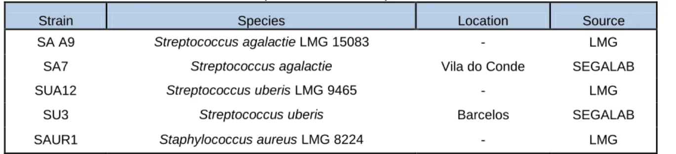
Five strains listed in Table I and previously characterized by Almeida et al., 2013 were used as controls.
Table I - Bacterial strains used as controls (Almeida et al., 2013)
Strain Species Location Source
SA A9 Streptococcus agalactie LMG 15083 - LMG
SA7 Streptococcus agalactie Vila do Conde SEGALAB
SUA12 Streptococcus uberis LMG 9465 - LMG
SU3 Streptococcus uberis Barcelos SEGALAB
SAUR1 Staphylococcus aureus LMG 8224 - LMG
LMG- Belgian Co-ordinated collections of microorganisms, Gent, Belgium.
2. Bacterial culture conditions and DNA extraction
All the samples identified as S.agalactiae or S. uberis by the VITEK 2 system were cultured in Brain Heart Infusion (BHI) (biolab®, Hungary) at 37ºC. After growth in BHI medium, all isolates were stores at -80ºC in 20% glycerol.
DNA was extracted from pure cultures using the E.Z.N.A bacterial DNA purification Kit (Omega Bio Tek, Norcross, GA), following the manufacturer’s instructions. The Qubit 2.0 Flurometer HS Assay (Invitrogen, Carlsbad, CA) was used to quantify the extracted DNA, and the quality was assessed by electrophoresis using 1% agarose gels stained with GelRed (Biotum). Gel images were obtained using a Gel-Doc system (Bio Rad).
12
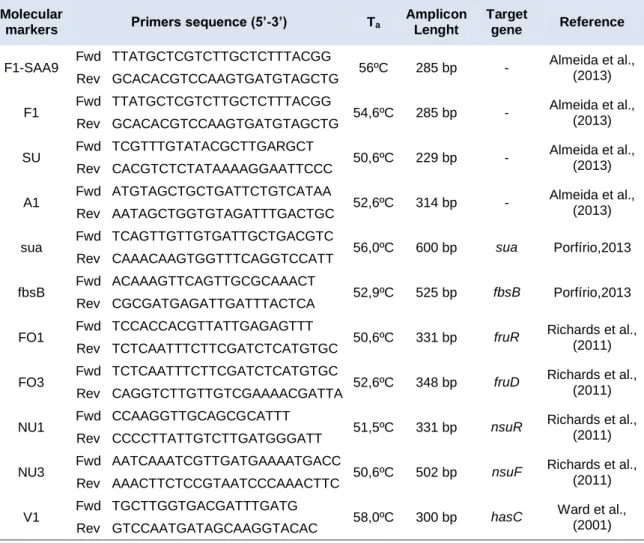
3. PCR amplification
For amplification of the cfb (CAMP factor) and pauA genes, primer-pairs were designed using the Vector NTI software (Invitrogen, Carlsbad,CA.) and synthetized by STABVida (Lisbon, Portugal). Amplicon specificity was confirmed using the BLAST software (Basic Local Alignment Search Tool) (Altschul,1990) (BLAST, http://blast.ncbi.nlm.nih.gov/).
The PCR master mix contained 1x Dream Taq buffer, containing 1.5mM of MgCl2
(Fermentas, Ontario, Canada), 0.2mM of each dNTP (Fermentas), 0.2µM of each primers (forward and reverse) and 1U of DreamTaq DNA polymerase (Fermentas). 25ng of bacterial DNA from pure bacterial cultures were used in each reaction. The PCR conditions were as follows: an initial denaturation of 95ºC for 5 min, 35 cycles at 90ºC for 30s, 55ºC for 30s, 72ºC for 45s, followed by a final extension of 10min at a temperature of 72ºC.
PCR products were visualized in 1.5% agarose gels stained with 3µl of GelRed (Biotum®) the expected bands were cut using a scalpel and purified using the Ilustra® GFX™ PCR DNA and Gel Band Purification Kit (GE Healthcare, Buckinghamshire, UK). To confirm their identity these amplicons were sequenced by STABVida (Lisbon, Portugal).
Table II- Taxa specific and functional markers and PCR primers sequences used in this study.
Molecular
markers Primers sequence (5’-3’) Ta
Amplicon Lenght
Target
gene Reference
F1-SAA9 Fwd TTATGCTCGTCTTGCTCTTTACGG 56ºC 285 bp - Almeida et al., (2013) Rev GCACACGTCCAAGTGATGTAGCTG
F1 Fwd TTATGCTCGTCTTGCTCTTTACGG 54,6ºC 285 bp - Almeida et al., (2013) Rev GCACACGTCCAAGTGATGTAGCTG
SU Fwd TCGTTTGTATACGCTTGARGCT 50,6ºC 229 bp - Almeida et al., (2013) Rev CACGTCTCTATAAAAGGAATTCCC
A1 Fwd ATGTAGCTGCTGATTCTGTCATAA 52,6ºC 314 bp - Almeida et al., (2013) Rev AATAGCTGGTGTAGATTTGACTGC
sua Fwd TCAGTTGTTGTGATTGCTGACGTC 56,0ºC 600 bp sua Porfírio,2013 Rev CAAACAAGTGGTTTCAGGTCCATT
fbsB Fwd ACAAAGTTCAGTTGCGCAAACT 52,9ºC 525 bp fbsB Porfírio,2013 Rev CGCGATGAGATTGATTTACTCA
FO1 Fwd TCCACCACGTTATTGAGAGTTT 50,6ºC 331 bp fruR Richards et al.,
(2011) Rev TCTCAATTTCTTCGATCTCATGTGC
FO3 Fwd TCTCAATTTCTTCGATCTCATGTGC 52,6ºC 348 bp fruD Richards et al.,
(2011) Rev CAGGTCTTGTTGTCGAAAACGATTA
NU1 Fwd CCAAGGTTGCAGCGCATTT 51,5ºC 331 bp nsuR Richards et al.,
(2011) Rev CCCCTTATTGTCTTGATGGGATT
NU3 Fwd AATCAAATCGTTGATGAAAATGACC 50,6ºC 502 bp nsuF Richards et al.,
(2011) Rev AAACTTCTCCGTAATCCCAAACTTC
V1 Fwd TGCTTGGTGACGATTTGATG 58,0ºC 300 bp hasC Ward et al.,
(2001) Rev GTCCAATGATAGCAAGGTACAC
13
V2 Fwd GCTCCTGGTGGAGATGATGT 55,0ºC 189 bp gapC Reinoso et al.,
(2011) Rev GTCACCAGTGTAAGCGTGGA
V3 Fwd GGCCTAACCAAAACGAAACA 54,0ºC 419 bp oppF Smith et al.,
(2002) Rev GGCTCTGGAATTGCTGAAAG
CAMP Fwd GGATTCAACTGAACTCCAACAGCA 57,0ºC 614 bp cfb/cfu This study Rev CATGCTGATCAAGTGACAACTCCA
pauA Fwd TTTTGGGAATATTTGGTTGTGC 55,0ºC 427 bp pauA This study Rev TCAACCCGTTTTCTGAGAATAA
4. Dot-Blot screening
Purified PCR products were labelled with digoxigenin to obtain DNA probes, using the DIG-High Prime labelling kit (Roche, Basel, Switzerland) according to the manufacturer´s instructions. For marker F1, a family-specific marker previously validated (Almeida et al., 2013), a new probe was obtained using as template DNA from S. agalactiae SAA9.
To perform the Dot-Blot hybridization 100ng of heat-denatured DNA from each bacterial strain was spotted into a nylon membrane optimized for the transfer of nucleic acids (Amersham Hybond™-N GE Healthcare, Buckimghamshire,UK), using a Bio-Dot apparatus (Bio Rad) (Tables III, IV and V). Hybridization was carried out over night at 68ºC, using 100ng/ml as the final probe concentration. Washing and detection of the membranes were performed according the recommendations of the DIG system (Roche). DIG-labeled nucleic acids were detected by chemiluminescence using X-ray films (GE, Healthcare) and a Molecular Imager Chemi-Doc system (Bio Rad).
In order to analyze the obtained results, an image processing algorithm was used. This software uses as references both the positive and negative controls present in the membranes and, calculates the probability of each dot being a positive signal. The exposure time in the Chemi-Doc system was adjusted to ensure that all dots were below pixel saturation (Albuquerque et al.,2011).
Table III- Layout of the first Streptococcus agalactiae membrane used in the dot-blot hybridization assay
1 2 3 4 5 6 7 8 9 10 11 12
A SA7 SA253 SA329 SA335 SA254 SA322 SA332 SA344 SA202 SA203 SA204 SA7
B TE SA205 SA326 SA327 SA328 SA257 SA330 SA331 SA336 SA337 SA154 TE
C SA194 SA195 SA196 SA197 SA323 SA324 SA325 SA258 SA319 SA334 SA346 SA255 D SA320 SA321 SA333 SA345 SA318 SA343 SU112 SA38 SA275 SA276 SA277 SA221 E SA222 SA283 SA284 SA285 SA286 SA67 SA68 SA232 SA233 SA270 SA271 SA272 F SA243 SA244 SA295 SA35 SA223 SA296 SA249 SA250 SA251 SA252 SA307 SA308 G TE SA309 SA310 SU16 SA58 SA218 SA219 SA266 SA267 SA234 SA306 TE
H SA7 SU113 SA56 SA245 SA246 SA247 SA248 SA311 SA312 SA313 SA314 SA7 SU112, SU16 and SU113- negative controls.
14
Table IV- Layout of the second Streptococcus agalactiae membrane used in dot blot hybridization assay.
1 2 3 4 5 6 7 8 9 10 11 12
A SA7 SA207 SA260 SA224 SA225 SA226 SA227 SA302 SA303 SA304 SA305 SA7
B TE SA50 SA237 SA238 SA239 SA240 SU52 SU58 SA57 SA300 SU16 TE
C TE SA59 SA241 SA242 SA280 SA281 SA282 SU90 SA43 SA214 SA301 TE
D SA7 TE TE TE TE TE TE TE TE TE TE SA7
SU52, SU58, SU16 and SU90- negative controls.
Table V- Layout of the Streptococcus uberis membrane in the dot blot hybridization assays
1 2 3 4 5 6 7 8 9 10 11 12
A SU3 SU57 SU91 SU114 SU52 SU53 SU58 SU59 SU16 SU112 SU90 SU3
B TE SU113 SA283 SU76 SU80 SU72 SU73 SU89 SU70 SU86 SU63 TE
C SU64 SU65 SU82 SU83 SU67 SU103 SA284 SU40 SU62 SU79 SU99 SU66
D TE SU81 SU105 SU45 SU101 SU42 SU43 SU69 SU85 SU48 SU49 TE
E SU3 SU68 SU87 SU88 SU104 SA285 SU60 SU61 SU84 SU98 SA286 SU3 .SA283, SA284, SA285 and SA286- negative controls.
5. CAMP test
The CAMP test is a presumptive identification test of S. agalactiae (Lancefield group B). (Phillps et al., 1980, Ratner et al., 1986). 5% sheep blood agar was used as cultured medium. This test consists in a synergistic lysis of sheep erythrocytes between Staphylococcus aureus and a protein from group B S. agalactiae, the CAMP factor (pathogenicity factor) (Gase et
al.,1999). A positive result is verified by the presence of a halo which confirms β-hemolysis. To
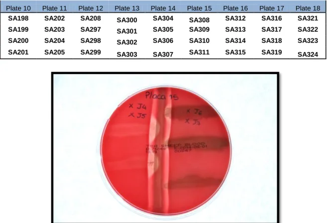
perform this assay, a strain of S. aureus LMG 8224 was placed in the center of a culture plate, and four strains of S.agalactiae two on each side were placed 3 mm perpendicularly to the strain, without direct contact. Streptococcus uberis, Bacillus subtillis and Klebsiella pneumonia were used as negative controls. The plates were incubated overnight at 37ºC. A total of seventy two strains were tested in eighteen petri dishes (Table VI).
Only one strain (1.4%) presented a negative result (SA311) (Figure 1). Sequencing of the 16S rRNA gene allowed identifying SA311 as Staphylococcus epidermidis. (Weiseburg et
al., 1991) This analysis showed that this strain was misidentified as S.agalactiae by the VITEK
system.
Table VI- Scheme used for plating the strains in petri dishes.
Plate 1 Plate 2 Plate 3 Plate 4 Plate 5 Plate 6 Plate 7 Plate 8 Plate 9
SA214 SA232 SA223 SA211 SA246 SA238 SA43 SA190 SA194
SA215 SA233 SA252 SA94 SA247 SA239 SA59 SA191 SA195
SA216 SA207 SA218 SA56 SA50 SA241 SA68 SA192 SA196
15
Plate 10 Plate 11 Plate 12 Plate 13 Plate 14 Plate 15 Plate 16 Plate 17 Plate 18
SA198 SA202 SA208 SA300 SA304 SA308 SA312 SA316 SA321
SA199 SA203 SA297 SA301 SA305 SA309 SA313 SA317 SA322
SA200 SA204 SA298 SA302 SA306 SA310 SA314 SA318 SA323
SA201 SA205 SA299 SA303 SA307 SA311 SA315 SA319 SA324
Figure 1 – Example of the CAMP test plates. The central strain is Staphylococcus aureus LMG 8224, and the arrow head shaped hemolytic halo associated to the Streptococcus agalactiae isolates is indicative of a positive result. The isolated cultured in the right side of the petri plates middle is a negative control, and the strain in the right bottom is a CAMP factor negative (XJ7- corresponding to isolate SA311).
RESULTS
1. Mastitis frequency in herds
Milk quality programs have been increasingly implemented on farms in order to reduce clinical mastitis and decrease the CCS in milk. To accomplish this task, veterinarians are expected to visit frequently the farms, in order to evaluate the general health state of cows and to collect milk and bulk tank samples to search for pathogenic agents causing clinical and sub-clinical infections.
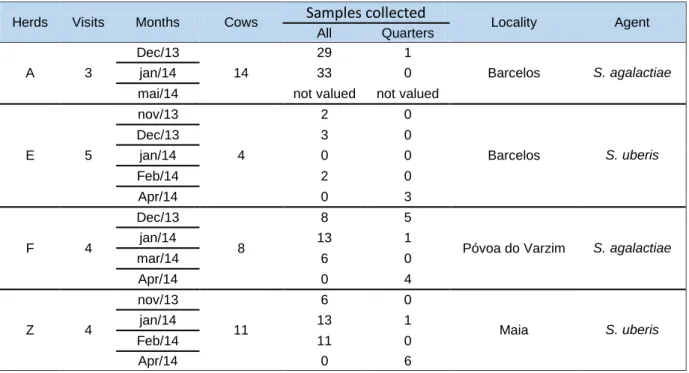
In this study, several visits to the four selected herds (A, E, F and Z) were carried out in order to select cows diagnosed with mastitis caused by S.agalactiae and S. uberis in consecutive visits. The results obtained from this study showed that, from a total of 154 isolates, only 3.9% were responsible for clinical mastitis. Additionally, it was shown, that the average somatic cell count was higher in farms prevalent for S.uberis, than in farms where mastitis was predominantly caused by S.agalactiae. Regarding data analysis, S.agalactiae infections presented both the highest and the lowest SCC. The lowest SCC are easily unnoticed, as the threshold of detection of mastitis is 200.000 somatic cells per milliliter (Table VIII). Concerning
16
bacterial virulence patterns, it was possible to observe that for each pathogenic agent, both
S.agalactiae and S.uberis, there are some virulence patterns that are more common amongst
cows, and that there are some virulence patterns that cause an elevated count of SCC, which translates in a more severe mammary gland infection (Table IX). From the 4 herds studied, 37 cows were diagnosed with mastitis in repeated visits, from which, 22 cows infected with
S.agalactiae and 15 with S.uberis (Table VII).
Table VII- Grid showing the selected cows for this study.
Herds Visits Months Cows Samples collected Locality Agent All Quarters A 3 Dec/13 14 29 1 Barcelos S. agalactiae jan/14 33 0
mai/14 not valued not valued
E 5 nov/13 4 2 0 Barcelos S. uberis Dec/13 3 0 jan/14 0 0 Feb/14 2 0 Apr/14 0 3 F 4 Dec/13 8 8 5
Póvoa do Varzim S. agalactiae
jan/14 13 1 mar/14 6 0 Apr/14 0 4 Z 4 nov/13 11 6 0 Maia S. uberis jan/14 13 1 Feb/14 11 0 Apr/14 0 6 nv- not valued
Table VIII- Data analysis of simple and composed samples.
Agent Statistics AD AE PD PE ALL
S. uberis Mean 1416,5 5945,5 1568,7 883,2 1071,3 St. Deviation 1206,69 4917,64 1691,27 372,10 889,80 Maximum 3782 12914 5498 1156 3457 Minimum 172 223 165 138 135 S. agalactiae Mean 2121,3 435,7 1496,4 1420,2 1175,6 St. Deviation 3128,13 394,97 1921,68 1315,35 2441,19 Maximum 10077 1575 5725 4913 9153 Minimum 106 104 85 60 50
17
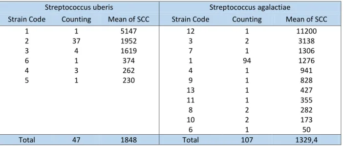
Table IX- Data analysis showing strain code, number of isolates and respective mean of SCC.
Streptococcus uberis Streptococcus agalactiae
Strain Code Counting Mean of SCC Strain Code Counting Mean of SCC
1 1 5147 12 1 11200 2 37 1952 3 2 3138 3 4 1619 7 1 1306 6 1 374 1 94 1276 4 3 262 4 1 941 5 1 230 9 1 828 13 1 427 11 1 355 8 2 282 10 2 173 6 1 50 Total 47 1848 Total 107 1329,4
2. Preliminary tests
In order to assess the efficiency of taxonomic DNA markers for rapid identification of mastitis isolates, all the strains collected in this work were tested by dot blot assays.
Results corresponding to markers- V1, V2,V3 and SU showed that all these markers gave positive hybridization signals with all S.uberis strains tested. Marker NU3, which is specific for S.uberis, as expected, was negative for the tested S.agalactiae strains. Due to inconsistent results obtained with marker F1, a new F1 probe obtained from a S. agalactiae strain (SAA9) was labeled and tested. This new labelled taxonomic marker previously reported to be genus-specific for Streptococcus, provided positive results with all tested strains (data not shown).
3. Assays with selected strains
3.1- Taxonomic markers
Concerning the taxonomic and virulence analyses of all selected strains, three types of dot-blot membranes were made, two for S.agalactiae, due to its higher number of isolates, and one for S.uberis. The markers selected for testing with the S.agalactiae membranes were F1SAA9, A1, CAMP, fbsB, FO3 and FO1. For S.uberis membrane F1SAA9, CAMP, NU1, pauA,
sua and ermB were the markers used for the hybridization assays.
Regarding the probability values of hybridization outputted by the image analysis software used, a color code was employed to evaluate the obtained results 0 to 0.25 - low probability, represented in red, 0.25 to 0.75 -average probability, represented in yellow and 0.75 to 1.00-high probability, represented in green.
A total of 110 isolates of S.agalactiae and 48 isolates of S.uberis were tested. For
18
were used as negative controls (these strains were also tested in S.uberis membranes). For
S.uberis membrane, the S.agalactiae isolates used negative controls were SA283, SA284,
SA285 and SA286, which were also evaluated in S.agalactiae membranes.
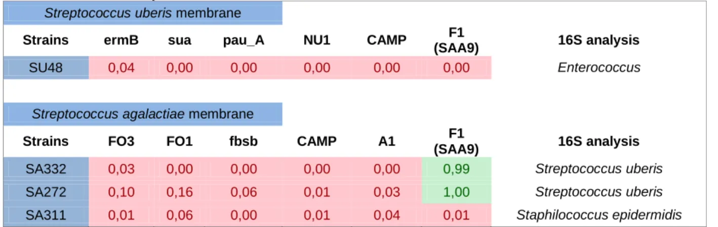
A preliminary analysis of the obtained hybridization data revealed that the results obtained with three isolates previously identified by the VITEK system as S.agalactiae (SA332, SA272 and SA311) and one isolate identified as S.uberis (SU48), were indicative of a misidentification, as shown in Table X. In fact, 16S rRNA gene sequencing of the isolates, showed that these bacteria were incorrectly identified by the VITEK system and were withdrawn from further analysis.
Table X- Isolates with atypical results in Dot-Blot membranes, that after 16S gene sequencing analysis, were withdrawn from this study.
Streptococcus uberis membrane
Strains ermB sua pau_A NU1 CAMP F1
(SAA9) 16S analysis
SU48 0,04 0,00 0,00 0,00 0,00 0,00 Enterococcus
Streptococcus agalactiae membrane
Strains FO3 FO1 fbsb CAMP A1 F1
(SAA9) 16S analysis
SA332 0,03 0,00 0,00 0,00 0,00 0,99 Streptococcus uberis
SA272 0,10 0,16 0,06 0,01 0,03 1,00 Streptococcus uberis
SA311 0,01 0,06 0,00 0,01 0,04 0,01 Staphilococcus epidermidis
Regarding Streptococcus specific taxonomic marker F1 SAA9, dot blot hybridization showed a specificity of 99.1% (106/107) in S.agalactiae membranes, with only one isolate presenting a very low probability value of hybridization. However, when tested for marker A1, a taxonomic marker specific for this species, i.e. S.agalactiae, the result was a positive hybridization allowing to confirm the identity of this isolate.
Marker A1, had a specificity of 92.5% (99/107), with eight isolates, 7.5% (8/107), presenting an average hybridization probability.
In S.uberis membrane, the only taxonomic marker tested, F1 SAA9, present a specificity of 98% (46/47).
3.2- Virulence markers
A survey of the literature, allowed to select several virulence factors that seem to play an important role during mammary gland infections. In the present study, regions related to adhesion and invasion, toxin production, ability to growth in milk or in the environment and production of bacteriocins were selected for analysis.
19
In S.agalactiae membranes, concerning adhesion and invasion to the epithelium, the fibrinogen binding protein (fbsB) probe was selected. In S.uberis membrane the adhesion molecule (Sua) was tested.
Concerning toxin production, the CAMP factor was tested for both Streptococcus species.
The ability of S.uberis to grow in milk/ environment was assessed using the pauA factor probe, and in S.agalactiae, the fructose operons FO1 and FO3 were selected. For the study of bacteriocins, NU1, was chosen.
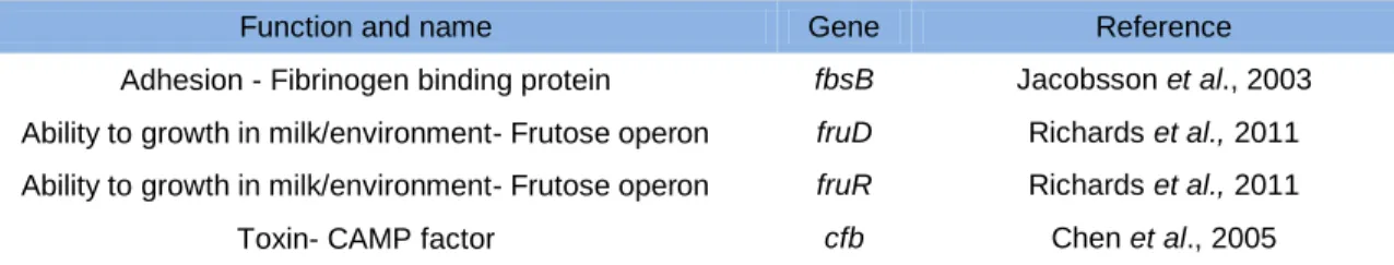
Table XI- Virulence factors for S.agalactiae used in this study.
Function and name Gene Reference
Adhesion - Fibrinogen binding protein fbsB Jacobsson et al., 2003 Ability to growth in milk/environment- Frutose operon fruD Richards et al., 2011 Ability to growth in milk/environment- Frutose operon fruR Richards et al., 2011 Toxin- CAMP factor cfb Chen et al., 2005
Table XII- Virulence factors for S.uberis used in this study.
Function and name Gene Reference
Adhesion and invasion- S.uberis adhesion molecule sua Almeida et al., 2006 Ability to growth in milk/environment- Plasminogen activator pauA Rosey et al., 1999; Ward &
Leight, 2002 Toxin- CAMP factor cfu Reinoso et al., 2011 Bacteriocin- Nisin U nsu Wirawan et al., 2006
In S.agalactiae membranes, (Table XI), the results obtained with the fbsB probe revealed that this gene was present in 95.3% (102/107) of the strains. Three strains (2.8%) have an average probability to have the gene, and two isolates (1.9%) were negative suggesting that these isolates do not have the fbsB gene.
Results from the CAMP factor marker, showed that 95.3% (102/107) of the isolates were positive for this gene, four (3.7%) have an average probability and one isolate (0.9%) was negative for this gene.
Markers from fructose operons revealed that for FO1, 92.5% (99/107) of the tested isolates have the gene, four isolates (3.7%) have an average probability and four isolates (3.7%) were negative for the presence of FO1. For the markers FO3, 98.1% of the isolates studied (105/107) were positive for the presence of this gene with high probability values and 1.9% have an average probability.
20
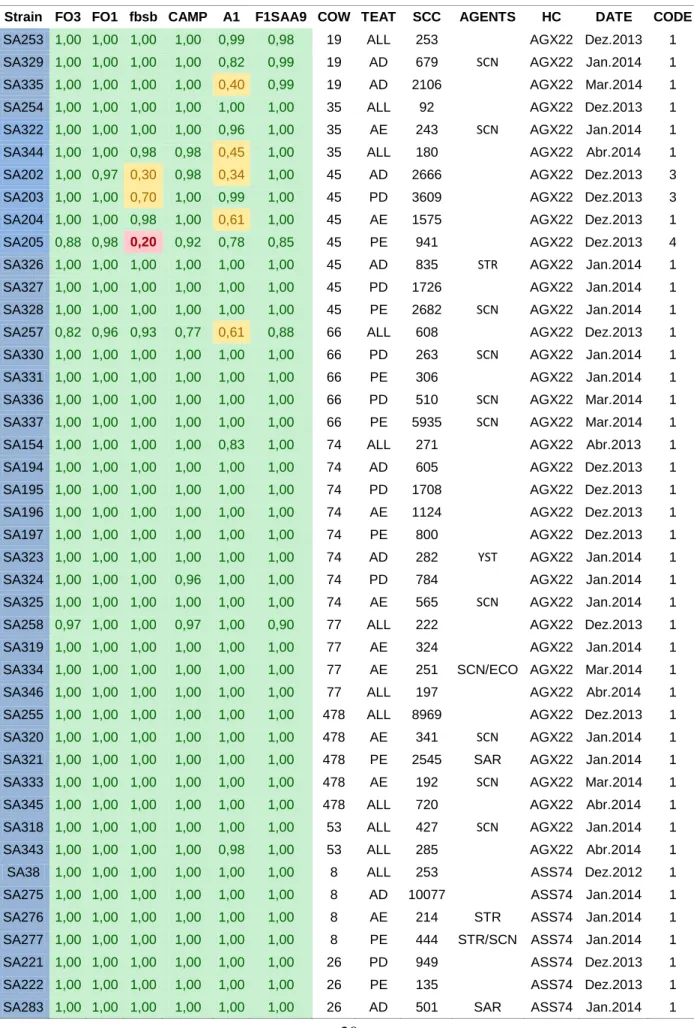
Table XIII- Probability values of the results obtained from the Dot-Blot assays from S.agalactiae membranes
Strain FO3 FO1 fbsb CAMP A1 F1SAA9 COW TEAT SCC AGENTS HC DATE CODE
SA253 1,00 1,00 1,00 1,00 0,99 0,98 19 ALL 253 AGX22 Dez.2013 1 SA329 1,00 1,00 1,00 1,00 0,82 0,99 19 AD 679 SCN AGX22 Jan.2014 1 SA335 1,00 1,00 1,00 1,00 0,40 0,99 19 AD 2106 AGX22 Mar.2014 1 SA254 1,00 1,00 1,00 1,00 1,00 1,00 35 ALL 92 AGX22 Dez.2013 1 SA322 1,00 1,00 1,00 1,00 0,96 1,00 35 AE 243 SCN AGX22 Jan.2014 1 SA344 1,00 1,00 0,98 0,98 0,45 1,00 35 ALL 180 AGX22 Abr.2014 1 SA202 1,00 0,97 0,30 0,98 0,34 1,00 45 AD 2666 AGX22 Dez.2013 3 SA203 1,00 1,00 0,70 1,00 0,99 1,00 45 PD 3609 AGX22 Dez.2013 3 SA204 1,00 1,00 0,98 1,00 0,61 1,00 45 AE 1575 AGX22 Dez.2013 1 SA205 0,88 0,98 0,20 0,92 0,78 0,85 45 PE 941 AGX22 Dez.2013 4 SA326 1,00 1,00 1,00 1,00 1,00 1,00 45 AD 835 STR AGX22 Jan.2014 1 SA327 1,00 1,00 1,00 1,00 1,00 1,00 45 PD 1726 AGX22 Jan.2014 1 SA328 1,00 1,00 1,00 1,00 1,00 1,00 45 PE 2682 SCN AGX22 Jan.2014 1 SA257 0,82 0,96 0,93 0,77 0,61 0,88 66 ALL 608 AGX22 Dez.2013 1 SA330 1,00 1,00 1,00 1,00 1,00 1,00 66 PD 263 SCN AGX22 Jan.2014 1 SA331 1,00 1,00 1,00 1,00 1,00 1,00 66 PE 306 AGX22 Jan.2014 1 SA336 1,00 1,00 1,00 1,00 1,00 1,00 66 PD 510 SCN AGX22 Mar.2014 1 SA337 1,00 1,00 1,00 1,00 1,00 1,00 66 PE 5935 SCN AGX22 Mar.2014 1 SA154 1,00 1,00 1,00 1,00 0,83 1,00 74 ALL 271 AGX22 Abr.2013 1 SA194 1,00 1,00 1,00 1,00 1,00 1,00 74 AD 605 AGX22 Dez.2013 1 SA195 1,00 1,00 1,00 1,00 1,00 1,00 74 PD 1708 AGX22 Dez.2013 1 SA196 1,00 1,00 1,00 1,00 1,00 1,00 74 AE 1124 AGX22 Dez.2013 1 SA197 1,00 1,00 1,00 1,00 1,00 1,00 74 PE 800 AGX22 Dez.2013 1 SA323 1,00 1,00 1,00 1,00 1,00 1,00 74 AD 282 YST AGX22 Jan.2014 1 SA324 1,00 1,00 1,00 0,96 1,00 1,00 74 PD 784 AGX22 Jan.2014 1 SA325 1,00 1,00 1,00 1,00 1,00 1,00 74 AE 565 SCN AGX22 Jan.2014 1 SA258 0,97 1,00 1,00 0,97 1,00 0,90 77 ALL 222 AGX22 Dez.2013 1 SA319 1,00 1,00 1,00 1,00 1,00 1,00 77 AE 324 AGX22 Jan.2014 1 SA334 1,00 1,00 1,00 1,00 1,00 1,00 77 AE 251 SCN/ECO AGX22 Mar.2014 1 SA346 1,00 1,00 1,00 1,00 1,00 1,00 77 ALL 197 AGX22 Abr.2014 1 SA255 1,00 1,00 1,00 1,00 1,00 1,00 478 ALL 8969 AGX22 Dez.2013 1 SA320 1,00 1,00 1,00 1,00 1,00 1,00 478 AE 341 SCN AGX22 Jan.2014 1 SA321 1,00 1,00 1,00 1,00 1,00 1,00 478 PE 2545 SAR AGX22 Jan.2014 1 SA333 1,00 1,00 1,00 1,00 1,00 1,00 478 AE 192 SCN AGX22 Mar.2014 1 SA345 1,00 1,00 1,00 1,00 1,00 1,00 478 ALL 720 AGX22 Abr.2014 1 SA318 1,00 1,00 1,00 1,00 1,00 1,00 53 ALL 427 SCN AGX22 Jan.2014 1 SA343 1,00 1,00 1,00 1,00 0,98 1,00 53 ALL 285 AGX22 Abr.2014 1 SA38 1,00 1,00 1,00 1,00 1,00 1,00 8 ALL 253 ASS74 Dez.2012 1 SA275 1,00 1,00 1,00 1,00 1,00 1,00 8 AD 10077 ASS74 Jan.2014 1 SA276 1,00 1,00 1,00 1,00 1,00 1,00 8 AE 214 STR ASS74 Jan.2014 1 SA277 1,00 1,00 1,00 1,00 1,00 1,00 8 PE 444 STR/SCN ASS74 Jan.2014 1 SA221 1,00 1,00 1,00 1,00 1,00 1,00 26 PD 949 ASS74 Dez.2013 1 SA222 1,00 1,00 1,00 1,00 1,00 1,00 26 PE 135 ASS74 Dez.2013 1 SA283 1,00 1,00 1,00 1,00 1,00 1,00 26 AD 501 SAR ASS74 Jan.2014 1
21
SA284 1,00 1,00 1,00 1,00 1,00 1,00 26 PD 330 ASS74 Jan.2014 1 SA285 1,00 1,00 1,00 0,95 1,00 1,00 26 AE 223 ASS74 Jan.2014 1 SA286 1,00 1,00 1,00 1,00 1,00 1,00 26 PE 1393 SCN ASS74 Jan.2014 1 SA67 1,00 1,00 1,00 1,00 1,00 1,00 29 AD 15719 ASS74 Dez.2012 1 SA68 1,00 1,00 1,00 1,00 1,00 1,00 29 ALL 9153 ASS74 Dez.2012 1 SA232 1,00 1,00 1,00 1,00 1,00 1,00 29 AE 896 ASS74 Dez.2013 1 SA233 1,00 1,00 1,00 1,00 1,00 1,00 29 PE 83 ASS74 Dez.2013 1 SA270 1,00 1,00 1,00 1,00 1,00 1,00 29 AD 57 ASS74 Jan.2014 1 SA271 1,00 1,00 1,00 1,00 1,00 1,00 29 PD 4888 STR ASS74 Jan.2014 1 SA243 0,94 1,00 1,00 1,00 1,00 0,99 30 PD 201 ASS74 Dez.2013 1 SA244 1,00 1,00 1,00 1,00 1,00 1,00 30 PE 621 ASS74 Dez.2013 1 SA295 1,00 1,00 1,00 1,00 1,00 1,00 30 PE 2299 ASS74 Jan.2014 1 SA35 1,00 1,00 1,00 1,00 1,00 1,00 44 ALL 56 ASS74 Dez.2012 1 SA223 1,00 1,00 1,00 1,00 1,00 1,00 44 PE 6545 ASS74 Dez.2013 1 SA296 1,00 1,00 1,00 1,00 1,00 1,00 44 PE 3281 ASS74 Jan.2014 1 SA249 1,00 1,00 1,00 1,00 1,00 1,00 57 AD 616 SCN/YST ASS74 Dez.2013 1 SA250 1,00 1,00 1,00 1,00 1,00 1,00 57 PD 248 ASS74 Dez.2013 1 SA251 1,00 1,00 1,00 1,00 1,00 1,00 57 AE 200 ASS74 Dez.2013 1 SA252 1,00 1,00 1,00 1,00 1,00 1,00 57 PE 1134 ASS74 Dez.2013 1 SA307 1,00 1,00 1,00 1,00 1,00 1,00 57 AD 2641 SCN ASS74 Jan.2014 1 SA308 1,00 1,00 1,00 1,00 1,00 1,00 57 PD 341 ASS74 Jan.2014 1 SA309 1,00 1,00 1,00 1,00 1,00 1,00 57 AE 287 ASS74 Jan.2014 1 SA310 0,94 1,00 0,99 0,96 1,00 0,91 57 PE 1857 SCN ASS74 Jan.2014 1 SA218 1,00 1,00 1,00 1,00 1,00 1,00 81 AE 410 ASS74 Dez.2013 1 SA219 1,00 1,00 1,00 1,00 1,00 1,00 81 PE 239 ASS74 Dez.2013 1 SA266 1,00 1,00 1,00 1,00 1,00 1,00 81 AE 810 ASS74 Jan.2014 1 SA267 1,00 1,00 1,00 1,00 1,00 1,00 81 PE 3637 ASS74 Jan.2014 1 SA234 1,00 1,00 1,00 1,00 1,00 1,00 121 PE 138 ASS74 Dez.2013 1 SA306 1,00 1,00 1,00 1,00 1,00 1,00 121 PE 558 SCN/STR ASS74 Jan.2014 1 SA56 1,00 1,00 1,00 1,00 1,00 1,00 866 ALL 328 ASS74 Dez.2012 1 SA245 1,00 1,00 1,00 1,00 1,00 1,00 866 AD 106 ASS74 Dez.2013 1 SA246 1,00 1,00 1,00 1,00 1,00 1,00 866 PD 44 ASS74 Dez.2013 1 SA247 1,00 1,00 1,00 1,00 1,00 1,00 866 AE 115 ASS74 Dez.2013 1 SA248 1,00 1,00 1,00 1,00 1,00 1,00 866 PE 69 FUN ASS74 Dez.2013 1 SA312 1,00 1,00 1,00 1,00 1,00 1,00 866 PD 126 SCN ASS74 Jan.2014 1 SA313 1,00 1,00 1,00 1,00 1,00 1,00 866 AE 93 SCN ASS74 Jan.2014 1 SA314 1,00 1,00 1,00 1,00 1,00 1,00 866 PE 50 ASS74 Jan.2014 1 SA207 0,76 0,24 0,73 0,87 0,32 0,75 714 ALL 50 ASS74 Dez.2013 6 SA260 1,00 0,98 0,98 1,00 0,97 1,00 714 AE 317 ASS74 Jan.2014 1 SA224 0,71 0,44 0,75 0,88 0,82 0,90 901 AD 1306 ASS74 Dez.2013 7 SA225 1,00 0,96 0,99 1,00 0,92 1,00 901 PD 492 ASS74 Dez.2013 1 SA226 0,98 0,94 0,98 1,00 0,97 0,93 901 AE 221 ASS74 Dez.2013 1 SA227 0,99 0,85 0,99 1,00 0,91 0,97 901 PE 1012 ASS74 Dez.2013 1 SA302 1,00 0,98 0,86 0,98 0,98 0,93 901 AD 136 ASS74 Jan.2014 1 SA303 1,00 0,99 0,98 0,99 0,63 1,00 901 PD 118 ASS74 Jan.2014 1 SA304 0,85 0,97 0,88 1,00 0,97 0,94 901 AE 74 ASS74 Jan.2014 1