Isolation and characterization of a serine
proteinase with thrombin-like activity from
the venom of the snake Bothrops asper
A.V. Pérez
1,2, A. Rucavado
1, L. Sanz
3, J.J. Calvete
3and J.M. Gutiérrez
11
Instituto Clodomiro Picado, Facultad de Microbiología, Universidad de Costa Rica, San José, Costa Rica
2Facultad de Medicina, Universidad de El Salvador, San Salvador, El Salvador
3
Laboratorio de Proteinómica Estructural, Instituto de Biomedicina de Valencia, Valencia, España
Correspondence to: J.M. Gutiérrez, Instituto Clodomiro Picado, Facultad de Microbiología,
Universidad de Costa Rica, San José, Costa Rica
Fax: +506-292-0485. E-mail: [email protected]
A serine proteinase with thrombin-like activity was isolated from the venom of the Central American pit viper Bothrops asper. Isolation was performed by a combination of affinity chromatography on aminobenzamidine-Sepharose and ion-exchange chromatography on DEAE-Sepharose. The enzyme accounts for approximately 0.13% of the venom dry weight and has a molecular mass of 32 kDa as determined by SDS-PAGE, and of 27 kDa as determined by MALDI-TOF mass spectrometry. Its partial amino acid sequence shows high identity with snake venom serine proteinases and a complete identity with a cDNA clone previously sequenced from this species. The N-terminal sequence of the enzyme is VIGGDECNINEHRSLVVLFXSSGFL CAGTLVQDEWVLTAANCDSKNFQ. The enzyme induces clotting of plasma (minimum coagulant dose = 4.1 µg) and fibrinogen (minimum coagulant dose = 4.2 µg) in vitro, and promotes defibrin(ogen)ation in vivo (minimum defibrin(ogen)ating dose = 1.0 µg). In addition, when injected intravenously in mice at doses of 5 and 10 µg, it induces a series of behavioral changes, i.e., loss of the righting reflex, opisthotonus, and intermittent rotations over the long axis of the body, which closely resemble the ‘gyroxin-like’ effect induced by other thrombin-like enzymes from snake venoms.
Key words: Snake venom; Bothrops asper; Serine proteinase; Thrombin-like serine proteinase; Defibrin(ogen)ation
Research supported by NeTropica (No. 2-N-2005), CYTED (No. 206AC0281), Ministerio de Educación y Ciencia, Madrid, España (No. BFU2004-01432), and Vicerrectoría de Investigación, Universidad de Costa Rica (No. 741-A6-505).
This study was carried out in partial fulfillment of the requirements for the M.Sc. degree of A.V. Pérez at the University of Costa Rica.
Received May 3, 2007. Accepted September 20, 2007
Viperid snakes of the genus Bothrops sp inflict the vast majority of snakebites in Central and South America (1,2). In Central America and parts of northern South America, the species Bothrops asper is responsible for the majority of cases (2). Envenomation by this species is associated with prominent local pathological effects, i.e., myonecro-sis, hemorrhage, edema, dermonecrosis and blistering (3), as well as with systemic alterations characterized by coagulopathy, hemorrhage, cardiovascular shock, and acute renal failure (2,4,5). Alterations in hemostasis lead-ing to thrombocytopenia, platelet hypoaggregation, defibrin(ogen)ation and disseminated intravascular
co-agulation are often observed in victims of B. asper bite envenomation (5-7). Such complex hemostatic distur-bances potentiate the profuse bleeding initiated by the disruptive action of hemorrhagic metalloproteinases in the microvasculature (7,8).
laboratory clotting test parameters, such as whole blood clotting time, prothrombin time and activated partial throm-boplastin time (6,9). Procoagulant components in Bothrops sp venoms include zinc-dependent metalloproteinases that activate factor X and prothrombin, as well as serine protein-ases that directly convert fibrinogen into fibrin (10,11). In the case of B. asper, previous investigations have described a prothrombin activator, which is a class P-III metalloprotein-ase named basparin A (12), and a thrombin-like serine proteinase, named asperase (13,14); however, the charac-terization of the latter is limited to molecular mass estimation and amino acid composition, with no sequence information available. On the other hand, this venom is devoid of factor X activators (15). Owing to the pathophysiological relevance of coagulant enzymes in this medically relevant snake venom, in the present paper we describe the isolation of a thrombin-like serine proteinase, the determination of its partial amino acid sequence, and the characterization of its in vitro coagu-lant and in vivo defibrin(ogen)ating activities.
Serine proteinase was purified by a two-step chromato-graphic protocol. Samples of 250 mg B. asper venom, obtained from adult specimens collected in the Pacific re-gion of Costa Rica, were dissolved in 5 mL 50 mM Tris-HCl, 0.4 M NaCl, pH 9.0. After centrifugation at 500 g for 10 min, the supernatant was applied to a benzamidine-Sepharose 4 Fast Flow affinity chromatography column (3 x 12 cm; Amer-sham Biosciences, Uppsala, Sweden), previously equili-brated with the same buffer. The sample was passed sev-eral times through the column in order to maximize binding. After elution of the unbound fraction, a 0.1 M glycine-HCl, pH 3.0, buffer was applied to the column and the absorbance of the eluting material was monitored at 280 nm. Fractions of 3 mL were collected into tubes containing 0.5 mL 0.5 M Tris buffer, pH 8.8. Throughout fractionation, fibrinogen-clotting activity was assessed in order to identify fractions with thrombin-like activity. To this end, 200 µL of a 4 mg/mL bovine fibrinogen (Sigma-Aldrich, St. Louis, MO, USA) in 0.12 M NaCl, 40 mM phosphate, pH 7.2 (PBS) was incu-bated with 100 µL dilutions of the fractions. Proteins from the peaks showing fibrinogen-clotting activity were diafiltered and concentrated by ultrafiltration against 50 mM Tris-HCl, 50 mM KCl buffer, pH 9.0. The concentrated material was applied to a 3 x 8 cm DEAE-Sepharose column previously equilibrated with the dialysis buffer. After elution of the unbound material, a linear KCl gradient (50 mM to 0.75 M) in the same Tris buffer was developed in a total volume of 500 mL. Active peaks were diafiltered against distilled water and lyophilized. Homogeneity was assessed by reverse-phase HPLC on a C4 column (Vydac; 250 x 4.6 mm; flow rate 1.0 mL/min) using an Agilent 1100 instrument (Tokyo, Japan) and a linear gradient from 0 to 60% acetonitrile in 0.1%
trifluoroacetic acid.
For molecular mass analysis and amino acid sequenc-ing, the fibrinogen-clotting protein purified by DEAE-Sepha-rose chromatography was submitted to reverse-phase HPLC using an ETTAN™ LC HPLC system (Amersham Biosciences, Piscataway, NJ, USA) and a Lichrosphere RP100 C18 column (250 x 4 mm, 5 µm particle size) eluted at 1 mL/min with a linear gradient of 0.1% TFA in water (solution A) and acetonitrile (solution B) employing the following chromatographic conditions: isocratically (5% B) for 10 min, followed by linear gradients of 5-15% B over 20 min, 15-45% B over 120 min, and 45-70% B over 20 min. Protein detection was at 215 nm. Fractions were collected manually and dried in a Speed-Vac instrument (Savant, Ramsey, MN, USA). The molecular mass of the purified protein was determined by SDS-PAGE (16), run under reducing conditions on 12% polyacrylamide gels and by MALDI-TOF mass spectrometry (using a Voyager DE-Pro™ instrument, Applied Biosystem, Foster City, CA, USA, and sinapinic acid as the matrix) and electrospray ioniza-tion mass spectrometry using an Applied Biosystem Qtrap™ mass spectrometer (17) operated in Enhanced Multiple Charge mode in a 600-1700-m/z range.
The purified protein was subjected to N-terminal se-quence analysis using a Procise instrument (Applied Bio-systems) following manufacturer instructions. For internal sequence determination, the protein band of ~30 kDa was excised from a Coomassie brilliant blue-stained SDS-PAGE and subjected to automated reduction with DTT and alky-lation with iodoacetamide, and in-gel digestion with se-quencing grade bovine pancreas trypsin (Roche, San Cugart del Vallés, Barcelona, Spain) using a ProGest digestor (Genomic Solutions, Cambridgeshire, UK) follow-ing manufacturer instructions. A 0.65-µL amount of the tryptic peptide mixtures (total volume of ~20 µL) was spotted onto a MALDI-TOF sample holder, mixed with an equal volume of a saturated solution of α
-cyano-4-hy-droxycinnamic acid (Sigma) in 50% acetonitrile containing 0.1% TFA, dried, and analyzed with a Voyager-DE Pro MALDI-TOF mass spectrometer (Applied Biosystems) op-erated in delayed extraction and reflector modes. A tryptic peptide mixture of Cratylia floribunda seed lectin (SwissProt accession code P81517) prepared and previously charac-terized in our laboratory was used as mass calibration standard (mass range, 450-3300 Da).
se-lected peptides from the MALDI-TOF mass fingerprint spectra were analyzed in Enhanced Resolution MS mode and the monoisotopic ions were fragmented using the Enhanced Product Ion tool with Q0 trapping. Enhanced Resolution was performed at 250 amu/s across the entire mass range. Settings for MS/MS experiments were as follows: Q1 - unit resolution; Q1 to Q2 collision energy, 30-40 eV; Q3 entry barrier, 8 V; linear ion trap Q3 fill time, 250 ms, and Q3 scan rate, 1000 amu/s. Collision-induced dissociation spectra were interpreted manually or using the on-line form of the MASCOT program at http://www. matrixscience. com.
The coagulant activity of fractions and purified enzyme was assessed on citrated human plasma and on a 4 mg/mL solution of bovine fibrinogen, dissolved in PBS, as described above. Samples of 100 µL fractions or various concentra-tions of the enzyme were added to 200 µL plasma or fibrinogen, previously incubated at 37°C. Clotting times were recorded, and the minimum coagulant doses (MCD) for plasma or fibrinogen were determined; MCD corresponds to the amount of enzyme that induces clotting in 60 s (18). In vivo alterations were assessed by intravenous (iv ) injection of various amounts of the enzyme dissolved in 100 µL PBS, in a group of four CD-1 mice (18-20 g body weight); controls received the same volume of PBS under otherwise identical conditions. One hour after injection, mice were bled under anesthesia. Blood was placed in dry clean glass tubes and left at room temperature; the formation of clots was regularly assessed by tilting the tubes, and clotting time was recorded (18). In addition, mice injected iv were observed for behav-ioral changes occurring during the first minutes after injec-tion. Experiments involving mice were approved by the Institutional Committee for the Care and Use of Laboratory Animals (CICUA) of the University of Costa Rica.
Fractionation of B. asper venom by affinity chromatog-raphy on benzamidine-Sepharose yielded a peak, which bound to the gel, having fibrinogen-clotting activity (Figure 1A). This fraction was separated into five peaks on a DEAE-Sepharose column eluted with a linear KCl gradient (Figure 1B). Only peak 1 showed fibrinogen-clotting activ-ity. When analyzed by reverse-phase HPLC, this peak had one main component (Figure 1C). The yield of this enzyme was low, as only 2 mg was recovered from an initial amount of 1.5 g venom, thus corresponding to approximately 0.13% of B. asper venom. The apparent molecular mass of this thrombin-like enzyme was 32 kDa (non-reduced)/24 kDa (reduced), as determined by SDS-PAGE (Figure 1C, in-set). MALDI-TOF mass spectrometry evidenced three molecular species of molecular masses 27,067, 27,356, and 27,649 Da (± 27 Da). The mass difference of 290-293 Da may indicate that the purified enzyme is a mixture of
glycoforms differing in their degree of sialylation (mass increment due to sialylation = 292 Da).
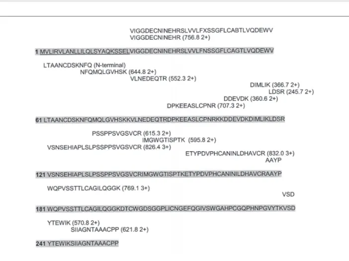
N-terminal sequence analysis (Figure 2) revealed 48 residues with high identity with previously characterized thrombin-like serine proteinases from viperid snake ven-oms (19). Moreover, the internal sequences of 14 peptides resulting from tryptic digestion also showed characteristic sequences of venom serine proteinases. All the sequences determined showed 100% identity with a deduced amino acid sequence of a cDNA clone derived from RNA isolated from the venom gland of B. asper from Costa Rica (UniProtKB/TrEMBL entry Q072L6; Figure 2). Therefore, we may conclude that this sequence corresponds to the thrombin-like serine proteinase described in this commu-nication. The cDNA-deduced sequence of B. asper serine proteinase exhibits a single potential N-glycosylation site at position 20 of the mature protein. The mass difference between the cDNA-deduced sequence (isotope-averaged molecular mass = 25308 Da, assuming that its 12 cys-teines are involved in the formation of disulfide bonds) and the MALDI-TOF estimated molecular masses support the hypothesis that each of the three purified thrombin-like enzyme glycoforms bears a single di-, mono-, or non-syalilated, fucosylated complex-type dianntenary glycan chain (calculated mass increments of 2352.1, 2060.1, and 1768.1 Da, respectively). On the other hand, the similarity in molecular mass with asperase, a thrombin-like serine proteinase isolated from this venom by Ortiz and Gubensek (13), suggests that the enzyme described here is likely to correspond to asperase.
Figure 1. Figure 1. Figure 1. Figure 1.
Figure 1. Isolation of a thrombin-like serine proteinase from Bothrops asper venom. A, Venom (250 mg) was dissolved in 50 mM Tris-HCl, 0.4 M NaCl, pH 9.0, and applied to a benzamidine-Sepharose 4 Fast Flow column. After elution of unbound material, a 0.1 M glycine-HCl buffer, pH 3.0, was applied (arrow) and the eluted material was collected into tubes containing 0.5 M Tris buffer, pH 8.8. B, The benzamidine-binding fraction isolated in A was further fractionated by ion-exchange chromatography on a DEAE-Sepharose column, equilibrated with 50 mM Tris-HCl, 50 mM KCl buffer, pH 9.0. After elution of unbound material, a linear gradient (G) of KCl from 50 mM to 0.75 M was developed in the same Tris buffer. Only the first peak collected after the onset of the gradient (discontinuous arrow) had thrombin-like activity. C, Reverse-phase HPLC analysis of the active peak after DEAE-Sepharose separation, carried out on a C4 column at a flow rate of 1.0 mL/min, using a linear gradient from 0 to 60% acetonitrile in 0.1% trifluoroacetic acid. The inset shows an SDS-PAGE of the purified protein run under reducing (R) and non-reducing (NR) conditions. D, MALDI-TOF mass spectrum of the purified thrombin-like enzyme.
the venom (15). Clearly, therefore, metalloproteinases con-stitute the most important coagulant components of B. asper venom.
Upon iv injection in mice, the serine proteinase induced defibrin(ogen)ation. Blood collected from control mice in-jected with PBS clotted within 2 min, whereas blood col-lected from mice injected with 10, 5, 2.5, and 1.25 µg of the enzyme were unclottable; blood from mice receiving 0.5 µg showed normal clotting. The minimum defibrin(ogen)ating
pre-dominant role of metalloproteinases over serine proteinases in B. asper-induced coagulopathy.
Intravenous administration of doses of 10 and 5 µg of the serine proteinase induced a series of behavioral changes in mice within the first 2-3 min after injection. These were characterized by loss of the righting reflex, opisthotonus, and intermittent rotations over the long axis of the body. These effects persisted during approximately 10 min, after which the animals apparently recovered. No such effect was observed with doses below 5 µg. This effect is very similar to that described for gyroxin, a throm-bin-like serine proteinase isolated from Crotalus durissus terrificus venom (20). Similar observations were made with thrombin-like serine proteinases from other venoms (20). It is likely that this ‘gyroxin-like’ effect is typical of thrombin-like serine proteinases from viperid snake venoms, al-Figure 2.
Figure 2.Figure 2. Figure 2.
Figure 2. Alignment of the N-terminal sequence of Bothrops asper serine proteinase determined by automated Edman degradation (N-terminal), and internal amino acid sequences gathered by collision-induced dissociation tandem mass spectrometry of doubly- and triply-charged tryptic peptides (the m/z value is indicated at the end of each peptide), with the deduced amino acid sequence of a cDNA clone derived from RNA isolated from the venom gland of B. asper (highlighted in gray) (UniProtKB/TrEMBL entry Q072L6). The underlined residues correspond to the signal peptide. There is a 100% identity between the sequenced peptides and the deduced sequence.
though the underlying mechanism remains unknown. In conclusion, a thrombin-like serine proteinase, pres-ent in low concpres-entration, was isolated from the venom of B. asper from Costa Rica. It is likely that this enzyme corre-sponds to asperase, isolated from this venom (13,14), and to a previously described sequence (UniProtKB/TrEMBL entry Q072L6). The enzyme displays fibrinogen-clotting activity in vitro and defibrin(ogen)ating activity in vivo, and is likely to play a role, albeit not a predominant one, in the coagulopathy characteristic of envenomation inflicted by B. asper bites.
ACKNOWLEDGMENTS
ACKNOWLEDGMENTS
ACKNOWLEDGMENTS
ACKNOWLEDGMENTS
ACKNOWLEDGMENTS
REFERENCES
REFERENCES
REFERENCES
REFERENCES
REFERENCES
1. Fan HW, Cardoso JL. Clinical toxicology of snake bites in South America. In: Meier J, White J (Editors), Handbook of clinical toxicology of animal venoms and poisons. Boca Raton: CRC Press; 1995. p 667-688.
2. Gutiérrez JM. Clinical toxicology of snakebite in Central America. In: Meier J, White J (Editors), Handbook of clinical toxicology of animal venoms and poisons. Boca Raton: CRC Press; 1995. p 645-665.
3. Gutiérrez JM, Lomonte B. Efectos locales en el envenena-miento ofídico en América Latina. In: Cardoso JLC, França FOS, Wen FH, Málaque CMS, Haddad V (Editors), Animais peçonhentos no Brasil. Biologia, clínica e terapêutica dos acidentes. São Paulo: Sarvier; 2003. p 310-323.
4. Bolaños R. Las serpientes venenosas de Centroamérica y el problema del ofidismo. Primera parte. Aspectos zoológi-cos, epidemiológicos y biomédicos. Rev Costarric Cienc Med 1982; 3: 165-184.
5. Warrell DA. Epidemiology, clinical features and manage-ment of snake bites in Central and South America. In: Campbell JA, Lamar WW (Editors), Venomous reptiles of the Western hemisphere. Vol. 2. Ithaca: Cornell University Press; 2004. p 709-761.
6. Barrantes A, Solis V, Bolaños R. Alterations in the coagula-tion mechanisms of patients bitten by Bothrops asper (Terciopelo). Toxicon 1985; 23: 399-407.
7. Rucavado A, Soto M, Escalante T, Loría GD, Arni R, Gutiér-rez JM. Thrombocytopenia and platelet hypoaggregation induced by Bothrops asper snake venom. Toxins involved and their contribution to metalloproteinase-induced pulmo-nary hemorrhage. Thromb Haemost 2005; 94: 123-131. 8. Gutiérrez JM, Rucavado A, Escalante T, Díaz C.
Hemor-rhage induced by snake venom metalloproteinases: bio-chemical and biophysical mechanisms involved in micro-vessel damage. Toxicon 2005; 45: 997-1011.
9. White J. Snake venoms and coagulopathy. Toxicon 2005; 45: 951-967.
10. Markland FS. Snake venoms and the hemostatic system. Toxicon 1998; 36: 1749-1800.
11. Serrano SM, Maroun RC. Snake venom serine proteinases: sequence homology vs substrate specificity, a paradox to be solved. Toxicon 2005; 45: 1115-1132.
12. Loría GD, Rucavado A, Kamiguti AS, Theakston RD, Fox JW, Alape A, et al. Characterization of ‘basparin A’, a pro-thrombin-activating metalloproteinase, from the venom of the snake Bothrops asper that inhibits platelet aggregation and induces defibrination and thrombosis. Arch Biochem Biophys 2003; 418: 13-24.
13. Ortiz FA, Gubensek F. Isolation and some properties of blood clotting enzyme from the venom of Bothrops asper. Bull Inst Pasteur 1976; 74: 145-148.
14. Aragón F, Gubensek F. Characterization of thrombin-like proteinase from Bothrops asper venom. In: Rosenberg P (Editor), Toxins: animal, plant and microbial. Oxford: Perga-mon Press; 1978. p 107-111.
15. Rucavado A, Escalante T, Gutiérrez JM. Effect of the metal-loproteinase inhibitor batimastat in the systemic toxicity in-duced by Bothrops asper snake venom: understanding the role of metalloproteinases in envenomation. Toxicon 2004; 43: 417-424.
16. Laemmli UK. Cleavage of structural proteins during the
assembly of the head of bacteriophage T4. Nature 1970; 227: 680-685.
17. Le Blanc JC, Hager JW, Ilisiu AM, Hunter C, Zhong F, Chu I. Unique scanning capabilities of a new hybrid linear ion trap mass spectrometer (Q TRAP) used for high sensitivity pro-teomics applications. Propro-teomics 2003; 3: 859-869. 18. Theakston RD, Reid HA. Development of simple standard
assay procedures for the characterization of snake venom. Bull World Health Organ 1983; 61: 949-956.
19. Sant’Ana CD, Ticli FK, Oliveira LL, Giglio JR, Rechia CG, Fuly AL, et al. BjussuSP-I: A new thrombin-like enzyme isolated from Bothrops jararacussu snake venom. Comp Biochem Physiol A Mol Integr Physiol 2007 (in press). 20. Alexander G, Grothusen J, Zepeda H, Schwartzman RJ.