Involvement of Nitric Oxide on
Bothropoides
insularis
Venom Biological Effects on Murine
Macrophages
In Vitro
Ramon R. P. P. B.de Menezes1*, Clarissa P. Mello2, Dânya B. Lima2, Louise D. Tessarolo2, Tiago Lima Sampaio1, Lívia C. F. Paes1, Natacha T. Q. Alves1, Eudmar M. Assis Junior3,
Roberto C. P. Lima Junior1, Marcos H. Toyama4, Alice M. C. Martins2
1Department of Physiology and Pharmacology, School of Medicine, Federal University of Ceará, Fortaleza, Ceará, Brazil,2Department of Clinical and Toxicological Analysis, Federal University of Ceará, Fortaleza, Ceará, Brazil,3University of Bristol, School of Biochemistry, Bristol, United Kingdom,4São Vicente Unit,
Paulista Coastal Campus, São Paulo State University (UNESP), São Paulo, Brazil
*ramonppessoa@gmail.com
Abstract
Viperidaevenom has several local and systemic effects, such as pain, edema, inflamma-tion, kidney failure and coagulopathy. Additionally, bothropic venom and its isolated compo-nents directly interfere on cellular metabolism, causing alterations such as cell death and proliferation. Inflammatory cells are particularly involved in pathological envenomation mechanisms due to their capacity of releasing many mediators, such as nitric oxide (NO). NO has many effects on cell viability and it is associated to the development of inflammation and tissue damage caused byBothropsandBothropoidesvenom.Bothropoides insularis
is a snake found only in Queimada Grande Island, which has markedly toxic venom. Thus, the aim of this work was to evaluate the biological effects ofBothropoides insularisvenom (BiV) on RAW 264.7 cells and assess NO involvement. The venom was submitted to colori-metric assays to identify the presence of some enzymatic components. We observed that BiV induced H2O2production and showed proteolytic and phospholipasic activities. RAW
264.7 murine macrophages were incubated with different concentrations of BiV and then cell viability was assessed by MTT reduction assay after 2, 6, 12 and 24 hours of incubation. A time- and concentration-dependent effect was observed, with a tendency to cell prolifera-tion at lower BiV concentraprolifera-tions and cell death at higher concentraprolifera-tions. The cytotoxic effect was confirmed after lactate dehydrogenase (LDH) measurement in the supernatant from the experimental groups. Flow cytometry analyses revealed that necrosis is the main cell death pathway caused by BiV. Also, BiV induced NO release. The inhibition of both prolifer-ative and cytotoxic effects with L-NAME were demonstrated, indicating that NO is important for these effects. Finally, BiV induced an increase in iNOS expression. Altogether, these results demonstrate thatB.insularisvenom have proliferative and cytotoxic effects on mac-rophages, with necrosis participation. We also suggest that BiV acts by inducing iNOS expression and causing NO release.
OPEN ACCESS
Citation:Menezes RRPPBde, Mello CP, Lima DB, Tessarolo LD, Sampaio TL, Paes LCF, et al. (2016) Involvement of Nitric Oxide onBothropoides insularis
Venom Biological Effects on Murine MacrophagesIn Vitro. PLoS ONE 11(3): e0151029. doi:10.1371/ journal.pone.0151029
Editor:Luis Eduardo M Quintas, Universidade Federal do Rio de Janeiro, BRAZIL
Received:June 26, 2015
Accepted:February 23, 2016
Published:March 14, 2016
Copyright:© 2016 de Menezes et al. This is an open access article distributed under the terms of the
Creative Commons Attribution License, which permits unrestricted use, distribution, and reproduction in any medium, provided the original author and source are credited.
Data Availability Statement:All relevant data are within the paper.
Funding:This work was supported by "Conselho Nacional de Desenvolvimento Científico e Tecnológico" (CNPq) and "Coordenação de Aperfeiçoamento Pessoal de Nível Superior (CAPES).
Introduction
Snake venoms consist of 95% proteins, with or without enzymatic activities, such as neurotox-ins, cardiotoxneurotox-ins, lecithneurotox-ins, disintegrneurotox-ins, L-amino acid oxidases (svLAAOs) natriuretic peptides, metalloproteases (svMPs), phospholipases A2(svPLA2) and phosphodiesterases [1–3]. These
proteins are the main responsible agents for the toxic effects observed in animal and human envenomations.
BothropsandBothropoidesvenoms show a variety of local and systemic effects. Main local effects include intense pain, edema, local hemorrhage, inflammation and tissue necrosis [4], while the most important systemic effects are acute kidney failure, coagulopathy, arterial hypo-tension, hemodynamic alterations and intravascular hemolysis [5]. Although the severity of these effects is associated with many factors, their qualitative and quantitative composition is essential to determine the type and intensity of their toxicity [6].
These venoms, as well as their proteins, have shown marked cytotoxic effects on many cell types [7,8]. It has been postulated that the study of this cytotoxicity may help to elucidate the pathological mechanisms of toxicity in snakebites [9]. Events such as cell death, proliferation and expression of mediators can interfere with both their function and tissue response to the venom-induced lesion [8,10].
Moreover, it is known that inflammation plays an important role in the development of snake venom toxicity. It is closely associated to the onset of local and systemic toxicity induced by bothropic venom and its fractions, as described by several authors [11–13]. Additionally, some proteins in these venoms have shown effects on cells associated to inflammation, such as macrophages [14–17] and neutrophils [15,18]. However, the mechanisms involved in these effects are not well known, as well as whether these effects may contribute to the clinical out-comes in snakebites.
Bothropoides insularisis a native snake from Queimada Grande Island, located in São Paulo, Brazil. Authors suggest that this species was differentiated fromB.jararaca, due to the geographic isolation of the island [19]. Its venom shows marked toxicity, as described by sev-eral authors [20,21].
Therefore, the aim of the present work was to study the biological effects induced by Bothro-poides insularis(BiV) venom on cultured RAW 264.7 cells and evaluate the participation of NO in its action mechanism.
Material and Methods
Venom and chemicals
Bothropoides insularisvenom (BiV) was kindly donated by the Biochemistry Department of Universidade Estadual de São Paulo (UNESP). For all experiments, the venom was diluted in sterile Phosphate-Buffered Saline (PBS), at a pH of 7.4.
Azocasein, o-phenylenediamine, 4-nitro-3-octanoyloxy-benzoic acid (4N3OBA) and perox-idase were purchased from Sigma-Aldrich (St. Louis, USA).
In vitro
enzymatic assays
Proteolytic assay. In order to evaluate the presence of metalloproteinases in BiV, its pro-teolytic activity was assessed as described by Rucavadoet al. [22], at the concentrations used in our biological activity experiments. Briefly, BiV was mixed with azocasein (5 mg/mL) diluted in reaction buffer (containing 25 mM Tris-HCl, 150 mM NaCl, 5 mM CaCl2, pH 7.4) in
supernatants were transferred to new plates. Finally, 0.5 M NaOH was added and the plates were read at 450 nm (Asys UV Expert Plus1, Cambridge, UK). One unit of proteolytic activity was considered as an increase of 0.01 on absorbance [23].
Evaluation of hydrogen peroxide production. The presence of L-amino acid oxidase was analyzed by a colorimetric assay [24]. The reaction mixture (50μg/mL o-phenylenediamine,
50μg/mL L-leucine and 0.81 U/mL peroxidase, diluted in 0.1 M Tris-HCl buffer, pH 7.4) was
mixed with BiV and incubated for 1 hour at 37°C. The reading was performed at 492 nm after addition of H2SO4. H2O2concentrations were calculated by extrapolation from H2O2standard
curve.
PLA2 assay. BiV was mixed with reaction buffer (10 mM Tris-HCl, 10 mM CaCl2, 100
mM NaCl, pH 8.0) containing 4-nitro-3-octanoyloxy-benzoic acid (2 mM 4N3OBA,). Absor-bance at 425 nm was read after 20 minutes of incubation at 37 ºC [25].
Cell maintenance and experimental design
RAW 264.7 cells, a murine macrophage cell line obtained from the peritoneal cavity, were pur-chased from Rio de Janeiro Cell Bank, and cultured in sterile plastic flasks, using Dulbecco’s Modified Eagle Medium (DMEM) supplemented with antibiotics and fetal bovine serum (10% FBS) in a CO2incubator (Tecnal, São Paulo, Brazil) at 37°C until confluence was reached. For
the experiments, confluent cultures were washed with PBS, dislocated with trypsin/EDTA, cen-trifuged (4000 rpm, 5 minutes) and quantified in a Neubauer Chamber, using trypan blue as a vital stain. Finally, the cells were plated at 1 × 10⁵cells/mL in 24 or 96-well flat bottom sterile plates. Sterile PBS was used as negative control. After 24 hours of incubation, BiV was added at several concentrations to perform the methods described below.
Viability assays
MTT reduction assay. Cell viability was assessed as described by Mosmann [26], using MTT (3-(4,5-dimethylthiazol-2-yl)-2,5-diphenyltetrazolium bromide), a yellow tetrazole dye, which is reduced to formazan by cytoplasmic and mitochondrial reductases present in viable cells. For this purpose, 96-well plates containing RAW 264.7 cells treated with BiV (200, 100, 50, 25, 12.5 and 6.25μg/mL) for 2, 6, 12 or 24 hours were treated with MTT 2.5 mg/mL
(Sigma-Aldrich, St. Louis, USA) for 4 hours in the dark. The resulting formazan was solubilized by adding 10% sodium dodecyl sulphate (SDS). After 17 hours, the plates were read at 570 nm.
Trypan blue method. The cells were harvested in 24-well plates and treated with BiV for 24 hours. The experimental groups were dislocated, centrifuged and the pellet was diluted in trypan blue 0.1%. The number of viable cells was determined by counting in Neubauer Chamber.
Lactate dehydrogenase (LDH) release measurement. LDH is a cytoplasmic enzyme, which is released when plasmatic membrane damage occurs [27]. Thus, in order to investigate cell lysis induced byB.insularisvenom, the supernatant of the MTT assay experimental groups was removed to determine LDH activity using a colorimetric kit (Roche, Mannheim, Ger-many). The method is based on the NAD+/NADH+H conversion induced by the enzyme. NADH+H is used do convert a tetrazolium salt into a red product, read at 490 nm.
Nitrate/nitrite measurement
(Nω-Nitro-L-arginine methyl ester, Sigma-Aldrich) for 2 hours before the treatment with BiV
[29], to evaluate the efficacy of NO production blockage by L-NAME.
Flow cytometry
After 24 hours of treatment, RAW 264.7 macrophages were submitted to flow cytometry analy-sis, to determinate cell death mechanisms involved in BiV cytotoxic effect. The cells were dislo-cated, centrifuged and washed twice with binding buffer (10mM Hepes, 140 mM NaCl, 2.5 mM CaCl2, pH 7.4). Treated and untreated cells were labeled with annexin V-PE and
7-ami-noactinomycin D (7-AAD) for 15 minutes in the dark. Finally, the experimental groups were analyzed in FACSCalibur flow cytometry device (BD Biosciences, New Jersey, USA) using the CellQuest Pro1software.
In vitro
blockage of NO production
To determine whether NO was responsible for BiV biological effects, RAW 263.7 cells were treated with 25μM L-NAME for 2 hours, treated with BiV and submitted to MTT assay after
24 hours. These results were compared to the effect of BiV without the pre-treatment with L-NAME.
Inducible Nitric Oxide Synthase (iNOS) expression evaluation
iNOS expression was assessed by western blotting, as performed by Chaeet al. [30], with modi-fications. Briefly, cells were collected and washed twice with cold PBS. Pellets were resuspended and lyzed with RIPA buffer (Tris 50mM; NaCl 150mM; EDTA 1mM; Triton X-100 1%; PMSF 1mM) and centrifuged. We measured total protein in the supernatant and added the volume, which had 20μg of wild protein with 1:1 sample buffer (Glycerol; Tris;β-mercaptoethanol;
Bromophenol Blue; SDS). After that, the mixture was heated in a water bath up to 100°C for 5 minutes and separated in a polyacrylamide SDS-page gel (Bio-Rad, Hercules, CA, USA). Sep-arated proteins were transferred to nitrocellulose membranes, which were incubated with 5% non-fat milk in TBS-T overnight to block the nonspecific binding site. On the next day, the membranes were washed thrice and incubated with primary antibody for 2 hours (1:800 5% non-fat milk TBS-T; NOS2 Antibody M-19 by Santa Cruz Biotechnology1). After that, the membranes were again washed thrice and incubated with secondary antibody for 1 hour (1:1000 5% non-fat milk TBS-T; Anti-Rabbit Antibody by Santa Cruz Biotechnology1), again washed thrice and incubated with the Western Blotting Luminol Reagent (Santa Cruz Biotech-nology1), following the manufacturer's recommendations, and finally placed in a radiography frame together with a radiographic film for mammography (FUJI AD-M) for 5 minutes, and the films were developed in an automatic processor and the images were analyzed using Ima-geJ1program.
Statistical analysis
All experiments were performed in triplicate (n = 3) and the data were expressed as mean ± Standard Error Mean (SEM). ANOVA was used for comparison of the results with Dunnett’s post-test, with significance being set at p<0.05. The analyses were carried out with
GraphPad Prism1software, version 5.0 and Microsoft Excel12010.
Results
activities, and results in H2O2production. These findings suggest the presence of
metallopro-teases (svMPs), PLA2and LAAO in BiV, respectively (Fig 1). It is important to highlight that
these assays were performed at the same concentrations used in the other experiments, aiming to identify possible fractions responsible for BiV biological effects.
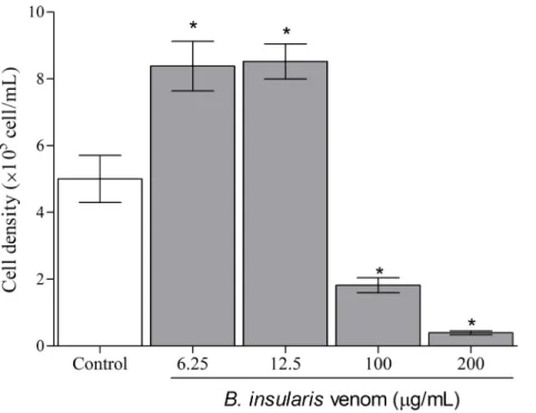
In the present study, macrophages exposed to BiV showed a concentration- and time-dependent effect when submitted to MTT assay. After 2 hours (Fig 2A), all experimental groups treated with the venom showed an increase in cell viability, suggesting a possible prolif-erative effect. The groups exposed for 6, 12 and 24 hours (Fig 2B, 2C and 2D) showed dual effects, with cell death at high concentrations and increased cell viability at lower concentra-tions of BiV. It was also observed an increase on the number of cells treated with BiV at lower concentrations, when analyzed by counting with trypan blue (Fig 3). This cytotoxic effect was confirmed by the determination of LDH activity in the supernatant of experimental groups (Fig 4).
Fig 1. Enzymatic activities exhibited by BiV.[A] Proteolytic activity, assessed by azocasein cleavage method; [B] phospholipasic assay, determined using 4N3OBA as the lipidic substrate; and [C] H2O2production induced by BiV, suggestive of the presence of LAAO.*p<0.05 vs control group (absence of
venom).
In order to investigate the cell death mechanism induced by BiV, treated and untreated cells were collected, washed and labeled with PI and Annexin V-FITC. As result, it was observed that BiV induced an increase in PI+cells (Fig 5). These results indicate that BiV induced necro-sis in the analyzed cells.
In the present study, we observed thatB.insularisvenom causes not only cell death of mac-rophages, but also have a potential proliferative effect. It is known that inflammatory cells, such as macrophages, can respond to damaging effects through cell activation and production of inflammatory mediators. These mediators are partially responsible by both local and sys-temic alterations observed inViperidaeenvenomation.
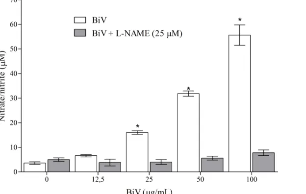
Consequently, aiming to identify the participation of nitric oxide on BiV biological effects, the supernatants from experimental groups treated for 24 hours were collected to determine nitrate/nitrite concentrations as markers of NO production. As shown inFig 6, increases in nitrate/nitrite levels in the groups treated with BiV (25, 50 and 100μg/mL) were observed.
Also, NO release was successfully blocked by the addition of L-NAME (25μM), suggesting the
importance of iNOS pathway in its production. L-NAME is a nonselective inhibitor of NO synthase, which blocks NO production. Therefore, we suggest that NO is released by macro-phages after incubation with BiV by activation of L-arginine-nitric oxide-cGMP pathway.
Fig 2. Time- and concentration-dependent biological effect caused by BiV on RAW 264.7 cells.Cell viability was determined after [A] 2 hours, [B] 6 hours, [C] 12 hours and [D] 24 hours of incubation with BiV, using MTT reduction assay.*p<0.05 vs. control group.
To investigate how NO influences BiV effect on RAW 264.7 cell viability, we performed MTT assays in cells treated with BiV for 24 hours in the presence of L-NAME (25μM).
L-NAME was able to reduce the cytotoxic and proliferative effects of BiV (Fig 7A), suggesting that NO is associated to both pathways. Also, it was demonstrated that BiV induced an increase in iNOS expression (Fig 7B and 7C).
Fig 3. Cell number in machophages treated with BiV.The cell density was determined by counting in Neubauer chamber.*p<0.05 vs. negative control group.
doi:10.1371/journal.pone.0151029.g003
Fig 4. LDH release in macrophages treated with BiV.The percentage of enzymatic release was calculated using a group treated with 1% Triton X-100 as total cell lysis control (data not shown).*p<0.05 vs. negative control group.
Discussion
Pathological events associated with snakebites are complex and have several variables, such as the bite site, amount of venom injected and, specially, venom composition. Therefore, research groups worldwide have sought to elucidate the biological mechanisms involved in these effects, as well the main proteins responsible for the toxicity. This strategy may contribute to the devel-opment of effective treatments and adequate clinical procedures.B.insularisvenom is consid-ered an important source of bioactive and/or toxic proteins, being valuable as a
pharmacological tool.
Although many studies have demonstrated inflammatory influence on the clinical outcomes in snakebites, the direct effects ofViperidaevenoms on cells associated with inflammatory response are not well known, as well as the role of these effects on the consequences of envenomation.
The enzymatic activities observed in this work have been corroborated by other authors. Liraet al. [31] demonstrated that BiV has important proteolytic and phospholipasic activities. Also, many authors have isolated and characterized these proteins in other bothropic venoms, such as svMPs isolated fromB.alternatusandB.pauloensisvenoms [32,33]; svPLA2, identified
inB.pauloensisandB.leucurus[34,35]; and LAAOs, which has been identified in several ven-oms, such asB.jararacussu,B.marajoensisandB.pirajai[36–38].
Valenteet al. [39] found out that almost 1/4 of the total proteins in BiV correspond to svMPs, which may explain its high myotoxicity and cytotoxicity [40]. Different svPLA2
iso-forms and a svLAAO have also been identified.
Fig 5. Necrotic potential of BiV on murine macrophagesin vitro.[A-C] Dot plot diagrams representing respectively control and groups treated with BiV at
50μg/mL and 100μg/mL. [D] Increase in the percentage of PI-labeled cells, suggestive of cell death by necrosis.*p<0.05 vs. control group.
In the present paper, we demonstrate that BiV causes both cell death and proliferation on macrophages, depending of concentration and exposure time. These results are supported by the MTT assay, which verifies the metabolic ability of cells, and by trypan blue method, which assess membrane integrity and number of viable cells. DifferentBothropsandBothropoides
venoms have influence on cell viability. In a previous study, our group showed a similar effect usingBothropoides lutzivenom [41]. Recently, we also demonstrated that BiV has a marked cytotoxic effect on kidney cells, as demonstrated by Mello et al. [42]. In this work, BiV showed high toxicity at 200μg/mL, which is also the concentration with the highest proteolytic and
phospholipasic activities.
This corroborates other studies, as bothropic venom cytotoxicity is commonly associated with svMPs and svPLA2. For instance, svPLA2isolated fromB.alternatusandB.jararacussu
show cytotoxic effect on muscle cells [43,44]. svMPs with cytotoxic effect include the ones iso-lated fromB.asper[45,46]. The effects observed inBothropsandBothropoidesvenoms are probably due to a synergic and/or additive effect between these and other fractions with cyto-toxic effect [43]. An important aspect ofB.insularisvenom is the presence of an Asp49 PLA2,
which presents high enzymatic activity when compared to svPLA2 present in other snake ven-oms, as suchB.jararacaandB.jararacussu[47]. This might be related to its high toxicity.
Cyototoxicity was also confirmed by LDH measurement, which is an important marker of cell lysis, both in clinical and experimental samples. It is released specially after loss of mem-brane integrity, a characteristic of cell death by necrosis.B.atroxvenom causes an increase in LDH release in mice [48]; svMP fromB.neuwiedicauses LDH release in endothelial cells [49].
Melloet al. (2014) demonstrated That BiV induced necrosis and late apoptosis in kidney tubular cells. Additionally, svPLA2fromB.leucurusacts mainly by necrosis, with Ca++
Fig 6. Analyses of nitrate/nitrite concentrations in macrophages after treatment with BiV with and without L-NAME.The analysis was performed using Griess method with modifications.*p<0.05 vs. control group.
involvement and mitochondrial depolarization [7]. Necrosis is also induced byB.pauloensis,B.
diporusandB.pirajaivenoms against tumor cells [50]. Necrosis is a cell death pathway that involves release of intracellular content and inflammatory response. Therefore, direct cytotox-icity might be related to local or systemic effects observed in envenomation, such as platelet activation and hemorrhage observed in svMPs fromB.leucurus[51].
Local and systemic toxicity of animal venoms are dependent of the concomitant effect of its several bioactive proteins. For instance, it was demonstrated that blood flow maintaining is necessary to the establishment of muscular damage induced by svMPs [5]. This might be facili-tated by vasodilatation promoted through NO production in endothelial cells and macro-phages stimulated by svLAAOs and svPLA2[33].
Cytotoxicity can also be induced by svLAAOs, as previously reported [38]. Important func-tional alterations are caused by svLAAOs, such as DNA damage [52], mitochondrial dysfunc-tion [53] and apoptosis [54].
We decided to investigate the role of nitric oxide on the biological effects found in this work. NO is an important mediator of cell viability and proliferation. The proliferative effect of NO on endothelial cells has been demonstrated [55,56]. Moreover, NO is associated with the cytotoxic mechanism of some drugs [57] and hepatic lesions [58]. In macrophages, NO is pro-duced by activated macrophages as a cytotoxic molecule, aiming at the neutralization of micro-organisms [59].
Fig 7. Involvement of NO in BiV biological effects.[A] L-NAME (25μM) inhibited both cytotoxic and proliferative effects of BiV, assessed by MTT method. [B] Western blotting diagrams representing the expression ofβ-actin and iNOS in RAW 264.7 cells in control group and treated with BiV (50μg/mL). [C] Relative expression of iNOS, correlated withβ-actin expression.*p<0.05 vs. control group.
Inflammation induced by bothropic venoms has been associated with NO in several cases. Jararhagin, a svMP isolated fromB.jararacavenom, causes endothelial dysfunction, NO and prostacyclin production, leukocytosis and hemorrhage [60]. It is also important for the devel-opment of edema and inflammatory infiltrate in myonecrosis induced byB.aspervenom [61].
However, it is still unclear whether NO influences the tissue lesion found after bothropic snakebite. Chaveset al. (61) demonstrated that NO production blockage did not change the muscular lesion induced byB.aspervenom. In this way, it seems that NO is involved in hemor-rhage and recruitment of inflammatory cells after lesion, but not directly over tissue lesion.
Beside these results, in vivo blockage of NO production may still be important on manage-ment of envenomation, since this mediator is related to establishmanage-ment of inflammation micro-environment. High tissue NO levels and its metabolites, as such as peroxynitrite, contributes to pathological damage observed in inflammatory site [30]. In contrast, low levels of NO exert protective effect by modulating the immune system [62]. NO management can also be useful in the control of nephrotoxicity induced be snake venoms. It has been suggested that enhance on machophage iNOS expression in kidney after injury aggravates the lesion [63].
Altogether, these results demonstrate the cytotoxic and proliferative effects of BiV on murine macrophages through a necrotic pathway. This result seems to be related mainly to the proteolytic and phospholipasic activities and NO is partially responsible for these effects. We also observed that the venom causes iNOS expression induction.
Author Contributions
Conceived and designed the experiments: RRPPBM AMCM. Performed the experiments: RRPPBM CPM DBL LDT TLS LCFP NTQA. Analyzed the data: RRPPBM TLS. Contributed reagents/materials/analysis tools: MHT RCPLJ EMAJ. Wrote the paper: RRPPBM AMCM.
References
1. Grishin EV. Natural Toxins 2 [Internet]. Advances in experimental medicine and biology. 1996. 231– 236 p. Available:http://www.springerlink.com/index/10.1007/978-1-4613-0361-9
2. Matsui T, Fujimura Y, Titani K. Snake venom proteases affecting hemostasis and thrombosis. Biochim Biophys Acta—Protein Struct Mol Enzymol. 2000; 1477(1–2):146–56.
3. Rajendra W, Armugam A, Jeyaseelan K. Toxins in anti-nociception and anti-inflammation. Toxicon. 2004; 44(1):1–17. PMID:15225557
4. Warrell D a. Snake bite. Lancet [Internet]. Elsevier Ltd; 2010; 375(9708):77–88. Available:http://dx.doi. org/10.1016/S0140-6736(09)61754-2doi:10.1016/S0140-6736(09)61754-2PMID:20109866 5. Gutiérrez JM, Núñez J, Escalante T, Rucavado A. Blood flow is required for rapid endothelial cell
dam-age induced by a snake venom hemorrhagic metalloproteinase. Microvasc Res. 2006; 71(1):55–63. PMID:16337973
6. Jorge RJB, Monteiro HS a., Gonçalves-Machado L, Guarnieri MC, Ximenes RM, Borges-Nojosa DM, et al. Venomics and antivenomics of Bothrops erythromelas from five geographic populations within the Caatinga ecoregion of northeastern Brazil. J Proteomics [Internet]. Elsevier B.V.; 2015; 114:93–114. Available:http://linkinghub.elsevier.com/retrieve/pii/S187439191400534Xdoi:10.1016/j.jprot.2014.11. 011PMID:25462430
7. de Morais ICO, Torres AFC, Pereira GJDS, Pereira TP, Pessoa Bezerra de Menezes RRDP, Mello CP, et al. Bothrops leucurus venom induces nephrotoxicity in the isolated perfused kidney and cultured renal tubular epithelia. Toxicon [Internet]. Elsevier Ltd; 2013; 61(1):38–46. Available:http://dx.doi.org/ 10.1016/j.toxicon.2012.10.005
8. Nunes ES, Souza MAA, Vaz AFM, Silva TG, Aguiar JS, Batista AM, et al. Cytotoxic effect and apopto-sis induction by Bothrops leucurus venom lectin on tumor cell lines. Toxicon [Internet]. Elsevier Ltd; 2012; 59(7–8):667–71. Available:http://dx.doi.org/10.1016/j.toxicon.2012.03.002doi:10.1016/j. toxicon.2012.03.002PMID:22445823
10. Ramos OHP, Terruggi CHB, Ribeiro JU, Cominetti MR, Figueiredo CC, Bérard M, et al. Modulation of in vitro and in vivo angiogenesis by alternagin-C, a disintegrin-like protein from Bothrops alternatus snake venom and by a peptide derived from its sequence. Arch Biochem Biophys. 2007; 461(1):1–6. PMID:17428438
11. Monteiro-Machado M, Tomaz MA, Fonseca RJC, Strauch MA, Cons BL, Borges PA, et al. Occurrence of sulfated fucose branches in fucosylated chondroitin sulfate are essential for the polysaccharide effect preventing muscle damage induced by toxins and crude venom from Bothrops jararacussu snake. Tox-icon [Internet]. 2015; 98:20–33. Available:http://linkinghub.elsevier.com/retrieve/pii/
S0041010115000495doi:10.1016/j.toxicon.2015.02.010PMID:25702961
12. Saturnino-Oliveira J, Santos DDC, Guimarães AG, Santos Dias A, Tomaz MA, Monteiro-Machado M, et al.Abarema cochliacarposExtract Decreases the Inflammatory Process and Skeletal Muscle Injury Induced byBothrops leucurusVenom. Biomed Res Int [Internet]. 2014; 2014:1–9. Available:http:// www.hindawi.com/journals/bmri/2014/820761/
13. Wanderley CWS, Silva CMS, Wong DVT, Ximenes RM, Morelo DFC, Cosker F, et al. Bothrops jarara-cussu snake venom-induces a local inflammatory response in a prostanoid- and neutrophil-dependent manner. Toxicon [Internet]. 2014; 90:134–47. Available:http://linkinghub.elsevier.com/retrieve/pii/ S0041010114002244doi:10.1016/j.toxicon.2014.08.001PMID:25127849
14. Furtado JL, Oliveira GA, Pontes AS, Setúbal SDS, Xavier CV, Lacouth-Silva F, et al. Activation of J77A.1 macrophages by three phospholipases A2 isolated from Bothrops atrox snake venom. Biomed Res Int. 2014; 2014(i).
15. Setubal SDS, Pontes AS, Nery NM, Bastos JSF, Castro OB, Pires WL, et al. Effect of Bothrops bilineata snake venom on neutrophil function. Toxicon [Internet]. Elsevier Ltd; 2013; 76:143–9. Available:http:// dx.doi.org/10.1016/j.toxicon.2013.09.019doi:10.1016/j.toxicon.2013.09.019PMID:24080355 16. Cristina Giannotti K, Leiguez E, Moreira V, Nascimento NG, Lomonte B, Gutiérrez JM, et al. A Lys49
phospholipase A2, isolated from Bothrops asper snake venom, induces lipid droplet formation in mac-rophages which depends on distinct signaling pathways and the C-terminal region. Biomed Res Int. 2013;2013.
17. Rueda AQ, Rodríguez IG, Arantes EC, Setúbal SS, Calderon LDA, Zuliani JP, et al. Biochemical char-acterization, action on macrophages, and superoxide anion production of four basic phospholipases Afrom Panamanian Bothrops asper snake venom. Biomed Res Int. 2013; 2013:1–9.
18. Moreira V, Gutiérrez JM, Amaral RB, Lomonte B, Purgatto E, Teixeira C. A phospholipase A2 from Bothrops asper snake venom activates neutrophils in culture: Expression of cyclooxygenase-2 and PGE2 biosynthesis. Toxicon [Internet]. Elsevier Ltd; 2011; 57(2):288–96. Available:http://dx.doi.org/ 10.1016/j.toxicon.2010.12.004doi:10.1016/j.toxicon.2010.12.004PMID:21147147
19. Grazziotin FG, Monzel M, Echeverrigaray S, Bonatto SL. Phylogeography of the Bothrops jararaca complex (Serpentes: Viperidae): Past fragmentation and island colonization in the Brazilian Atlantic Forest. Mol Ecol. 2006; 15(13):3969–82. PMID:17054497
20. Cogo JC, Lilla S, Souza GHMF, Hyslop S, de Nucci G. Purification, sequencing and structural analysis of two acidic phospholipases A2 from the venom of Bothrops insularis (jararaca ilhoa). Biochimie. 2006; 88(12):1947–59. PMID:17140721
21. Oliveira-Carvalho AL, Guimarães PR, Abreu PA, Dutra DLS, Junqueira-de-Azevedo ILM, Rodrigues CR, et al. Identification and characterization of a new member of snake venom thrombin inhibitors from Bothrops insularis using a proteomic approach. Toxicon. 2008; 51(4):659–71. doi:10.1016/j.toxicon. 2007.11.026PMID:18221976
22. Rucavado A, Henríquez M, García J, Gutiérrez JM. Assessment of metalloproteinase inhibitors clodro-nate and doxycycline in the neutralization of hemorrhage and coagulopathy induced by Bothrops asper snake venom. Toxicon. 2008; 52(7):754–9. doi:10.1016/j.toxicon.2008.08.009PMID:18824013 23. Gomes MSR, Naves de Souza DL, Guimaraes DO, Lopes DS, Mamede CCN, Gimenes SNC, et al.
Biochemical and functional characterization of Bothropoidin: the first haemorrhagic metalloproteinase from Bothrops pauloensis snake venom. J Biochem [Internet]. 2014; 157(3):137–49. Available:http:// jb.oxfordjournals.org/cgi/doi/10.1093/jb/mvu058doi:10.1093/jb/mvu058PMID:25261583
24. Braga MDM, Martins AMC, Amora DN, de Menezes DB, Toyama MH, Toyama DO, et al. Purification and biological effects of l-amino acid oxidase isolated from Bothrops insularis venom. Toxicon. 2008; 51(2):199–207. PMID:17983639
25. Cotrim CA, De Oliveira SCB, Diz Filho EBS, Fonseca FV, Baldissera L, Antunes E, et al. Quercetin as an inhibitor of snake venom secretory phospholipase A2. Chem Biol Interact [Internet]. Elsevier Ireland Ltd; 2011; 189(1–2):9–16. Available:http://dx.doi.org/10.1016/j.cbi.2010.10.016doi:10.1016/j.cbi. 2010.10.016PMID:21056032
27. Lobner D. Comparison of the LDH and MTT assays for quantifying cell death: Validity for neuronal apo-ptosis? J Neurosci Methods. 2000; 96(2):147–52. PMID:10720679
28. Tsikas D. Analysis of nitrite and nitrate in biological fluids by assays based on the Griess reaction: Appraisal of the Griess reaction in the l-arginine/nitric oxide area of research. J Chromatogr B Anal Technol Biomed Life Sci. 2007; 851(1–2):51–70.
29. Souza JM, Alvarez MN, López GV, Paul RS. Macrophage activation induces formation of the anti-inflammatory lipid cholesteryl-nitrolinoleate. 2012; 417(1):223–34.
30. Chae H-S, Song H-H, Kim Y-M, Lee H-K, Oh S-R, Chin Y-W. Euphorbia supina inhibits inflammatory mediators in mouse bone marrow-derived mast cells and macrophages. Int Immunopharmacol [Inter-net]. Elsevier B.V.; 2015; 29(2):966–73. Available:http://linkinghub.elsevier.com/retrieve/pii/ S1567576915301119doi:10.1016/j.intimp.2015.09.008PMID:26386544
31. Lira MS, Furtado MF, Martins LMP, Lopes-Ferreira M, Santoro ML, Barbaro KC. Enzymatic and immu-nochemical characterization of Bothrops insularis venom and its neutralization by polyspecific Bothrops antivenom. Toxicon. 2007; 49(7):982–94. PMID:17382362
32. de Paula FFP, Ribeiro JU, Santos LM, de Souza DHF, Leonardecz E, Henrique-Silva F, et al. Molecular characterization of metalloproteases from Bothrops alternatus snake venom. Comp Biochem Physiol Part D Genomics Proteomics [Internet]. Elsevier Inc.; 2014; 12:74–83. Available:http://linkinghub. elsevier.com/retrieve/pii/S1744117X14000471doi:10.1016/j.cbd.2014.09.001PMID:25463060 33. Naves de Souza DL, Gomes MSR, Ferreira FB, Rodrigues RS, AchêDC, Richardson M, et al.
Bio-chemical and enzymatic characterization of BpMP-I, a fibrinogenolytic metalloproteinase isolated from Bothropoides pauloensis snake venom. Comp Biochem Physiol—B Biochem Mol Biol [Internet]. Else-vier Inc.; 2012; 161(2):102–9. Available:http://dx.doi.org/10.1016/j.cbpb.2011.10.002doi:10.1016/j. cbpb.2011.10.002PMID:22008900
34. Sucasaca-monzón G, Randazzo-moura P, Rocha T, Torres-huaco FD, Vilca-quispe A, Ponce-soto LA, et al. Bp-13 PLA 2 : Purification and Neuromuscular Activity of a New Asp49 Toxin Isolated from Bothrops pauloensis Snake Venom. 2015; 2015.
35. Marangoni FA, Ponce-soto LA, Marangoni S, Cristina E, Landucci T. Unmasking Snake Venom of Bothrops leucurus : Puri�cation and Pharmacological and Structural Characterization of New PLA Bleu TX-III. 2013; 2013.
36. Ullah A, De Souza TDACB, Masood R, Murakami MT, Arni RK. Purification, crystallization and prelimi-nary X-ray diffraction analysis of a class P-III metalloproteinase (BmMP-III) from the venom of Bothrops moojeni. Acta Crystallogr Sect F Struct Biol Cryst Commun. International Union of Crystallography; 2012; 68(10):1222–5.
37. Costa Torres AF, Dantas RT, Toyama MH, Filho ED, Zara FJ, Rodrigues de Queiroz MG, et al. Antibac-terial and antiparasitic effects of Bothrops marajoensis venom and its fractions: Phospholipase A 2 and l-amino acid oxidase. Toxicon. 2010; 55(4):795–804. doi:10.1016/j.toxicon.2009.11.013PMID: 19944711
38. Izidoro LF, Ribeiro MC, Souza GR, Sant'Ana CD, Hamaguchi A, Homsi-Brandeburgo MI, et al. Bio-chemical and functional characterization of an l-amino acid oxidase isolated from Bothrops pirajai snake venom. Bioorganic Med Chem. 2006; 14(20):7034–43.
39. Valente RH, Guimarães PR, Junqueira M, Neves-Ferreira AGC, Soares MR, Chapeaurouge A, et al.
Bothrops insularis venomics: A proteomic analysis supported by transcriptomic-generated sequence data. J Proteomics [Internet]. Elsevier B.V.; 2009; 72(2):241–55. Available:http://dx.doi.org/10.1016/j. jprot.2009.01.001doi:10.1016/j.jprot.2009.01.001PMID:19211044
40. Cogo JC, Prado-Franceschi J, Cruz-Hofling MA, Corrado AP, Rodrigues-Simioni L. Effect of Bothrops insularis venom on the mouse and chick nerve-muscle preparation. Toxicon. 1993; 31(10):1237–47. PMID:8303718
41. Menezes RRPPB, Torres AFC, Silva TSJ da, Sousa DF de, Lima DB, Norjosa DB, et al. Antibacterial and Antiparasitic Effects of Bothropoides lutzi venom. Nat Prod Commun. 2012; 7(1):71–4. PMID: 22428250
42. Mello CP, Morais ICO, Menezes RRPPB, Pereira GJS, Torres AFC, Lima DB, et al. Bothropoides insu-laris venom cytotoxicity in renal tubular epithelia cells. Toxicon [Internet]. 2014; 88:107–14. Available: http://linkinghub.elsevier.com/retrieve/pii/S0041010114001433doi:10.1016/j.toxicon.2014.05.009 PMID:24874890
44. Bonfim VL, De Carvalho DD, Ponce-Soto LA, Kassab BH, Marangoni S. Toxicity of phospholipases A2 D49 (6–1 and 6–2) and K49 (Bj-VII) from Bothrops jararacussu venom. Cell Biol Toxicol. 2009; 25 (6):523–32. doi:10.1007/s10565-008-9106-6PMID:18975118
45. Brenes O, Muñóz E, Roldán-Rodríguez R, Díaz C. Cell death induced by Bothrops asper snake venom
metalloproteinase on endothelial and other cell lines. Exp Mol Pathol [Internet]. Elsevier Inc.; 2010; 88 (3):424–32. Available:http://dx.doi.org/10.1016/j.yexmp.2010.02.002doi:10.1016/j.yexmp.2010.02. 002PMID:20219457
46. Lomonte B, Tarkowski A, Hanson LA. Host response to Bothrops asper snake venom. Analysis of edema formation, inflammatory cells, and cytokine release in a mouse model. Inflammation. 1993; 17 (2):93–105. PMID:8491517
47. Gonçalves-Machado L, Pla D, Sanz L, Jorge RJB, Leitão-De-Araújo M, Alves MLM, et al. Combined
venomics, venom gland transcriptomics, bioactivities, and antivenomics of two Bothrops jararaca popu-lations from geographic isolated regions within the Brazilian Atlantic rainforest. J Proteomics [Internet]. Elsevier B.V.; 2015;1–17. Available:http://linkinghub.elsevier.com/retrieve/pii/S1874391915002213 48. de Souza CA, Kayano AM, Setúbal SS, Pontes AS, Furtado JL, Kwasniewski FH, et al. Local and
sys-temic biochemical alterations induced by Bothrops atrox snake venom in mice. J Venom Res [Internet]. 2012; 3:28–34. Available:http://www.pubmedcentral.nih.gov/articlerender.fcgi?artid=3595125&tool= pmcentrez&rendertype=abstractPMID:23487552
49. Baldo C, Tanjoni I, León IR, Batista IFC, Della-Casa MS, Clissa PB, et al. BnP1, a novel P-I metallopro-teinase from Bothrops neuwiedi venom: Biological effects benchmarking relatively to jararhagin, a P-III SVMP. Toxicon. 2008; 51(1):54–65. PMID:17889921
50. Jorge RJB, Martins AMC, Morais ICO, Ximenes RM, Rodrigues FAR, Soares BM, et al. In vitro studies on Bothrops venoms cytotoxic effect on tumor cells. J Exp Ther Oncol [Internet]. 2011; 9(3):249–53. Available:http://www.ncbi.nlm.nih.gov/pubmed/22070057PMID:22070057
51. Gomes MSR, De Queiroz MR, Mamede CCN, Mendes MM, Hamaguchi A, Homsi-Brandeburgo MI, et al. Purification and functional characterization of a new metalloproteinase (BleucMP) from Bothrops leucurus snake venom. Comp Biochem Physiol—C Toxicol Pharmacol [Internet]. Elsevier B.V.; 2011; 153(3):290–300. Available:http://dx.doi.org/10.1016/j.cbpc.2010.11.008doi:10.1016/j.cbpc.2010.11. 008PMID:21130897
52. Marcussi S, Stábeli RG, Santos-Filho NA, Menaldo DL, Silva Pereira LL, Zuliani JP, et al. Genotoxic effect of Bothrops snake venoms and isolated toxins on human lymphocyte DNA. Toxicon [Internet]. Elsevier Ltd; 2013; 65:9–14. Available:http://dx.doi.org/10.1016/j.toxicon.2012.12.020doi:10.1016/j. toxicon.2012.12.020PMID:23333649
53. Yang CA, Cheng CH, Lee JW, Lo CT, Liu SY, Peng KC. Monomeric l-amino acid oxidase-induced mito-chondrial dysfunction in rhizoctonia solani reveals a novel antagonistic mechanism of Trichoderma har-zianum ETS 323. J Agric Food Chem. 2012; 60(10):2464–71. doi:10.1021/jf203883uPMID:22352318 54. Naumann GB, Silva LF, Silva L, Faria G, Richardson M, Evangelista K, et al. Cytotoxicity and inhibition
of platelet aggregation caused by an l-amino acid oxidase from Bothrops leucurus venom. Biochim Bio-phys Acta—Gen Subj [Internet]. Elsevier B.V.; 2011; 1810(7):683–94. Available:http://dx.doi.org/10. 1016/j.bbagen.2011.04.003
55. Li W, Du D, Wang H, Liu Y, Lai X, Jiang F, et al. Silent information regulator 1 (SIRT1) promotes the migration and proliferation of endothelial progenitor cells through the PI3K / Akt / eNOS signaling path-way. 2015; 8(3):2274–87. PMID:26045735
56. Zhang W, Yan L, Li Y, Chen W, Hu N, Wang H, et al. Roles of miRNA-24 in regulating endothelial nitric oxide synthase expression and vascular endothelial cell proliferation. Mol Cell Biochem [Internet]. Springer US; 2015; 405(1–2):281–9. Available:http://link.springer.com/10.1007/s11010-015-2418-y doi:10.1007/s11010-015-2418-yPMID:25920448
57. Ash D, Subramanian M, Surolia A, Shaha C. Nitric oxide is the key mediator of death induced by fisetin in human acute monocytic leukemia cells. 2015; 5(2):481–97. PMID:25973292
58. Khan MW, Priyamvada S, Khan SA, Khan S, Gangopadhyay A, Yusufi A. Fish/flaxseed oil protect against nitric oxide-induced hepatotoxicity and cell death in the rat liver. Hum Exp Toxicol [Internet]. 2015;1–10. Available:http://het.sagepub.com/cgi/doi/10.1177/0960327115586207
59. Grzegorz B. Peroxinitrite mediator of the toxic actio of nitric oxide. Acta Biochimica Polonica. 1996. p. 645–60. PMID:9104501
61. Chaves F, Teixeira CFP, Gutiérrez JM. Role of nitric oxide in the local and systemic pathophysiological effects induced by Bothrops asper snake venom in mice. Inflamm Res. 2006; 55(6):245–53. PMID: 16955244
62. Hobbs AJ, Higgs A, Moncada S. Inhibition of nitric oxide synthase as a potential therapeutic target. Annu Rev Pharmacol Toxicol [Internet]. 1999; 39:191–220. Available:http://www.ncbi.nlm.nih.gov/ pubmed/10331082PMID:10331082
![Fig 1. Enzymatic activities exhibited by BiV. [A] Proteolytic activity, assessed by azocasein cleavage method; [B] phospholipasic assay, determined using 4N3OBA as the lipidic substrate; and [C] H 2 O 2 production induced by BiV, suggestive of the presence](https://thumb-eu.123doks.com/thumbv2/123dok_br/16424180.195483/5.918.73.821.101.709/enzymatic-activities-exhibited-proteolytic-phospholipasic-determined-production-suggestive.webp)

![Fig 5. Necrotic potential of BiV on murine macrophages in vitro. [A-C] Dot plot diagrams representing respectively control and groups treated with BiV at 50 μg/mL and 100 μg/mL](https://thumb-eu.123doks.com/thumbv2/123dok_br/16424180.195483/8.918.62.778.114.571/necrotic-potential-macrophages-diagrams-representing-respectively-control-treated.webp)
![Fig 7. Involvement of NO in BiV biological effects. [A] L-NAME (25 μM) inhibited both cytotoxic and proliferative effects of BiV, assessed by MTT method](https://thumb-eu.123doks.com/thumbv2/123dok_br/16424180.195483/10.918.300.808.116.586/involvement-biological-effects-inhibited-cytotoxic-proliferative-effects-assessed.webp)
