461 Tropical Plant Pathology 38 (6) November - December 2013
RESEARCH ARTICLE / ARTIGO
Virulence and molecular characterization of Costa Rican
isolates of
Rhizoctonia solani
from common bean
Floribeth Mora-Umaña1,2*, Natalia Barboza2*, Ricardo Alvarado3, Marcela Vásquez2, Graciela Godoy-Lutz4, James R. Steadman4 &Pilar Ramírez2,5
1Servicio Fitosanitario del Estado (SFE), Sabana Sur, 1521-1200 San José, Costa Rica; 2CIBCM, Universidad de Costa Rica (UCR), Ciudad de la Investigación, 11501-2060, San José, Costa Rica; 3Escuela de Estadística, UCR, San Pedro, 11501-2060, San José, Costa Rica; 4University of Nebraska-Lincoln, 406 Plant Sciences Hall, Lincoln, NE 68583-0722, USA; 5Escuela de Biología, UCR, 11501-2060, San José, Costa Rica
Author for correspondence: Natalia Barboza, e-mail: [email protected]
*These authors contributed equally to the work ABSTRACT
Web blight is one of the main diseases that affects bean (Phaseolus vulgaris) cultivation. It infects diverse organs at any growth
stage of the plant and can be present at different altitudes in a humid tropical climate. The causal agent of this disease is Thanatephorus cucumeris in its sexual stage and Rhizoctonia solani in the anamorph. The objective of this investigation was to characterize molecular
isolates of R. solani obtained from bean plants from diverse production regions in Costa Rica and determine their virulence. Fifty-one
samples of symptomatic bean plants were collected using a global positioning system. Virulence was evaluated using the detached leaf technique. Isolates were identified using AG 1-IA, AG 1-IB, AG 1-IC, AG 1-ID, AG 2-2, AG 2-2IIIB, AG 2-2IV and AG 4 molecular markers. ITS sequences were obtained and analyzed with BLAST, aligned, and a phylogenetic tree was constructed. A high degree of virulence and genetic variability between isolates was identified and the anastomosis subgroups of isolates were independent of their geographical origin.
Key words: Phaseolus vulgaris, anastomosis group (AG), genetic variability, molecular characterization, sclerotium size, virulence.
INTRODUCTION
Common bean (Phaseolus vulgaris L.) is one of the
basic components of Costa Rican and Latin-American diets. It is a valuable legume that supplies a significant amount of nutrients in developing countries (FAO, 2004; Godoy-Lutz et al., 2008). Web blight (WB) is a common and destructive bean disease in tropical and subtropical regions of the American continent. The causal agent is the fungus
Thanatephorus cucumeris (Frank) Donk, the sexual stage of Rhizoctonia solani Kühn, a basidiomycete. It survives from
one season to the next mostly via sclerotia or mycelium in crop residues in the field (Godoy-Lutz et al.,2000; Polanco et al., 1996). It is endemic to Central America and the Caribbean, and causes defoliation and severe losses in seed quality and production (Godoy-Lutz et al., 2003). Crop losses above 20% have been reported in more than 200 cultivars around the world (Beaver et al.,2002). Chemical and cultural control are the most common management techniques (Beebe et al., 1991; Godoy-Lutz et al., 1996a; Polanco et al., 1996), but during the last decades the
biocontrol using different bacterial genus has been adopted as a sustainable approach for crop health management. However, they are not sufficient to avoid production losses (Solanki et al., 2013; Kanini et al., 2013).
A common bean line with high tolerance to WB in different geographical regions has not been obtained or identified (Beaver et al., 2002). Breeding efforts to create
a WB resistant common bean have been limited by fungal variability in varietal reactions and the lack of knowledge of pathogen variation, mating systems and disease resistance mechanisms (Godoy-Lutz et al., 2003; Godoy-Lutz et al., 2008).
R. solani is a complex, heterogeneous species
of several anastomosis groups (AG), which are defined as isolates with somatic or vegetative compatibility (Matsumoto, 2005). Worldwide, 14 AGs and numerous subgroups have been identified based on physical, chemical, pathogenic and molecular characteristics. Isolates can exist as multinucleate homokaryons or heterokaryons and present diverse mating strategies (homothallic or heterothallic) (González et al., 2012). Phaseolus vulgaris isolates belong
Tropical Plant Pathology 38 (6) November - December 2013 462
F. Mora-Umaña et al.
When evaluated on the same plant species, virulence between AGs varies from highly virulent to non-virulent. Godoy-Lutz et al. (1996b) observed discrepancies in the field since virulence patterns of isolates from different geographical regions may vary within the same subgroup, for example AG 4 or AG 1-IE (Godoy-Lutz et al., 2000). Awareness of the pathogenicity and genetic diversity of isolates helps to explain the variation in the cultivar reaction in pathogen-host interactions (Godoy-Lutz et al., 2003).
The development and progress of the disease is favored by high temperature and relative humidity. The pathogen can be spread as rain splashed sclerotia or mycelia, often in association with plant debris, airborne basidiospores, infested soil debris and asymptomatic infected seeds (Echandi, 1965; Godoy-Lutz et al., 1996a; Godoy-Lutz et al., 2003).
The classical hyphal fusion for identification and classification of Rhizoctonia spp. into anastomosis
groups has been widely used and is still valid. It has been genetically confirmed in recent years by the use of DNA-based molecular methods. One of the most useful tool is the analysis of sequences from rDNA ITS (Grosch et al. 2004, 2007). This has been used for identification of anastomosis groups. According to Pannecoucque & Höffe (2009), ITS regions evolve faster and have been used often for molecular identification of closely related fungi. Using this advantage, Godoy-Lutz et al. (2008) developed specific primers for each AG that affected bean. Recently, Wibberg et al. (2013) and Zheng et al. (2013) published the genome of R. solani
AG 1-IB and AG 1-IA, respectively. All this information can be used to learn about the structure and gene function and to develop phylogeny and diversity studies.
This investigation was conducted to determine the virulence of R. solani isolates infecting bean in different
climate regions of Costa Rica, and the intraspecific diversity of WB isolates using molecular markers specific for each AG group and ITS sequences.
MATERIALS AND METHODS
Sampling, isolation and morphological fungus identification
Leaf samples of bean plants with symptoms of web blight were collected in six climate regions in Costa Rica (South Pacific, Central Valley, North Region, Southern Mountain Region, North Pacific and Caribbean Region) during 2007 and 2009. A global positioning system (GPS) was used to mark the sampling sites. The pathogen was isolated and identified considering the hyphal morphological traits as branching at right angles with constriction at the base and presence of a septum near the point of origin. Monilioid cells and presence of multinucleated cells were checked out and verified (Naito, 1996). Isolates were stored at 4°C in Petri dishes on potato-dextrose-agar medium (PDA). A photographic database was established, with pictures and a morphological description
(mycelium color, size and shape of the sclerotia) of each isolate.
Virulence
Virulence of the 51 isolates was evaluated on two bean cultivars (Bribrí and Brunca) using the detached leaf method (Steadman et al., 1997). Severity was evaluated 24,
48, and 72 hours post inoculation (h.p.i) using the CIAT 1-9 severity scale, where 1= no visible symptoms of disease, 3= 5% of the foliar area infected, 5=10% of the area infected, 7=25% of the area infected and 9=>50% area infected (van Schoonhoven et al.,1987). In each evaluation, photographs were taken to determine the affected area with Scion Image alfa version 4.0.3.2.
Statistical analysis of data
Analysis of variance was used for isolate comparison, with time as a covariable, and isolate and variety as factors. The interaction between time and isolate was also included. Due to the high interaction between variables, the numerous samples and the low significance of cultivars, isolates were compared without considering cultivar. When differences were encountered, multiple Tukey comparisons were made.
Isolates were classified in three categories according to the virulence level: group A: isolates with average affected leaf areas less than 6.66 cm2; group B: isolates with average areas between 6.66 cm2 and 12.5 cm2; group C: isolates with averages higher than 12.5 cm2. Each evaluation was compared separately by time and group; however, only the 72 hour evaluations were reported because of their greater importance. Equality of variances and normality of residue, indispensable for the ANOVA, were determined. For the analysis a program was written in R language version 2.10.0.
DNA extraction and amplification
DNA was extracted from lyophilized mycelium of
R. solani, previously cultivated on AC agar (Sigma) with
120 µL/L (50 mg/mL) of ampicillin, according to the modified protocol by Pascual et al.(2000). Extracted DNA was treated with 5 µL RNase A (Fermentas, 10 mg/mL) and quantified with a spectrophotometer (Thermo Spectronic Helios Y).
463 Tropical Plant Pathology 38 (6) November - December 2013
a temperature profile of 95°C for 45s to denature the DNA, 45s at 58-62°C for annealing and 1 min at 72°C for extension. The third step consisted of 72°C for 10 min. The PCR products were separated by electrophoresis in a 1% agarose gel in TAE buffer for 90 min at 100V. The gel was dyed with ethidium bromide, observed under UV light and photographed with a Kodak EDAS 290 system. A molecular weight marker of 100 bp (50 ng/µL) (Fermentas) was used.
To confirm the relationships among the isolates, 35 isolates were selected at random and amplified using universal primers ITS4 and ITS5. A 25 µL PCR mixture was prepared using 0.5 µL of Taq DNA polymerase (Dreamtaq, Fermentas), 2.5 µL dNTPs mix (25mM), 2.5 µL master mix buffer (10x) Fermentas, 0.4 µL of each primer (10 µm) and 50 ng/ µL of the DNA sample. DNA was amplified with the oligonucleotides ITS4 (TCCTCCGCTTATTGATATGC) and ITS5 (GGAAGTAAAAGTCGTAACAAGG) (White et al., 1990) using a PTC-100 thermocycler (MJ Research) with a temperature profile of 94°C for 1 min, followed by 30 cycles of 94°C for 40 s, 55°C for 1 min, and 72°C for 1 min, and a final step at 72°C for 5 min. PCR products of 740 bp were visualized as indicated before. PCR products were purified with the QIAquick PCR Purification Kit (Qiagen) and sequenced using both primer strands by using the BigDyeTerminator v.3.1 cycle sequencing kit (Applied Biosystems) and an ABI Prism 3130 (Applied Biosystems). With the identification of each anastomosis subgroup and the GPS data, a map was constructed using Arcview GIS 3.2.
Sequence data for the complete ITS-5.8S rDNA of isolates of AG 1 and AG 2 and AG 4 was manually checked and edited using the Multiple Sequence Alignment of BioEdit software (Hall, 2007). Sequences of the complete ITS-5.8S rDNA region were compared with nine reference sequences (NCBI GenBank): AJ868444 (AG 1), AF308624 (AG 2-2), AF308623 (AG 2 WB), JF519829 (AG P), AB195928 (AG 1-IA), AB122139 (AG 1-IB), AB122141 (AG 1-IC), EF197798 (AG 1-ID), JF946728 (AG 1-IE) and JF946731 (AG 1-IF), JQ669932 (AG4).
All nucleotide substitutions were equally weighted and unordered. Alignment gaps were treated as missing data. The sequences were analyzed with BLAST for confirmation of the AG, aligned with the MAFFT algorithm in the GUIDANCE server and analyzed with MrBayes3.2 using 1,000,000 Markov chains (Ronquist et al., 2011) for phylogenetic tree construction with a GTR model. The tree was visualized in MEGA5.2 (Tamura et al., 2011). To ensure the robustness of the analysis additional trees were constructed using Neighbor joining (NJ) and Maximum likelihood (ML) methods with 5,000 bootstrap replications using MEGA 5.2.
RESULTS Sampling, isolation and identification
During 2007-2009, foliar bean tissue infected with R. solani was collected throughout the national
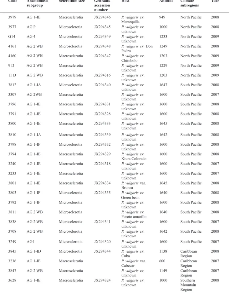
territory. Anastomosis subgroup, sclerotium size, GenBank accession, host, altitude, climate subregions and collecting year for each isolate are presented in Table 1. Fifty-one isolates of Rhizoctonia solani were obtained. The mycelium
coloration varied in intensity from a dark brown to beige and/or white. Sclerotia size varied from 5 to 20 mm, for macrosclerotia single or aggregated; and 1 mm for isolates with microsclerotia. Also all those mycelium presented multinucleated cells and near-right-angle branching pattern.
Virulence
The CIAT scale scores were compared with measured affected areas for each isolate and a linear correlation of 0.46 was obtained. Although significant, the value was lower than expected. Since the CIAT scale is subjective, these data were not used and conclusions were based on average measured affected leaf areas.
Group A was characterized by a number of heterogeneous isolates (p<0.0001, 27, 350) (Figure 1). Tukey comparisons showed differences among isolates when analyzed by pairs. In group B, significant differences in average areas were observed between isolates (p<0.0001, 16, 264) however a great homogeneity was observed. This group included 17 isolates and the Tukey comparisons showed differences between isolate 3 and those with lower averages.
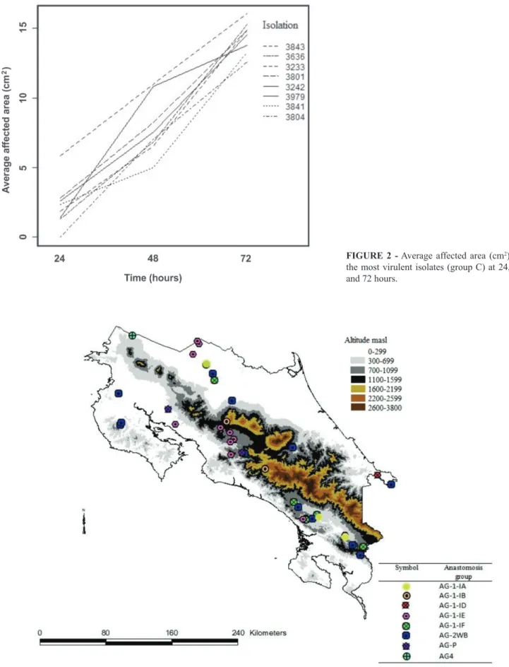
Group C was homogenous at every evaluation time, as observed in the Figure 2. The ANOVA confirmed that there was no significant difference between average areas of different isolates at the 72 hour evaluations (p=0.13, 7, 119). Isolates 3843, 3636, 3233, 3801, 3242, 3841 and 3804 are the members of this group. These seven isolates are AG 1, but belong to different subgroups and climate regions. Only isolate 3979 belongs to AG 2-2WB.
Molecular identification of the AGs
A high degree of variability between isolates was determined using specific primers for each AG and subgroup. AG groups AG 1, AG 2 and AG 4 were identified in different climate regions of Costa Rica using specific primers for each AG. Subgroups AG 1-IA, AG 1-IB, AG 1-ID, AG 1-IE, AG 1-IF, AG 2WB and AG 4 were identified. The AG was independent of geographical origin and climate region (Table 1).
A map with the distribution of the anastomosis subgroup was constructed. In the Figure 3 there is possible to observe that AG 1-IE subgroup was found with the greatest frequency in the different climate subregions, which are characterized by differences in altitude and precipitation. A single isolate of AG 1-1A was identified in a sample from the South Pacific region. AG 1-1B was found in the South Pacific, Central Valley and Northern regions.
Tropical Plant Pathology 38 (6) November - December 2013 464
F. Mora-Umaña et al.
Code Anastomosis subgroup
Sclerotium size GenBank accession number
Host Altitude Climate subregions
Year
3979 AG 1-IE Macrosclerotia JX294346 P. vulgariscv.
mantequilla
949 North Pacific 2008
3977 AG P Microsclerotia JX294345 P. vulgariscv.
unknown
1000 North Pacific 2008
G14 AG 4 Microsclerotia JX294349 P. vulgariscv.
unknown
1233 North Pacific 2009
4161 AG 2WB Microsclerotia JX294348 P. vulgariscv. Don
Pedro
1249 North Pacific 2008
4160 AG 2WB Macrosclerotia JX294347 P. vulgariscv.
Chimbolo
1203 North Pacific 2009
9 D AG 2WB Macrosclerotia P. vulgariscv.
unknown
1229 North Pacific 2009
11 D AG 2WB Macrosclerotia JX294316 P. vulgariscv.
unknown
1203 North Pacific 2009
3812 AG 1-IA Microsclerotia JX294340 P. vulgariscv.
unknown
1647 South Pacific 2008
3307 AG 2WB Macrosclerotia P. vulgariscv.
unknown
1600 South Pacific 2007
3796 AG 1-IE Macrosclerotia JX294331 P. vulgariscv.
unknown
1600 South Pacific 2008
3791 AG 1-IE Macrosclerotia JX294328 P. vulgariscv.
unknown
1600 South Pacific 2008
3800 AG 1-IE Macrosclerotia JX294333 P. vulgariscv.
unknown
1645 South Pacific 2008
3810 AG 1-IA Macrosclerotia JX294339 P. vulgariscv.
unknown
1642 South Pacific 2008
3798 AG 1-IF Microsclerotia JX294332 P. vulgariscv.
unknown
1600 South Pacific 2008
3794 AG 1-IE Macrosclerotia JX294329 P. vulgariscv.
Kiura Colorado
1600 South Pacific 2008
3240 AG 1-IE Macrosclerotia JX294318 P. vulgariscv.
unknown
1600 South Pacific 2007
3233 AG 1-IE Macrosclerotia P. vulgariscv.
unknown
1600 South Pacific 2007
3801 AG 1-IE Macrosclerotia JX294334 P. vulgarisvar.
Brunca
1645 South Pacific 2008
3803 AG 1-IF Microsclerotia JX294335 P. vulgariscv.
green bean
1640 South Pacific 2008
3792 AG 1-IF Microsclerotia P. vulgariscv.
unknown
1600 South Pacific 2008
3811 AG 2 WB Microsclerotia P. vulgariscv.
Poroto amarillo
1640 South Pacific 2008
3838 AG 2 WB Microsclerotia JX294341 P. vulgariscv.
unknown
1600 South Pacific 2007
3708 AG 2 WB Microsclerotia P. vulgariscv.
unknown
1642 South Pacific 2008
3249 AG4 Micrisclertoria JX294320 P. vulgariscv.
unknown
1600 South Pacific 2007
3845 AG 1-ID Microsclerotia JX294344 P. vulgariscv.
Cuba
1138 Caribbean
Region
2008
3236 AG 1-IE Macrosclerotia P. vulgarisvar.
Cabecar
600 Caribbean
Region
2007
3847 AG 2 WB Macrosclerotia P. vulgariscv.
unknown
1149 Caribbean
Region
2007
3628 AG 1-IE Macrosclerotia JX294324 P. vulgariscv.
unknown
1000 Southern
Mountain Region
2008
P. vulgaris
P. vulgaris
P. vulgaris
P. vulgaris
TABLE 1 - Code, anastomosis subgroup (AG), sclerotium size, GenBank accession, host, altitude (meters above sea level), climate subregions and collecting year of 51 isolates of Rhizoctonia solani collected in different geographic regions of Costa Rica and reference
accessions used for phylogenetic tree construction obtained from GenBank
Cont.
Mantequilla
AG 2 WB
AG 2 WB
AG 2 WB
AG 2 WB
Green bean
465 Tropical Plant Pathology 38 (6) November - December 2013
3629 AG P Microsclerotia JX294325 P. vulgarisvar.
Mexico 80
1125 Southern
Mountain
Region
2008
3668 AG 1-IB Macrosclerotia P. vulgariscv.
unknown
543 Southern
Mountain
Region
2008
3620 AG 2 WB Microsclerotia JX294321 P. vul gariscv.
unknown
1097 Southern
Mountain
Region
2008
3806 AG 1 IA Macrosclerotia JX294336 P. vulgariscv.
unknown
1107 Northern
Region
2008
3804 AG 1-IB Microsclerotia P. vulgariscv.
unknown
1214 Northern
Region
2008
3808 AG 1-IE Macrosclerotia JX294338 P. vulgariscv.
unknown
1202 Northern
Region
2008
3077 AG 1-IE Macrosclerotia P. vulgariscv. unknown
1000 Northern Region
2007
3093 AG 1-IE Macrosclerotia JX294317 P. vulgariscv.
unknown
1000 Northern
Region
2007
3840 AG 1-IF Microsclerotia P. vu lgariscv.
unknown
1236 Northern
Region
2008
3841 AG 1-IB Microsclerotia JX294342 P. vulgariscv. unknown
1123 Central Valley 2008
3843 AG 1-IE Macrosclerotia JX294343 P. vulgariscv.
unknown
600 Central Valley 2008
3973 AG 1-IE Macrosclerotia P. vulga riscv.
unknown
1042 Central Valley 2008
Cach AG 2 WB Microsclerotia P. vulgariscv.
unknown
1042 Central Valley 2008
3677 AG 1-IE Macrosclerotia JX294326 P. vulgariscv.
unknown
543 Central Valley 2008
3667 AG 1-IE Macrosclerotia P. vulgariscv.
Landrace
700 Central Valley 2008
3793 AG 1-IF Microsclerotia P. vulgariscv.
unknown
700 Central Valley 2008
3795 AG 1-IF Microsclerotia JX294330 P. vulgariscv.
unknown
1470 Central Valley 2008
3807 AG 2 WB Macrosclerotia JX294337 P. vulgariscv.
unknown
646 Central Valley 2008
3809 AG 2 WB Macrosclerotia P. vulgariscv.
unknown
646 Central Valley 2008
AG 1-IB AJ868444
AG 2-2 AF308624
AG 2 WB AF308623
AG P JF519829
AG 1 IA AB195928
AG 1 IB AB122139
AG 1 IC AB122141
AG 1 ID EF197798
AG 1 IE JF946728
AG 1 IF JF946731
AG 4 JQ669932
Code Anastomosis subgroup
Sclerotium size GenBank accession number
Host Altitude Climate subregions
Year
3628 AG 1-IE Macrosclerotia JX294324 P. vulgariscv. unknown
1000 Southern Mountain Region
2008
3626 AG 1-IE Macrosclerotia JX294323 P. vulgariscv. Landrace
1125 Southern Mountain Region
2008
3636 AG 1-IE Macrosclerotia P. vulgariscv. unknown
1000 Southern Mountain Region
2008
3242 AG 1-IF Microsclerotia JX294319 P. vulgariscv. unknown
1600 Southern Mountain Region
2007
3625 AG P Macrosclerotia JX294322 P. vulgariscv. Mantequilla
1000 Southern Mountain
2008
Region
Cont.
Cont.
-Tropical Plant Pathology 38 (6) November - December 2013 466
F. Mora-Umaña et al.
3629 AG P Microsclerotia JX294325 P. vulgarisvar.
Mexico 80
1125 Southern
Mountain
Region
2008
3668 AG 1-IB Macrosclerotia P. vulgariscv.
unknown
543 Southern
Mountain
Region
2008
3620 AG 2 WB Microsclerotia JX294321 P. vul gariscv.
unknown
1097 Southern
Mountain
Region
2008
3806 AG 1 IA Macrosclerotia JX294336 P. vulgariscv.
unknown
1107 Northern
Region
2008
3804 AG 1-IB Microsclerotia P. vulgariscv.
unknown
1214 Northern
Region
2008
3808 AG 1-IE Macrosclerotia JX294338 P. vulgariscv.
unknown
1202 Northern
Region
2008
3077 AG 1-IE Macrosclerotia P. vulgariscv.
unknown
1000 Northern
Region
2007
3093 AG 1-IE Macrosclerotia JX294317 P. vulgariscv.
unknown
1000 Northern
Region
2007
3840 AG 1-IF Microsclerotia P. vu lgariscv.
unknown
1236 Northern
Region
2008
3841 AG 1-IB Microsclerotia JX294342 P. vulgariscv. unknown
1123 Central Valley 2008
3843 AG 1-IE Macrosclerotia JX294343 P. vulgariscv.
unknown
600 Central Valley 2008
3973 AG 1-IE Macrosclerotia P. vulga riscv.
unknown
1042 Central Valley 2008
Cach AG 2 WB Microsclerotia P. vulgariscv. unknown
1042 Central Valley 2008
3677 AG 1-IE Macrosclerotia JX294326 P. vulgariscv.
unknown
543 Central Valley 2008
3667 AG 1-IE Macrosclerotia P. vulgariscv.
Landrace
700 Central Valley 2008
3793 AG 1-IF Microsclerotia P. vulgariscv. unknown
700 Central Valley 2008
3795 AG 1-IF Microsclerotia JX294330 P. vulgariscv.
unknown
1470 Central Valley 2008
3807 AG 2 WB Macrosclerotia JX294337 P. vulgariscv.
unknown
646 Central Valley 2008
3809 AG 2 WB Macrosclerotia P. vulgariscv. unknown
646 Central Valley 2008
AG 1-IB AJ868444
AG 2-2 AF308624
AG 2 WB AF308623
AG P JF519829
AG 1 IA AB195928
AG 1 IB AB122139
AG 1 IC AB122141
AG 1 ID EF197798
AG 1 IE JF946728
AG 1 IF JF946731
AG 4 JQ669932
Code Anastomosis subgroup
Sclerotium size GenBank accession number
Host Altitude Climate subregions
Year
3628 AG 1-IE Macrosclerotia JX294324 P. vulgariscv. unknown
1000 Southern Mountain Region
2008
3626 AG 1-IE Macrosclerotia JX294323 P. vulgariscv. Landrace
1125 Southern Mountain Region
2008
3636 AG 1-IE Macrosclerotia P. vulgariscv. unknown
1000 Southern Mountain Region
2008
3242 AG 1-IF Microsclerotia JX294319 P. vulgariscv. unknown
1600 Southern Mountain Region
2007
3625 AG P Macrosclerotia JX294322 P. vulgariscv. Mantequilla
1000 Southern Mountain
2008
Region 3629 AG P Microsclerotia JX294325 P. vulgarisvar.
Mexico 80
1125 Southern
Mountain
Region
2008
3668 AG 1-IB Macrosclerotia P. vulgariscv.
unknown
543 Southern
Mountain
Region
2008
3620 AG 2 WB Microsclerotia JX294321 P. vul gariscv.
unknown
1097 Southern
Mountain
Region
2008
3806 AG 1 IA Macrosclerotia JX294336 P. vulgariscv.
unknown
1107 Northern
Region
2008
3804 AG 1-IB Microsclerotia P. vulgariscv.
unknown
1214 Northern
Region
2008
3808 AG 1-IE Macrosclerotia JX294338 P. vulgariscv.
unknown
1202 Northern
Region
2008
3077 AG 1-IE Macrosclerotia P. vulgariscv.
unknown
1000 Northern
Region
2007
3093 AG 1-IE Macrosclerotia JX294317 P. vulgariscv.
unknown
1000 Northern
Region
2007
3840 AG 1-IF Microsclerotia P. vu lgariscv.
unknown
1236 Northern
Region
2008
3841 AG 1-IB Microsclerotia JX294342 P. vulgariscv.
unknown
1123 Central Valley 2008
3843 AG 1-IE Macrosclerotia JX294343 P. vulgariscv.
unknown
600 Central Valley 2008
3973 AG 1-IE Macrosclerotia P. vulga riscv.
unknown
1042 Central Valley 2008
Cach AG 2 WB Microsclerotia P. vulgariscv.
unknown
1042 Central Valley 2008
3677 AG 1-IE Macrosclerotia JX294326 P. vulgariscv.
unknown
543 Central Valley 2008
3667 AG 1-IE Macrosclerotia P. vulgariscv.
Landrace
700 Central Valley 2008
3793 AG 1-IF Microsclerotia P. vulgariscv.
unknown
700 Central Valley 2008
3795 AG 1-IF Microsclerotia JX294330 P. vulgariscv.
unknown
1470 Central Valley 2008
3807 AG 2 WB Macrosclerotia JX294337 P. vulgariscv.
unknown
646 Central Valley 2008
3809 AG 2 WB Macrosclerotia P. vulgariscv.
unknown
646 Central Valley 2008
AG 1-IB AJ868444
AG 2-2 AF308624
AG 2 WB AF308623
AG P JF519829
AG 1 IA AB195928
AG 1 IB AB122139
AG 1 IC AB122141
AG 1 ID EF197798
AG 1 IE JF946728
AG 1 IF JF946731
AG 4 JQ669932
Code Anastomosis subgroup
Sclerotium size GenBank accession number
Host Altitude Climate subregions
Year
3628 AG 1-IE Macrosclerotia JX294324 P. vulgariscv. unknown
1000 Southern Mountain Region
2008
3626 AG 1-IE Macrosclerotia JX294323 P. vulgariscv. Landrace
1125 Southern Mountain Region
2008
3636 AG 1-IE Macrosclerotia P. vulgariscv. unknown
1000 Southern Mountain Region
2008
3242 AG 1-IF Microsclerotia JX294319 P. vulgariscv. unknown
1600 Southern Mountain Region
2007
3625 AG P Macrosclerotia JX294322 P. vulgariscv. Mantequilla 1000 Southern Mountain 2008 Region Cont.
FIGURE 1 - Degree of virulence (average affected area) of Rhizoctonia solani
isolates inoculated in vivo
-467 Tropical Plant Pathology 38 (6) November - December 2013
FIGURE 2 - Average affected area (cm2) by the most virulent isolates (group C) at 24, 48 and 72 hours.
Tropical Plant Pathology 38 (6) November - December 2013 468
F. Mora-Umaña et al.
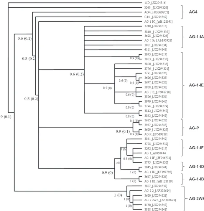
FIGURE 4 - Phylogenetic analysis using Bayesian inference based on ITS sequences, using MrBayes 3.2 with 1,000,000 Markov chains, with a multiple sequence alignment with the MAFFT algorithm on the GUIDANCE server.
previously been reported in P. vulgaris. The phylogenetic
tree (Figure 4) confirmed the grouping of isolates in two clades. The first clade contained accessions of AG 4 and AG 2 WB from diverse climate regions. Bootstrap values from 60-100% allowed subgroup separation. Accessions of the AG 1 subgroup were included in second clade. This was a heterogeneous group that included AG 1-ID, AG 1-IE, AG 1-IF and AG P from different geographical regions of the country. NJ and MP trees were constructed (data not shown) and similar results were found.
DISCUSSION
Fifty-one isolates of R. solani were obtained from
469 Tropical Plant Pathology 38 (6) November - December 2013
characteristics could be a key for distinguishing between isolates (Godoy-Lutz et al., 2003). The different phenotypes found in this study is indicative of the high variation that this fungi presents in our study.
Of the 51 isolates, 18 were from the South Pacific Region, and seven from the Northern Pacific Region (Table 1), both areas of major bean production in Costa Rica. The lowest number of isolates was found in the Caribbean, which is not a bean producing region and it is located near the Atlantic shore. The isolates with the highest virulence according to the CIAT scale and the measured area were placed in group C. Isolate 3843 was considered the most virulent at 24 h.p.i, because it caused the highest amount of affected foliar tissue in the shortest time period. At 48 hours there was no more healthy foliar tissue to infect, so at 72 hours, the affected area was similar to the other isolates. This isolate is characterized by macroesclerotia. Godoy-Lutz et al. (2003) mention that AG 1 macrosclerotia producing isolates were the most virulent, and AG 4 no if you read well is AG 2-2 the least virulent. Similar results have been observed in Latin America and the Caribbean, where virulence patterns vary between regions and genotypes (González-Vera et al., 2010).
Diverse levels of virulence between and within the identified groups were observed. The most virulent isolate (3843) was identified as AG 1-IE, and the least aggressive isolate was AG 2 WB. Isolate 3843 was from the Central Valley region, an area with precipitation from 1500 to 2500 mm3 and average temperatures between 19 and 22°C. With these data one might infer that these are favorable conditions for the disease development. Beaver et al. (2002) report differences in virulence on common bean using isolates of R. solani from Central America and the
Caribbean. Lee et al.(2006) mention that the virulence of R. solani can be affected by environmental conditions present
at the beginning of the infection.
No relation was observed between virulence, AG and geographical origin. Different AGs and subgroups were present in all of the regions sampled, regardless of edaphic and climatic conditions. This demonstrates the great adaptability and survival capacity of this pathogen, an important consideration in breeding P. vulgaris for
resistance. The variable genetic composition of the fungus can affect the dynamics of the pathogen-host relationship, as pathogens are able to adapt to specific ecological niches in diverse agrosystems (Araya, 2003).
Results of this study show that WB in common bean in Costa Rica is caused at least by four AGs, AG 1, AG 2, AG 4 and AG P, and diverse subgroups, AG 1-IA, AG 1-IB, AG 1-IC, AG 1-ID, AG 1-IE, AG 1-IF, AG 2-2WB and AG 4. The dominant groups in this study were AG 1 (65%) and the AG 1-IE subgroup (37%). AG 1-IE and AG 2 WB subgroups were found in every climate region sampled. AG 1-IE was the most frequent (Table 1). These results are consistent with reports in the literature that show a prevalence of this subgroup in field populations causing WB
in common bean in several Latin American and Caribbean countries (Echandi, 1965; Godoy-Lutz et al., 1996b).
The presence of different AGs found in this study in different geographical regions suggests that bean varieties are susceptible to a broad range of AG subgroups (Godoy-Lutz et al., 2008). The presence of AG P, not previously
reported in bean, in the North Pacific and Southern Mountain regions, suggests that new AGs may be appearing. AG P is a heterogeneous group, as shown by the percent similarity between isolates of this AG using MP and NJ methods. For some isolates, similarity ranged from 93% to 100%, but for others it was lower, from 90% to 94%. This AG may include several subgroups, but additional work with more isolates is needed to confirm the existence of AG P subgroups (Sharon et al., 2008).
Identification with specific primers aids in determining the prevalence of an AG or subgroup in a certain geographic area. Population genetics of this pathogenic fungus may be affected by diverse mechanisms not yet elucidated. Since in this study AGs were not related to place of origin, it appears that many mechanisms, such as mating systems, reproduction strategies and gene flow, could be contributing to pathogen dispersal (González et al., 2006). Bean programs in Costa Rica in collaboration with farmers associations have been done during the last years to provide certified seeds. However, the presence of anastomosis subgroups between regions could be an indicative that contaminated seed movement still spreads in the country. Godoy-Lutz et al. (2003) affirm that gene flow may be occurring through seed movement as the pathogen can be spread on asymptomatic seeds (Godoy-Lutz, 2003). One of the most important results of this research was to confirm the presence of different anastomosis subgroups in the country and their virulence. Systematical sampling of open field crops is necessary to generate recommendations for disease management such as use of crop rotations. According to Okubara et al. (2008) there are voids in the knowledge of pathogen dispersal, crop loss and the role of environmental conditions such as rain and temperature on pathogen populations.
The variable genetic composition of R. solani
can be affected by the dynamics of the host-pathogen relationship, genetic flexibility and the degree of adaptation to the ecological niche in diverse agroecosystems (Godoy-Lutz et al., 2008). Our phylogenetic tree shows that some anastomosis subgroups are not closely related to the controls. This may be an indication of the mentioned plasticity of the genetic composition of the R. solani AG subgroups since they present higher sequence
variability than other isolates of R. solani. AG P is closer
Tropical Plant Pathology 38 (6) November - December 2013 470
F. Mora-Umaña et al.
The support percentages of the Bayesian analysis ranged from 60% to 100%, most of the results were supported by a high value. This supports the tree structure for these isolates and their identification within an AG group or subgroup. Previous studies have shown that AG 4 and AG 2-WB have a close phylogenetical relationship, supporting our hypothesis presented on the tree. In this case, the sequences used were 5.8S rDNA with flanking sequences of ITS1 and ITS2. Sharon et al. (2008) affirm that there is a difference between ITS 1 and ITS2 in terms of variability. ITS 1 sequences are more variable than ITS2 among fungal isolates, but the combined 3 sequences ITS1 + 5.8S + ITS2 used in this study give more information than either sequence separately.
Proper deployment of molecular methods allows the identification and over time monitoring of subgroups associated with the disease in high epidemic risk areas and provides an important tool for analysis of isolate variation in the field and the possible generation of more pathogenic AG subgroups. Knowledge of virulence variation and distribution of the AGs in the country is useful in the assessment of management and breeding strategies in Costa Rica.
ACKNOWLEDGEMENTS
The authors would like to acknowledge the Consejo Nacional de Rectores (CONICIT), Vicerrectoría de Investigacion, Universidad de Costa Rica and University of Nebraska-Lincoln.
REFERENCES
Araya CM (2003) Coevolución de interacciones hospedante-patógeno en frijol común. Fitopatologia Brasileira 28:221-228. Beaver J, Godoy G, Rosas JC, Steadman JR (2002) Estrategia para la seleccionar frijol común con mayor resistencia a mustia hilachosa. Agronomía Mesoamericana 13:67-72.
Beebe SE, Pastor-Corrales MA (1991) Breeding for disease resistance. In: van Schoonhoven A, Voyest O (Eds.) Common beans: Research for crop improvement. Wallingford UK. CAB International. pp. 561-617.
Carling DE (1996) First report of powdery scab of potatoes in Alaska. Plant Disease 80:1208.
Carling DE, Pope EJ, Brainard KA, Carter DA (1999) Characterization of mycorrhizal isolates of Rhizoctonia solani
from an orchid, including AG 12, a new anastomosis group. Phytopathology 89:942-946.
Carling DE, Kuninaga S, Brainard KA (2002) Hyphal anastomosis reactions, rDNA-internal transcribed spacer sequences, and virulence levels among subsets of Rhizoctonia solani Anastomosis
Group 2 (AG 2) and AG BI. Phytopathology 92:43-50.
Echandi E (1965) Basidiospore infection by Pellicularia filamentosa (=Corticium microsclerotia), the incitant of web blight
of common bean. Phytopathology 55:698-99.
Food and Agriculture Organization - FAO (2004) Production Yearbook 2004. Rome Italy. FAO.
Godoy-Lutz G, Arias J, Steadman JR, Eskridge KM (1996a) The web blight pathogen: Its effect on common bean seed quality, germination and early disease development. Annual Report of the Bean Improvement Cooperative 39:152-153.
Godoy-Lutz G, Arias J, Saladin F, Steadman JR, Carling DE (1996b) Characterization of isolates of R. solani that cause web
blight of common beans in Central America and the Caribbean with implications for disease management. Annual Report of the Bean Improvement Cooperative 39:154-155.
Godoy-Lutz G, Steadman J, Powers R, Higgins B (2000) DNA variation and virulence among isolates causing web blight on common beans. Annual Report of the Bean Improvement Cooperative 43:72-73.
Godoy-Lutz G, Steadman J, Higgins B, Powers, K (2003) Genetic variation among isolates of the web blight pathogen of common bean based on PCR-RFLP of the ITS-rDNA Region. Plant Disease 87:766-771.
Godoy-Lutz G, Kuninaga S, Steadman R, Powers K (2008) Phylogenetic analysis of Rhizoctonia solani subgroups associated
with web blight symptoms on common bean based on ITS-5.8S rDNA.Journal of General Plant Pathology 74:32-40.
González D, Cubeta MA, Vilgalys R (2006) Phylogenetic utility of indels within ribosomal DNA and β-tubulin sequences from fungi in the Rhizoctonia solani species complex. Molecular
Phylogenetics and Evolution 40:459-470.
González-Vera AD, Bernardes-de-Assis J, Zala M, McDonald BA, Correa-Victoria F, Graterol-Matute EJ, Ceresini PC (2010) Divergence between sympatric rice- and maize-infecting populations of Rhizoctonia solani AG 1-IA from Latin America.
Phytopathology 100:172-182.
González N, Godoy-Lutz G, Steadman JR, Higgins R, Eskridge KM (2012) Assessing genetic diversity in the web blight pathogen
Thanatephorus cucumeris (anamorph = Rhizoctonia solani)
subgroups AG 1-IE and AG 1-IF with molecular markers. Journal of General Plant Pathology 78:85-98.
Grosch R, Schneider JHM, Kofoet A (2004) Characterisation of
Rhizoctonia solani anastomosis groups causing bottom rot in field
grown lettuce in Germany. European Journal of Plant Pathology 110:53-62.
Grosch R, Schneider JHM, Peth A, Waschke A, Franken P, Kofoet A, Jabaji-Hare SH (2007) Development of a specific PCR assay for the detection of Rhizoctonia solani AG 1-IB using SCAR
primers. Journal of Applied Microbiology 102:806-819.
Hall T (2007) BioEdit v7.0.9. Available at: http://www.mbio.ncsu. edu/BioEdit/ page2.html. Accessed on October 12, 2012.
Kanini GS, Katsifas EA, Savvides AL, Hatzinikolaou DG, Karagouni AD (2013) Greek indigenous streptomycetes as biocontrol agents against the soil-borne fungal plant pathogen
Rhizoctonia solani. Journal of Applied Microbiology
114:1468-1479.
Lee J, Bricker TM, Lefevre M, Pinson S, Oard J (2006) Proteomic and genetic approaches to identifying defence-related proteins in rice challenged with the fungal pathogen Rhizoctonia solani.
Molecular Plant Pathology 5:405-416.
471 Tropical Plant Pathology 38 (6) November - December 2013
anastomosis relationships of binucleate Rhizoctonia spp. from
strawberry roots. Phytopathology 78:379-384.
Naito S (1996) Basidiospore dispersal and survival. In: Sneh B, Jabaji-Hare S, Neate S, Dijst G (Eds.) Rhizoctonia especies:
Taxonomy, molecular, biology, ecology, pathology, and disease control. Dordrecht The Netherlands. Kluwer Academic Publishers. pp. 197-205.
Okubara PA, Schroeder KL, Paulitz TC (2008) Identification and quantification of Rhizoctonia solani and R. oryzae using real-time
polymerase chain reaction. Techniques 98:837-847.
Pannecoucque J, Höfte M (2009) Detection of rDNA ITS polymorphism in Rhizoctonia solani AG 2-1 isolates. Mycologia
101:26-33.
Pascual CB, Toda T, Raymondo AD, Hyakumachi M (2000) Characterization by conventional techniques and PCR of
Rhizoctonia solani isolates causing banded leaf sheath blight in
maize. Plant Pathology 49:108-118.
Polanco T, Rodríguez R, Beaver J (1996) Variabilidad entre aislados de Rhizoctonia solani en Puerto Rico. Journal of Agriculture of
the University of Puerto Rico 80:195-197.
Ronquist F, Teslenko M, Van Der Mark P, Ayres D, Darling A, Hohna S, Larget B, Liu L, Suchard M, Huelsenbeck J (2012) MRBAYES 3.2: Efficient Bayesian phylogenetic inference and model selection across a large model space. Systematics Biology 61:1-4.
Schneider JHM, Salazar O, Rubio V, Keijer J (1997) Identification of Rhizoctonia solani associated with field-grown tulips using ITS
rDNA polymorphism and pectic zymograms. European Journal of Plant Pathology 103:607-622.
Van Schoonhoven A, Pastor-Corrales A (1987) Sistema estándar para la evaluación de germoplasma de fríjol. Cali Colombia. CIAT.
TPP 2013-0108 - Received 21 January 2013 - Accepted 29 August 2013 Section Editor: Bernardo A. Halfeld-Vieira
Sharon M, Kuninaga S, Hyakumachi M, Naito S, Sneh B (2008) Classification of Rhizoctonia spp. using rDNA-ITS sequence
analysis supports the genetic basis of the classical anastomosis grouping. Mycoscience 49:93-114.
Solanki MK, Singh RK, Srivastava S, Kumar S, Kashyap PL, Srivastava AK, Arora DK (2013) Isolation and characterization of siderophore producing antagonistic rhizobacteria against Rhizoctonia solani. Journal of Basic
Microbiology e10.1002.
Steadman JR, Powers K, Higgins B (1997) Screening common bean for white mold resistance using detached leaves. Annual Report of the Bean Improvement Cooperative 40:140-141. Tamura K, Peterson D, Peterson N, Stecher G, Nei M, Kumar S (2011) MEGA5: Molecular evolutionary genetics analysis using Maximum Likelihood, Evolutionary Distance and Maximum Parsimony methods. Molecular Biology and Evolution 28:2731-2739.
White TJ, Bruns T, Lee S, Taylor J (1990) Amplification and direct sequencing of fungal ribosomal RNA genes for phylogenetics. In: Innis MA, Gelfand DH, Sninsky JJ, White TJ (Eds.) PCR protocols: A guide to methods and applications. San Diego CA, USA. Academic Press. pp. 315-322.
Wibberg D, Jelonek L, Rupp O, Kröber M, Eikmeyer FG, Goesmann A, Hartmann A, Borriss R, Grosch R, Pühler A, Schlüter A (2012) Establishment and interpretation of genome sequence of the phytopatogenic fungus Rhizoctonia solani AG