ORIGINAL
ARTICLE
Propolis derivatives inhibit the systemic inflammatory
response and protect hepatic and neuronal cells in
acute septic shock
Authors
Aida Abdelhamid Korish1 Maha Mohamed Arafa2
1PhD; Associate Professor of Physiology, Department of Physiology, Faculty of Medicine, King Saud University, Riyadh, Saudi Arabia
2KSUFSP; Assistant Professor of Pathology, Department of Pathology, Faculty of Medicine, King Saud University, Riyadh, Saudi Arabia
Submitted on: 01/09/2011 Approved on: 03/20/2011
Correspondence to: Aida Abdelhamid Korish King Saud University, Faculty of Medicine Department of Physiology 2925, Riyadh 11461, Saudi Arabia [email protected]
Financial Support: The study was supported by the College of Medicine Research Center, Faculty of Medicine King Saud University, grant #07-580.
We declare no conflict of interest.
©2011 Elsevier Editora Ltda. All rights reserved.
ABSTRACT
Background: Severe pathogenic infection triggers excessive release of cytokines as part of the massive inflammatory response associated with septic shock. Objectives: To investigate the protective effect of caffeic acid phenethye ester (CAPE) against lipopolysaccharide (LPS) induced endotoxemia, hepatic and neuronal damage and the associated systemic inflammatory response (SIR). Methods: Fifty male Wister rats were divided into: control, LPS, and CAPE+LPS groups. Plasma concentrations of various cytokines, including TNF-α, IL-1α, IL-1β, IL-6, IL-4, IL-10, and sICAM-1 were evaluated. In addition, the histopathological changes in the hepatic and neural cells were assessed. Results: The LPS group showed high inflammatory cytokines and sICAM-1 levels reflecting the presence of SIR. Hepatocyte necrosis, apoptosis, extensive hemorrhage and inflammatory cellular infiltration together with brain astrocytes swelling, early neuron injury and presence of inflammatory foci confirmed the toxic tissue damage. Use of CAPE decreased the inflammatory cytokines and increased the anti-inflammatory cy-tokines levels. This biochemical evidence of decreased SIR was confirmed histologically by decreased cellular infiltration in the liver and brain tissue which coincides with preserved structure and protec-tion of the liver and brain cells from the toxic effects of LPS. Conclusion: The ability of CAPE to allevi-ate the SIR, hepatic and neuronal cell damage induced by LPS and galactosamine could be attributed to its ability to reverse the imbalance of the pro- and anti-inflammatory cytokines which may lead to the inhibition of adhesion molecules’ expression. CAPE is a promising agent that could help in the prophylaxis and treatment of septic shock.
Keywords: lipopolysaccharides; septic shock; systemic inflammatory response syndrome; cytokines.
INTRODUCTION
Despite major advances in critical care medi-cine, derangement of the immune system with disturbed pro- and anti-inflammatory cy-tokines production and systemic inflammatory response syndrome (SIRS) remain significantly high, accounting to 40-70% of deaths in inten-sive care units.1 Septic shock is a whole body
massive inflammatory response triggered by systemic infection leading to uncontrolled in-flammatory response on the cellular and hu-moral defense mechanisms with excessive in-flammatory cytokines production, disordered vascular control manifested as hypotension and peripheral vasodilatation refractory to in-travascular volume replacement and vasopres-sor drugs. This leads to organ hypoperfusion with consequent multiple organ failure (MOF), and circulatory collapse.2
Lipopolysacharide (LPS) is an integral component of the outer membrane of Gram-negative bacteria and plays a major role in the pathogenesis of septic shock.3 The
com-bination of LPS and D-galactosamine (GalN) is used to induce experimental septic shock, and is also used frequently to induce liver inju-ry in rodents that simulate acute liver failure in clinic.3,4 D-galactosamine augments the
LPS-induced elevation of serum tumor necrosis factor-alpha (TNF-α), and sensitizes the ani-mal to it. Metabolism of GalN by the hepatic enzymes participating in the galactose path-way leads to consumption and rapid depletion of the uridine nucleotides in the liver, and to a decrease in RNA synthesis.5 LPS induced
congestion ensue resulting in rapid death within few hours of the combined injection of LPS and GalN.4 Under the
influ-ence of endotoxins, hepatic stellate cells (HSCs) can secrete a variety of chemokines and cytokines that contribute to he-patic damage.6 Several microbes and microbial products that
induce liver injury stimulate the transcription and synthesis of interleukin (IL)-1β which causes growth of fibroblasts, pro-liferation of lymphocytes, induction of adhesion molecules and stimulation ofother cytokines and inflammatory media-tors production in an autocrine and paracrine way.7
The pathophysiology of septic shock is complex, involv-ing multiple endogenous mediators as cytokines, nitric oxide (NO), and cycloxygenase 2 (COX2).8,9 Experimental
and clinical evidence indicate that patients with sepsis suf-fer from severe oxidative stress with overproduction of re-active oxygen species (ROS) and rere-active nitrogen species (RNS), which modulate cell adhesion and cause direct in-jury to the endothelium.10 The first line of defense against
infection is polymorphonuclear leucocytes (neutrophils and macrophages), which act as a double-sided sword; where its recruitment and activation localize, kill and clear the patho-gen;11 whereas uncontrolled sequestration and activation
pro-duce a large number of powerful pro-inflammatory media-tors that play a key role during the onset of sepsis through the development of extensive systemic inflammation and crucial tissue injury implicated in multi-organ damage and malfunc-tion.12 Several previous results suggest that LPS regulates nitric
oxide synthase (NOS) and prostaglandin E2 (PGE2) produc-tion from activated macrophages by controlling the activaproduc-tion of nuclear transcription factor kappa B (NF-κB). Binding of NF-κB to its binding sites on the promoter region of the gene encoding inducible NOS (iNOS) plays an important role in LPS-induced upregulation of iNOS gene.8 This high
expres-sion of iNOS contributes to the pathogenesis of septic shock.13
Despite better understanding of the pathophysiol-ogy of sepsis and although many treatment strategies have been used for septic shock, yet the mortality rate did not improve substantially,2 because of the complex interaction
of the inflammatory mediators with the contribution of multiple intracellular pathways.9 This leads researchers
in this field to have as a goal the search for a therapeutic measure to attenuate excessive acute inflammatory response without compromising essential host defense mechanisms. Caffeic acid phenethyl ester (CAPE), a natural derivative of the honeybee propolis,14 is a small lipid soluble potent
flavenoid with multiple biological effects that emerged in recent research as antioxidant15 and anti-inflammatory
agent.16 It passes rapidly into blood after intraperitoneal (IP)
injection and due to its lipophylic nature and small molecule size, it enters easily into cells by crossing cell membrane. This may explain the marked effects obtained with low dos-es.16 CAPE is a specific and potent inhibitor of the activation
of NF-κB,17 which predispose it to the proposed therapeutic
effect for treatment of inflammatory diseases.18 Several recent
studies investigated the effect of CAPE treatment in carbon tetrachloride (CCL4), cisplatin, and LPS induced toxicity.19-21
Where it was found to exert antioxidant and anti-inflamma-tory effects characterized by increased activity of the antioxi-dative enzymes, decrease ROS, and inflammatory cytokines production. This was reflected in better liver function tests and decreased signs of tissue damage and inflammatory cel-lular infiltration in diseased organs. But to our knowledge, the role of CAPE as a prophylactic measure against the SIRS and the hepatocellular and neuronal cell damage associated with endotoxic septic shock has not yet been investigated and needs to be studied. Therefore, the present study focused on the possible protective role of the prophylactic use of CAPE, when used two weeks before the exposure to LPS/GalN in-duced septic shock on hepatic and brain damage and on the associated inflammatory cytokines production.
METHODS
Animals and diet
Fifty adult (2-3 months old) male Wister rats (230-250 g) were kept in the animal house of the Faculty of Medicine, King Saud University (KSU) under standard laboratory conditions, with controlled temperature (23-25°C) and 12h light: dark cycles with free access to water, rodent chow ad libitum. The study design was approved by the College of Medicine Research Center, KSU and all the experimental procedures were done in accordance with the standard es-tablished guidelines of Laboratory Animal Care Committee of the Faculty of Medicine, KSU and Helsinki Declaration for experimental studies.
solution for histological examination. Plasma was separated by blood centrifugation at 2,000 g for 20 min at 4oC and was
stored at - 70oC until analyses.
Laboratory studies
Plasma concentrations of interleukins were measured with Specific Rat ELISA kits for IL-1α, IL-1β, IL-4, IL-6, IL-10 (R&D), and TNF-α (Biosource). Rat soluble inter-cellular adhesion molecule-1(sICAM-1) was measured by Quantikine ELISA kit (R&D) according to the manu-facturer instructions. The techniques were conducted using Automated Microplate Reader - model ELX800, serial number 191720, (Bio Tek Instruments Inc, Ver-mont, USA). The mean minimum detectable doses of the kits were 19.5 pg/mL for TNF-α, 10.0 pg/mL for IL-10, 21 pg/mL for IL-6, 5 pg/mL for IL-4, 1.24 pg/mL for IL-1α, 5 pg/mL for IL-1β and 2.0 pg/mL for sICAM-1.
Histopathological assessment
Control rat livers macroscopically look red in color but the livers of the LPS injected groups turned dark red to black in color and look very congested. Tissue speci-mens were embedded individually into paraffin blocks, then serially sectioned into 5-µm-thick samples, stained with hematoxylin-eosin, and examined under light mi-croscopy (Olympus BX 51) and pictures were taken by Olympus camera DP 72 at 4X, 10X, 40X and 60X. Brain tissue was investigated for astrocytes and neuron cell structural changes and liver samples were examined for hepatic stellate cells structural changes. In addition, the presence of hemorrhage, inflammatory cellular in-filtration, nuclear swelling, cytoplasmic vacuolization, focal necrotic islets, and single cell death was also ex-amined.
Statistical analysis
Results were analyzed using the SPSS package for statisti-cal analysis. Comparison between all groups was done by using one way ANOVA. A post Hoc LSD was used when ANOVA give significant differences. All data are present-ed as mean ± S.E.M. Results were considerpresent-ed significant when p < 0.05. Due to the death of some animals in both LPS and LPS+CAPE groups, the actual number included in the statistics was 12 rats in each of these groups.
RESULTS
Biochemical results
Pro-inflammatory cytokines
TNF-α
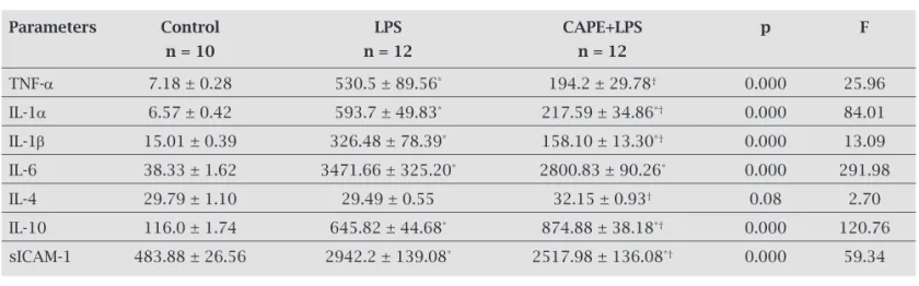
As shown in Table 1, there was significant increase in plasma TNF-α levels in untreated LPS rats in comparison to the control group (530.5 ± 89.56 vs. 7.18 ± 0.28 pg/mL, p = 0.000). CAPE+LPS rats showed significant decrease in their plasmatic TNF-α levels (194.2 ± 29.78 pg/mL) in com-parison to the untreated LPS rats (p = 0.000).
IL-1α
IL-1α levels increased significantly in LPS group reaching a mean value of 593.7 ± 49.83 pg/mL vs. 6.57 ± 0.42 pg/mL in the control group (p = 0.000). CAPE treatment caused marked significant decrease in IL-1α level to 217.59 ± 34.86 pg/mL in comparison to non-CAPE treated LPS rats (p = 0.000) (Table 1).
IL-1β
Plasma IL-1β increased significantly in LPS group in comparison to the control group (326.48 ± 78.39 vs. 15.01 ± 0.39 pg/mL, p = 0.000). CAPE treatment for two weeks
Table 1. Plasma cytokines and soluble intercellular adhesion molecule (sICAM-1) in all studied groups
Parameters Control LPS CAPE+LPS p F
n = 10 n = 12 n = 12
TNF-a 7.18 ± 0.28 530.5 ± 89.56* 194.2 ± 29.78† 0.000 25.96
IL-1a 6.57 ± 0.42 593.7 ± 49.83* 217.59 ± 34.86*† 0.000 84.01
IL-1b 15.01 ± 0.39 326.48 ± 78.39* 158.10 ± 13.30*† 0.000 13.09
IL-6 38.33 ± 1.62 3471.66 ± 325.20* 2800.83 ± 90.26* 0.000 291.98
IL-4 29.79 ± 1.10 29.49 ± 0.55 32.15 ± 0.93† 0.08 2.70
IL-10 116.0 ± 1.74 645.82 ± 44.68* 874.88 ± 38.18*† 0.000 120.76
sICAM-1 483.88 ± 26.56 2942.2 ± 139.08* 2517.98 ± 136.08*† 0.000 59.34
All data are presented as mean ± S.E.M. Results were considered significant when p < 0.05, LPS; acute septic shock group
receiving lipopolysacharide (LPS) 20 mg/kg (from Escherichea coli ,serotype O55:B5, Sigma-Aldrich) dissolved in PBS, and
D-galactosamine (GalN) 250 mg/kg, CAPE+LPS; septic shock group treated with caffeic acid phenethylester (CAPE) 10 μmol/kg.
* p < 0.05 versus control group;
significantly decreased plasma IL-1β levels in the CAPE+LPS group in comparison to the untreated LPS group with a mean value of 158.10 ± 13.3 pg/mL (p = 0.015) (Table 1).
IL-6
Untreated LPS rats showed tremendous increase in their plasma levels of IL-6 with a mean value of 3471.66 ± 325.20 pg/mL that was significantly higher than the control value (38.33 ± 1.62 pg/mL, p = 0.000). CAPE treatment decreased IL-6 level in the CAPE+LPS group to 2800.83 ± 90.26 pg/mL that was significantly lower than that of the non-CAPE treated LPS group (p = 0.004) (Table 1).
Anti-inflammatory cytokines
IL-4
Although IL-4 levels did not change significantly in LPS group in comparison to the control group (29.49 ± 0.55 vs. 29.79 ± 1.10 pg/mL, p = 0.08). CAPE treatment was effective in causing a significant increase in IL-4 levels (32.15 ± 0.93 pg/mL) in CAPE+LPS rats in compari-son to LPS group (p = 0.039), but this increase in IL-4 in CAPE+LPS group showed no significant difference from the control group (p = 0.07) (Table 1).
IL-10
IL-10 increased significantly in LPS group (645.82 ± 44.68 pg/mL) in comparison to the control rats (116.0 ± 1.74 pg/mL, p = 0.000). CAPE treatment caused further and significant increase in IL-10 level in CAPE+LPS group (874.88 ± 38.18 pg/mL, p = 0.000) in comparison to LPS group (Table 1).
Figure 1: 40X image of liver of control (left), untreated acute septic shock (middle) showing destruction of the hepatic lobules with marked infiltration by neutrophilic granulocytes, obstruction of sinusoids, endothelial cell damage and indistinct boundaries between the nucleus and cytoplasm, so much hemorrhage, hepatocyte necrosis. Acute septic shock group receiving caffeic acid phenethylester (right) showing normal hepatic lobules, hepatocytes are normal in shape, clearly visible margins between hepatocytes nucleus and cytoplasm, no apparent hemorrhage nor inflammatory cellular infiltration.
Figure 2: 40X image of brain tissue of control group (left), untreated acute septic shock group (middle) showing lymphocytic infiltration and swollen astrocytes and early neuron injury, while brain of the caffeic acid phenethyle ester treated septic shock group (right) normal astrocytes along the tracts, normal neurons structure, and absent inflammatory cellular infiltration.
sICAM-1
Soluble ICAM-1 levels increased significantly in LPS group reaching 2942.2 ± 139.08 pg/mL versus control value (483.88 ± 26.56 pg/mL). CAPE treated group showed signif-icantly lower levels of sICAM-1 (2517.98 ± 136.08 pg/mL) than LPS group (p = 0.007) (Table 1).
Histopathology
which may be a manifestation of brain edema. CAPE treat-ment preserved the histological picture of the brain with absent inflammatory cellular infiltration and intracellular edema (Figure 2, right).The brain tissue of the CAPE treat-ed rats looks almost like the control brain.
DISCUSSION
Septic shock is a complex pathophysiological process where a plethora of biologically active molecules play a role in its development.9 Therefore, therapeutic strategies directed
against a specific biomarker were not successful in con-trolling the problem, and the mortality rate remains unac-ceptably high. This raises the need for a therapeutic agent with multiple physiological and pharmacological activities. Pathogenic bacteria and their products, such as LPS, trigger the activation of NF-κB, which plays a central role in the in-duction of networks between cytokines and the inflammatory mediators, leading to the pathophysiology of septic shock.21
During inflammatory stimulation, translocation of NF-κB from the cytosol into the nuclei of cells induces the expres-sion of a large number of genes involved in inflammation; including those encoding cytokines (IL-1, IL-6 and TNF-α), adhesion molecules, acute phase proteins and enzymes such as NOS.22 Pro-inflammatory cytokines and
inflam-matory mediators stimulate increased production of Cox-2 in macrophages and endothelial cells which contribute to the edema and vasodilatation at the inflammation site.8
Activa-tion of NF-κB and its consequences occurs in all organs caus-ing organ damage that eventually lead to organ failure with increased morbidity and mortality of patients.22 This makes
NF-κB an attractive therapeutic target for the pharmacologi-cal control of endotoxemia. Among the multiple beneficial ef-fects reported for the natural honeybee propolis, CAPE – the active derivative of propolis – has an inhibitory effect on NF-κB activation with anti-inflammatory17 and antioxidant
activ-ities.15 This encourages us to investigate the potential
protec-tive effect of CAPE in the complex problem of septic shock with specific emphasis on its effect on the pro-inflammatory and anti-inflammatory cytokines production and the patho-logical changes induced by LPS/GalN in the liver and brain tissues during the process of endotoxemia.
Findings of the present study reveal marked increase in the pro-inflammatory cytokines IL-1α, IL-1β, IL-6 and TNF-α levels in animals with LPS endotoxemia. This coin-cides with death of 30% of the animals within six hours of LPS injection. This could be due to the circulatory collapse and organ hypoperfusion and damage associated with the acute endotoxemia.2 Locally, the liver was very much
con-gested and dark in color (with histological picture of hepatic necrosis, intra-hepatic hemorrhage and marked hepatocel-lular infiltration by neutrophils and macrophages). This could be explained by the ability of LPS and D-GalN to trigger activation of the immune cells as PMNL, monocytes
and Kupffer cells causing infiltration of the damaged liver with these cells. Activated macrophages send fluxes of cy-tokines, chemokines , superoxide [O(2)-], NO, hypochloric acid (HOCL), and hydrogen peroxide (H2O2) into the ECF
that induce oxidative stress and cause more cellular injury.8,23
Released pro-inflammatory cytokines subsequently induce inflammatory changes and release vasoconstrictive media-tors as endothlin-1 and thromboxane causing necrosis of endothelial cells with subsequent fibrin deposition in the si-nusoids.24 In contrast to healthy organs endotoxin perfused
liver cannot adapt to reduced portal blood flow by a com-pensatory increase in arterial perfusion, consequently, de-creased blood flow impairs the function of the hepatocytes and Kupffer cells.25
Increased plasma levels of the inflammatory cytokines IL-1α, IL-1β, IL-6, and TNF-α detected in the LPS group in the present study, reveals a picture of systemic inflammatory reaction. In addition, IL-10 which is known to have both in-flammatory and anti-inin-flammatory action26 was also found
to be elevated in the LPS group of animals in the current study. This elevation of IL-10 could be part of the SIR picture, but it may act as compensatory mechanism to inflammation, through its anti-inflammatory action. CAPE treatment was found to be associated with significant increase in IL-10 and IL-4 levels in the treated group. This may be explained by the potential anti-inflammatory effect of CAPE which decreases the release of the inflammatory cytokines from the inflam-matory cells and in the same time stimulates increased production of anti-inflammatory cytokines like IL-10 and IL-4. Inflammatory cellular infiltration of the hepatic tissue was reported to be associated with Fas/FasL pathway acti-vation with subsequent actiacti-vation of caspase-3 and 8 lead-ing to apoptosis of the liver cells.27 Adhesion molecules like
ICAM-1 regulate the interaction between T-lymphocytes and hepatocytes and this relation is deregulated in harmful inflammatory processes.28 Increased sICAM-1 levels were
verified in the plasma of the non-CAPE treated LPS group in the current study. The elevation of sICAM-1 was reported to trigger the extravasations of neutrophils in the liver paren-chyma producing more cytotoxic damage to hepatocytes.28
This together with accumulated intracellular metabolic end products functionally overloads the residual liver, impairs DNA synthesis and may inhibit liver regeneration.29 LPS
induced hepatotoxicity ruins liver function manifested by elevated liver transaminases and bilirubin levels and abnor-mal blood coagulation tests.21 Use of CAPE treatment in the
malondial-dehyde (MDA) and ROS was reported in ischemic injury16
at a concentration of 10 µmol/kg (similar to that used in our study), and was suggested to underlie the preventive effect of CAPE against these injuries. CAPE treatment also reduced the inflammation and lung damage induced by LPS in rats.29
ROS promote activities of pro-inflammatory redox sensi-tive nuclear factors, including NF-κB which is detected in various cell types and has been determined to have a critical function in immune and inflammatory responses.30,31
Acti-vation of NF-κB is associated with increased expression of matrix metaloprotinase-9 (MMP-9), IL-1β, and IL-8 that in turn enhances NF-κB activation in different cell systems.32
CAPE, being a specific inhibitor of the activation of NF-κB,17
blocked IL-1β-induced NF−κΒ promoter activation and was recently reported to be a potent inhibitor of NF-κB activity in several pathologies that were attributed to increased oxi-dative stress and neutrophil accumulation.33 These reported
actions of CAPE give us an explanation to the beneficial ef-fects obtained with CAPE treatment in the present study. Our findings clearly demonstrate that CAPE treatment in a dose of 10 µmol/kg for two weeks before the exposure to LPS and a single dose after LPS-induced endotoxemia markedly protect against LPS-induced hepatocellular and neural dam-age in treated rats together with decreased inflammatory cytokines production. This may be through its anti-inflam-matory16 and free radical scavenging properties15 that may
underlie the decreased pro-inflammatory cytokines levels (IL1-α, IL1-β, IL-6) and the TNF-α in the plasma of treated rats. CAPE treatment also decreased plasma sICAM-1 lev-els. This together with decreased inflammatory cellular infil-tration into the hepatic tissue as seen during the histological examination may partially contribute to the reduced hepatic necrosis and the decreased production of the inflammatory cytokines. These findings support previous reports about the beneficial effects of CAPE against other types of toxic induced liver failure19,20 that was attributed to the very
po-tent antioxidant activity of its major ingredients. Therefore, the antioxidant, anti-inflammatory and NF-κB inhibi-tory actions of CAPE could explain our finding of the protective effect of CAPE against LPS/D-GalN induced hepatocellular damage. CAPE treatment significantly reduced the neutrophilc infiltration and intrahepatic hemorrhagic and preserved architecture of the hepat-ic lobules in treated rats in the current study. This is consistent with previous reports that CAPE can inhibit the caspases 3 and 8 activities by inhibiting the Fas/FasL protein expression19 and, can protect against
Fas-medi-ated cell apoptosis.34 Neuronal cell damage induced by
LPS/GalN in the current study was found to be totally prevented in the CAPE+LPS group which showed pre-served astrocytes histological appearance with absent in-flammatory cellular infiltration, with no signs of edema or cytoplasmic swelling. The preventive effect of CAPE on the
inflammatory cellular infiltration into the hepatic tissue and brain could be directly attributed to its inhibitory effect on NF-κB activation. Decreased infiltration of the hepatic and brain tissue by inflammatory cells is expected to be a leading step to decreased pro-inflammatory cytokines production and other molecules like, ROS, NO and PGE2 that enhance the pathologic features of inflammation vice vasodilatation and edema.8,13
CONCLUSION
The ability of CAPE to alleviate the SIR, hepatic and neu-ronal cell damage induced by LPS/D-GalN could be attrib-uted to the antioxidant activity and/or inhibition of free rad-ical generation and the anti-inflammatory action of CAPE. This effect of CAPE was mainly through reversing the im-balance of the pro- and anti-inflammatory cytokines, which may lead to the inhibition of adhesion molecules expression. These results suggest that; the anti-inflammatory action of CAPE, the antioxidant effect, and its ability to inhibit NF-κB production; are the possible protective mechanisms. CAPE is a promising agent that could help in the prophylaxis and treatment of septic shock.
ACKNOWLEDGEMENTS
The research team is grateful to the College of Medicine Re-search Center (CMRC) for supporting this reRe-search by the grant number 07-580. We are also appreciative of the tech-nicians in the Physiology and Pathology Departments and the Animal Care Unit in King Khalid University for their expertise and support in accomplishing the practical part of this work.
REFERENCES
1. Martin GS, Mannino DM, Eaton S, Moss M. The epidemiology of sepsis in the United States from 1979 through 2000. N Engl J Med 2003; 348(16):1546-54.
2. Shapiro N, Howell MD, Bates DW, Angus DC, Ngo L, Talmor D. The association of sepsis syndrome and organ dysfunction with mortality in emergency department patients with sus-pected infection. Ann Emerg Med 2006; 48(5):583-90. 3. Morikawa A, Sugiyama T, KatoY, Koide N, Jiang GZ, Takahashi
K, Tamada Y, Yokochi T. Apoptotic cell death in the response of D-galactosamine-sensitized mice to lipopolysaccharide as an experimental endotoxic shock model. Infect Immun 1996; 64:737-8.
4. Galanos C, Freudenberg MA, Reutter W. Galactosamine-in-duced sensitization to the lethal effects of endotoxin. Proc Natl Acad Sci USA 1979; 76:5939-43.
5. Decker K, Keppler D. Galactosamine hepatitis: key role of the nucleotide deficiency period in the pathogenesis of cell injury and cell death. Rev Physiol Biochem Pharmacol 1974; 71:77- 106.
7. Hanada T, Yoshimura A. Regulation of cytokine signaling and inflammation. Cytokine Growth Factor Rev 2003; 13:413-21. 8. Mitchell JA, Larkin S, Williams TJ. Cyclooxygenase 2
regu-lation and relevance in inflammation. Biochem Pharmacol 1995; 50:1535-42.
9. Punyadeera C, Schneider EM, Schaffer D et al. A biomarker panel to discriminate between systemic inflammatory re-sponse syndrome and sepsis and sepsios severity. J Emerg Trauma Shock 2010; 3(10):26-35.
10. Gutteridge JMC, Mitchell J. Redox imbalance in the critically ill. Br Med Bull 1999;55(1):49-75.
11. Mercer-Jones MA, Heinzelmann M, Peyton JC, Wickel D, Cook M, Cheadle WG. Inhibition of neutrophil migration at the site of infection increases remote organ neutrophil seques-tration and injury. Shock 1997; 8(3):193-9.
12. Nathan C. Neutrophils and immunity: challenges and oppor-tunities. Nat Rev Immunol 2006; 6:173-82.
13. Thiemermann C. Nitric oxide and septic shock. Gen Pharma-col 1997; 29:159-66.
14. Nagai T, Inoue R, Inoue H, Suzuki N. Preparations and an-tioxidant properties of water extract of propolis. Food Chem 2003; 80:29-33.
15. Russo, Long, R, Vanella A. Antioxidant activity of propolis: role of caffeic acid phenethylester and galangin. Fitoterapia 2002; 73(Suppl1):S21-S29.
16. Yildiz Y, Serter M, Ek RO et al. Protective effects of caffeic acid phenethyl ester on intestinal ischemia-reperfusion injury. Dig Dis Sci 2009; 54(4):738-44.
17. Natarajan K, Singh S, Burke Jr. TR, Grunberger D, Aggarwal BB. Caffeic acid phenethyl ester is a potent and specific inhibi-tor of activation of nuclear transcription facinhibi-tor NF-kappa B. Proc Natl Acad Sci USA 1996; 93:9090-5.
18. Makarov SS, NF-κB as a therapeutic target in chronic inflam-mation: recent advances. Mol Med Today 2000; 6:441-8. 19. Lee KJ, Choi JH, Khanal T, Hwang YP, Chung YC, Jeong HG.
Protective effect of caffeic acid phenethyl ester against car-bon tetrachloride-induced hepatotoxicity in mice. Toxicology 2008; 248:18-24.
20. Iraz M, Ozerol E, Gulec M et al. Protective effect of caffeic acid phenethyl ester (CAPE) administration on cisplatin-induced oxidative damage to liver in rat. Cell Biochem Funct 2006; 24(4):357-61.
21. Korish AA. Effect of caffeic acid phenethyl ester on the hemo-static alterations associated with toxic-induced acute liver fail-ure. Blood Coag Fibrinol 2010; 21:158-63.
22. Liu SF, Malik AB, NF-κB activation as a pathological mecha-nism of septic shock and inflammation. Am J Physiol 2006; 290: L622-L45.
23. Victor VM, Espulgues JV, Hernández-Mijares A, Rocha M. Oxidative stress and mitochondrial dysfunction in sepsis: a potential therapy with mitochondria-targeted antioxidants. Infect Disord Drug Targets 2009; 9(4):376-89.
24. Yachida S, Kokudo Y, Wakabayashi H, Maeba T, Kaneda K, Maeta H. Morphological and functional alterations to sinu-soidal endothelial cells in early phase endotoxin-induced liver failure after partial hepatectomy in rats. Virchows Arch 1998; 433:173-81.
25. Secchi A, Ortanderl JM, Schmidt W, Gebhard MM, Martin E, Schmidt H. Effect of endotoximea on hepatic portal and sinusoidal blood flow in rats. J Surg Res 2000; 89:26-30. 26. Pils MC, Pisano F, Fasnacht N et al.
Monocytes/macrophag-es and/or neutrophils are the target of IL-10 in the LPS en-dotoxemia model. Eur J Immunol 2010; 40(2):443-8. 27. Doughty L, Clark RS, Kaplan SS, Sasser H, Carcillo J. sFas
and sFas ligand and pediatric sepsis-induced multiple or-gan failure syndrome. Pediatr Res 2002; 52(6):922-7. 28. Essani NA, Fisher MA, Farhood A, Manning AM, Smith
CW, Jaeschke H. Cytokine-induced upregulation of he-patic intercellular adhesion molecule-1messenger RNA expression and its role in the pathophysiology of murine endotoxin shock and acute liver failure. Hepatology 1995; 21:1632-9.
29. Antoniades CG, Berry PA, Wendon JA, Vergani D. The importance of immune dysfunction in determining out-come in acute liver failure. J Hepatol 2008; 49(5):845-61. 30. Koksel O, Ozdulger A, Tamer L et al. Effects of caffeic acid
phenethyl ester on lipopolysaccharide-induced lung in-jury in rats Pulm Pharmacol Ther 2006; 19(2):90-5. 31. Henderson Jr. WR, Chi EY, Teo JL, Nguyen C, Kahn M. A
small molecule inhibitor of redox-regulated NF kappa B and activator protein-1 transcription blocks allergic air-way inflammation in a mouse asthma model. J Immunol 2002; 169:5294-9.
32. Barnes PJ. Nuclear factor-kappa B. Int J Biochem Cell Biol 1997; 29:867-70.
33. Ozyurt H, Ozyurt B, Koca K, Ozgocmen S. Caffeic acid phenethyl ester (CAPE) protects rat skeletal muscle against ischemia-reperfusion-induced oxidative stress. Vascul Pharmacol 2007; 47(2-3):108-12.